Journal of Investigative Medicine High Impact Case Reports
October-December 2014: 1 –4 © 2014 American Federation for Medical Research
DOI: 10.1177/2324709614560907 hic.sagepub.com
Article
Introduction
Despite a decade of intense public education and medical advancement, stroke continues to represent a leading cause of long-term disability and death.1 The cost burden associated with ischemic stroke is also substantial compared with other diseases.2,3 According to multiple clinical studies, the cardio-embolic subtype of ischemic event has the most unfavorable prognosis, being associated with an increased risk of death or stroke recurrence, less favorable clinical outcomes including the risk of bleeding, and therapeutic benefits compared with other subtypes.4-6 Thus, it is critical for ischemic stroke patients to be rapidly evaluated and considered for possible cardioembolic sources to guide therapy as well as to reduce the physical and financial burden of poststroke manage-ment.7,8 Currently, transesophageal echocardiography (TEE) is the gold standard to diagnose cardiac thrombi in stroke and transient ischemic attack patients with undetermined cause.9,10 We present an evidence-based case of cerebral embolism when TEE did not show any cardiac source thrombus. Further investigation for cardioembolic stroke was initiated due to a positive risk marker, the increased plasma D-dimer level.
Case Report
A 79-year-old female presented for routine follow-up in our cardiology clinic. She had a 20-year history of an ostium
secundum atrial septal defect (ASD) for which she refused closure and had a prior cerebrovascular accident without sequel. The patient also had a history of hypertension and dyslipidemia. The patient reported having an episode of right upper limb weakness 6 days previously. The symptoms lasted for 20 minutes before completely resolving. On examination, her heart rate was 52/min, blood pressure 118/60 mm Hg, and the chest was clear to auscultation. The glucose level was 80 mg/dL. The patient did have a hyper-coagulability workup as follows: protein C activity (113%), protein S activity (124%), fibrinogen (313 mg/dL), ATIII (85%), APC ratio (3.0). Cardiac exam revealed fixed split-ting of the second heart sound. Her neurological exam was normal. A 12-lead electrocardiogram (EKG) showed minor anterolateral T-wave changes, less pronounced than on pre-vious EKG 6 months ago. There was no history or echocar-diographic findings to suspect valvular vegetation. Initially 560907HICXXX10.1177/2324709614560907Journal of Investigative Medicine High Impact Case ReportsSazonova et al
research-article2014
1
Georgia Regents University, Augusta, GA, USA
Corresponding Authors:
Irina Y. Sazonova.
Email: [email protected]; [email protected]
Vincent J. B. Robinson, Section of Cardiology, Department of Medicine, Georgia Regents University, 1120 15th Street (BBR6518), Augusta, GA 30912, USA.
Email: [email protected]
Embolic Stroke Diagnosed by Elevated
D-Dimer in a Patient With Negative TEE
for Cardioembolic Source
Irina Y. Sazonova, PhD
1, Roja Pondicherry-Harish, MD
1,
Nikhil Kadle, BS
1, Gyanendra K. Sharma, MD
1, Ramon E. Figueroa, MD
1,
and Vincent J. B. Robinson, MD
1Abstract
We report a case of cerebrovascular accident with thromboembolic stroke etiology in a patient who had atrial flutter and negative transesophageal echocardiography (TEE) results. The increased D-dimer levels (1877 ng/mL) initiated referral for magnetic resonance imaging and magnetic resonance angiography of the brain that showed classic recanalization of an embolic thrombus in the angular branch of the left middle cerebral distribution. The D-dimer level of this patient was normalized after 3 months of anticoagulation therapy. Although TEE is considered the gold standard for evaluation of cardiac source of embolism, exclusion of intracardiac thrombus with TEE alone does not eliminate the risk of thromboembolic events. This case highlights the utility of D-dimer as a potential adjunct in the decision-making process to guide investigation of thromboembolism, determine subsequent therapy, and hence reduce the risk of embolic stroke recurrence.
Keywords
2 Journal of Investigative Medicine High Impact Case Reports
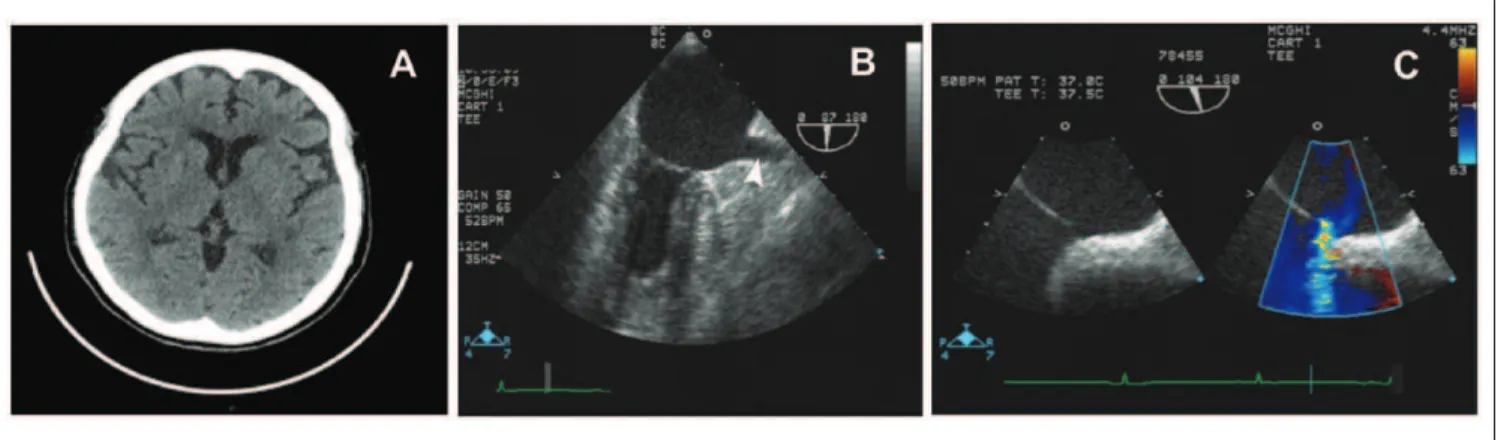
the patient was diagnosed with a transient ischemic attack. The computed tomography (CT) of the head without con-trast found no evidence of an acute intracranial process (Figure 1A). CT angiography of chest/pelvis/abdomen found no evidence of pulmonary embolus. A TEE was per-formed and showed no evidence of thrombus in the heart including the left atrial appendage (Figure 1B), no valvular vegetations, normal left ventricular function, and a small secondum ASD (Figure 1C).
The patient had impaired left atrial appendage function (LAAV = 39 cm/s), which is a potential risk for thromboem-bolism.11 The carotid duplex examination showed minimal plaque at both bifurcations but was otherwise unremarkable. Her D-dimer level was elevated with a critical value of 1877 ng/mL (reference range <250 ng/mL). A venous Doppler of lower extremities revealed no evidence of deep venous
thrombosis, and likelihood of deep vein thrombosis was hence significantly reduced. In view of the high thromboem-bolic risk (significantly elevated D-dimer level and impaired LAAV), the patient was referred to magnetic resonance imaging (MRI) and magnetic resonance angiography (MRA) of her head. The MRI showed a small acute left motor cortex infarct with spotty subcortical white matter ischemic lesions suggesting hypoperfusion injury or small embolic lesion with rapid fragmentation of thrombus, consistent with the patient’s presentation (Figure 2A). The MRA further sug-gested the presence of thromboembolism by identifying irregular segmental narrowing of the angular branch of the left middle cerebral artery (MCA) consistent with thrombus recanalization with either spontaneous fragmentation or thrombolyzed thrombus fragmentation (Figure 2B). Based on MRA findings and positive D-dimer level, the patient was Figure 1. (A) CT head without contrast found no evidence of acute intracranial process such as thrombus, hemorrhage, mass effect, or hydrocephalus. (B, C) TEE images showed no evidence of thrombus in the left atrial appendage nor valvular vegetations. It showed a normal left ventricular function with ejection fraction of greater than 55% and secondum ASD (8.2 mm size with qp:qs ratio of 1.6:1).
Sazonova et al 3
reclassified with cardioembolic stroke, most likely paradoxi-cal emboli via her ASD. As a result of the high cardioembolic risk and CHADS 3 status, the patient was started on Dabigatran therapy. Event monitoring for 30 days was requested by the neurology consultant. After percutaneous closure of her atrial septal defect, the event monitor results were obtained, showing no pacemaker requiring bradycardia with heart rates in excess of low 40s. However, she did have episodes of atrial flutter with controlled ventricular response and hence had to be continued on oral anticoagulation. After 3 months of anticoagulant therapy, the level of her plasma D-dimer was normalized to 218 ng/mL. Thus, the elevated D-dimer level and the impaired LAAV expedited the diagno-sis of cardioembolic etiology of stroke and the implementa-tion of appropriate management.
Discussion
Lack of conclusive findings to suggest cardioembolic source on negative TEE, hematology tests, or other routine diagnos-tics generally results in delaying anticoagulation therapy or stopping further workup. Although a negative TEE obviates the need for prolonged anticoagulation prior to cardiover-sion, it does not eliminate the presence of intracardiac thrombi that may generate stroke in about 1% of patients.10,12 Considering a 15% risk of cardioembolic stroke recurrence, this case underlines the diagnostic challenge in a cost- and time-benefit algorithm of stroke classification. Prothrombotic markers, including D-dimer, were suggested to assess patients at high risk of thrombogenesis.13,14 Although D-dimer is widely used for pulmonary and deep vein throm-bosis,15-18 it is still not applied in the routine practice for diagnosis of cardiac thrombi. Recent literature suggests D-dimer levels are significantly higher in the cardioembolic group of ischemic stroke than in the atherothrombotic and lacunar groups19,20 and are related to infarction volume and functional outcomes.21 D-dimer has also has been proposed for prediction of subsequent thromboembolic cardiovascular events in atrial fibrillation patients during oral anticoagulant therapy.14,22,23 However, the stratification value of routine D-dimer for acute cardioembolic management remains unclear. Our case describes an emerging role of the cost-effective D-dimer test in combination with cardiac morpho-logical data in evaluating the cardioembolic etiology of stroke. This presents a unique example of how thromboem-bolic risk may be underestimated in patients who undergo TEE with negative results10 and how to develop further investigations, treatment, and prevention strategies. Our patient suffered recent neurologic impairment, but TEE showed no evidence of thrombus or vegetation, so a cardiac source of emboli could not be strongly imputed. However, MRA did show irregular segmental narrowing of the left MCA with recanalization, which may be consistent with an embolic-type minor stroke pattern rather than an in situ ath-erothrombotic event. An isolated lesion in a distal segment of
the MCA is unlikely to be atheromatous in the absence of similar disease in more proximal segments. Vasospasm also would be unlikely in the patient clinical setting, especially in the absence of subarachnoid hemorrhage or meningitis (two of the most frequent triggers of vasospasm). The most plau-sible interpretation is, thus, an embolic lesion with thrombus fragmentation, which explains all the findings. The D-dimer test was crucial in pointing us toward the diagnosis of throm-boembolic stroke likely secondary to paradoxical embolism, and allowing the patient to undergo the necessary preventive measures, including the closure of the ASD and anticoagula-tion therapy. It is also important to note that the patient did not have evidence of deep vein thrombosis, as a potential source of the increased D-dimer level. Therefore, it is possi-ble that the cardioembolic subtype of stroke (due to a small cardiac thrombus or already embolized thrombus) is more common than has been previously recognized because of the difficulty of making this complex diagnosis. More clinical experience with D-dimer is needed, in order to reevaluate patients with high risk of cardioembolism and guide further investigations as become necessary. Such an approach could prevent future stroke recurrence, thus decreasing morbidity and associated health care cost. Thus, this case highlights the value of D-dimer and reviewing LAAV data when the TEE is negative to support the additional workup for the diagnosis of stroke etiology.
Authors’ Note
The authors obtained the patient’s permission to study and publish the results.
Declaration of Conflicting Interests
The author(s) declared no potential conflicts of interest with respect to the research, authorship, and/or publication of this article.
Funding
The author(s) disclosed receipt of the following financial support for the research, authorship, and/or publication of this article: The work was supported by the Cardiology Research Program, GRU, and NINDS00072-R21 to IYS, and the Cardiovascular Discovery Institute Award, GRU, to VJBR.
References
1. Lloyd-Jones D, Adams RJ, Brown TM, et al. Executive sum-mary: heart disease and stroke statistics—2010 update: a
report from the American Heart Association. Circulation.
2010;121:948-954.
2. Cohen JW, Krauss NA. Spending and service use among people with the fifteen most costly medical conditions, 1997.
Health Aff (Millwood). 2003;22:129-138.
3. Brown DL, Boden-Albala B, Langa KM, et al. Projected
costs of ischemic stroke in the United States. Neurology.
2006;67:1390-1395.
4 Journal of Investigative Medicine High Impact Case Reports
multicenter clinical trial. TOAST. Trial of Org 10172 in Acute
Stroke Treatment. Stroke. 1993;24:35-41.
5. Paci M, Nannetti L, D’Ippolito P, Lombardi B. Outcomes from ischemic stroke subtypes classified by the Oxfordshire
Community Stroke Project: a systematic review. Eur J Phys
Rehabil Med. 2011;47:19-23.
6. Kirshner HS. Differentiating ischemic stroke subtypes: risk
factors and secondary prevention. J Neurol Sci. 2009;279:
1-8.
7. Dangayach NS, Kane K, Moonis M. Paroxysmal atrial
fibril-lation in cryptogenic stroke. Ther Clin Risk Manag. 2011;7:
33-37.
8. Ionita CC, Xavier AR, Kirmani JF, Dash S, Divani AA, Qureshi AI. What proportion of stroke is not explained by classic risk
factors? Prev Cardiol. 2005;8:41-46.
9. Meenan RT, Saha S, Chou R, et al. Cost-effectiveness of echo-cardiography to identify intracardiac thrombus among patients
with first stroke or transient ischemic attack. Med Decis
Making. 2007;27:161-177.
10. Klein AL, Grimm RA, Murray RD, et al. Use of transesopha-geal echocardiography to guide cardioversion in patients with
atrial fibrillation. N Engl J Med. 2001;344:1411-1420.
11. Sakurai K, Hirai T, Nakagawa K, et al. Left atrial appendage function and abnormal hypercoagulability in patients with
atrial flutter. Chest. 2003;124:1670-1674.
12. Fatkin D, Kelly RP, Feneley MP. Relations between left atrial appendage blood flow velocity, spontaneous
echocardio-graphic contrast and thromboembolic risk in vivo. J Am Coll
Cardiol. 1994;23:961-969.
13. Lip GY, Lowe GD, Rumley A, Dunn FG. Increased markers of thrombogenesis in chronic atrial fibrillation: effects of warfarin
treatment. Br Heart J. 1995;73:527-533.
14. Sadanaga T, Sadanaga M, Ogawa S. Evidence that D-dimer levels predict subsequent thromboembolic and cardiovascular
events in patients with atrial fibrillation during oral
anticoagu-lant therapy. J Am Coll Cardiol. 2010;55:2225-2231.
15. Stein PD, Hull RD, Patel KC, et al. D-dimer for the exclusion of acute venous thrombosis and pulmonary embolism: a
sys-tematic review. Ann Intern Med. 2004;140:589-602.
16. Kruip MJ, Leclercq MG, van der Heul C, Prins MH, Büller HR. Diagnostic strategies for excluding pulmonary embolism
in clinical outcome studies. A systematic review. Ann Intern
Med. 2003;138:941-951.
17. Bates SM, Kearon C, Crowther M, et al. A diagnostic strategy involving a quantitative latex D-dimer assay reliably excludes
deep venous thrombosis. Ann Intern Med. 2003;138:787-794.
18. Mohamad A, Tanna S, Sharma G, et al. Utility and cost analysis of the D-dimer assay in diagnosis of pulmo-nary embolism using computerized tomographic
angiog-raphy in the emergency department. J Am Coll Cardiol.
2011;57:E1540-E1540.
19. Montaner J, Perea-Gainza M, Delgado P, et al. Etiologic diag-nosis of ischemic stroke subtypes with plasma biomarkers.
Stroke. 2008;39:2280-2287.
20. García-Berrocoso T, Fernández-Cadenas I, Delgado P, Rosell A, Montaner J. Blood biomarkers in cardioembolic stroke.
Curr Cardiol Rev. 2010;6:194-201.
21. Matsumoto M, Sakaguchi M, Okazaki S, et al. Relationship between plasma (D)-dimer level and cerebral infarction volume
in patients with nonvalvular atrial fibrillation. Cerebrovasc
Dis. 2013;35:64-72.
22. Ageno W, Finazzi S, Steidl L, et al. Plasma measurement of D-dimer levels for the early diagnosis of ischemic stroke
sub-types. Arch Intern Med. 2002;162:2589-2593.
23. Sadanaga T, Kohsaka S, Ogawa S. D-dimer levels in combina-tion with clinical risk factors can effectively predict subsequent thromboembolic events in patients with atrial fibrillation