Specific RNA Interference in
Caenorhabditis
elegans
by Ingested dsRNA Expressed in
Bacillus subtilis
Marco Lezzerini, Koen van de Ven, Martijn Veerman, Stanley Brul, Yelena V. Budovskaya*
Swammerdam Institute for Life Sciences, University of Amsterdam, Amsterdam, The Netherlands
Abstract
In nematodes, genome-wide RNAi-screening has been widely used as a rapid and efficient method to identify genes involved in the aging processes. By far the easiest way of inducing RNA interference (RNAi) inCaenorhabditis elegansis by feedingEscherichia colithat ex-presses specific double stranded RNA (dsRNA) to knockdown translation of targeted mRNAs. However, it has been shown that E. coli is mildly pathogenic toC.elegansand this pathogenicity might influence aging and the accuracy of the RNAi-screening during aging may as well be affected. Here, we describe a novel system that utilizes the non-pathogenic bacteriumBacillus subtilis, to express dsRNA and therefore eliminates the effects of bacte-rial pathogenicity from the genetic analysis of aging.
Introduction
Aging is a fundamental event in all animals and is a major risk factor for most diseases. Several model organisms are currently being used to study the aging process including yeast, fruit flies, nematodes, and mice [1]. Of these, the nematodeC.elegansis one of the most attractive organisms to study longevity.C.elegansnormally has a relatively short lifespan of two weeks, enabling one to rapidly assess the effects of different mutations or treatments on the life expec-tancy. There is no somatic cell division in adult worms and a cell that is inadvertently lost is not replaced by a new cell division. Therefore, aging inC.elegansis entirely post-mitotic, re-flecting the gradual loss of function in somatic cells as they grow old [2].
We know very little about how worms grow old and why they die in old age it is therefore important to understandC.elegansaging at the molecular level. The vast majority of aging re-search inC.elegansis based on finding and characterizing conditions or mutations that affect longevity. The RNAi technology is widely used to identify many aging related mutations on a genome wide scale. During RNAi, double-stranded RNA is introduced intoC.elegans her-maphrodites leading to the rapid and sequence-specific degradation of the targeted endoge-nous mRNA of corresponding sequence [3]. This technique has become an extremely
important tool for studying gene functionin vivo. Initial studies with RNAi showed that injec-tion of dsRNA in any part of the animal resulted in a robust RNAi effect in all tissues, implying OPEN ACCESS
Citation:Lezzerini M, van de Ven K, Veerman M, Brul S, Budovskaya YV (2015) Specific RNA Interference inCaenorhabditis elegansby Ingested dsRNA Expressed inBacillus subtilis. PLoS ONE 10(4): e0124508. doi:10.1371/journal.pone.0124508
Academic Editor:Myon-Hee Lee, East Carolina University, UNITED STATES
Received:October 7, 2014
Accepted:March 15, 2015
Published:April 30, 2015
Copyright:© 2015 Lezzerini et al. This is an open access article distributed under the terms of the
Creative Commons Attribution License, which permits unrestricted use, distribution, and reproduction in any medium, provided the original author and source are credited.
Data Availability Statement:All relevant data are within the paper and its Supporting Information files.
Funding:This research was funded my MacGillavry fellowship awarded to Yelena Budovskaya at the University of Amsterdam, Netherlands. The funder had no role in study design, data collection and analysis, decision to publish, or preparation of the manuscript.
that interference can cross over cellular boundaries. Later studies demonstrated that in addition to injections, RNAi can be performed by soaking worms in a solution of dsRNA, or by feeding RNAse III-deficient bacteria expressing dsRNA toC.elegans[3–5]. All three methods (injec-tion, soaking, and feeding) can be effectively used in large-scale genome-wide studies. Howev-er, because feeding RNAi to the worms is far less labor-intensive and considerably less expensive, performing RNAi by feeding has become the preferred method for conducting ge-nome-wide RNAi screens in aging studies. To date, scientists have identified approximately 300C.elegansmutants that show either extended or shortened lifespan [6]. However, in any organism, includingC.elegans, genetics is not the only factor influencing longevity. A great volume of recent data points towards the importance of the environment and diet in age-regulation.
Historically,C.eleganshas been grown on theE.coliOP50 strain, which is a uracil auxo-troph derivative ofE.coli[7]. Although this strain has been widely used byC.elegans research-ers since the 1970s, it remains poorly characterized. OP50 is considered to be non-pathogenic, however there is evidence of OP50 colonization and blockage of the worm intestine during old age which consequently triggers disruption of the intestinal lumen and eventually the entry of bacteria into adjacent tissues [8–10]. Therefore, we argue thatE.colimight not be the best food source forC.elegans, especially in aging studies. A key question herein is: how closely does a laboratory diet ofE.coliresemble the natural diet of nematodes.
In nature,C.eleganslives predominantly on decomposing plant material where it encoun-ters a wide variety of different microbes, such as Bacilli (B.subtilisand related bacteria), Staph-ylococci, Streptomyces,Micrococcus luteus,Comamonasand manyPseudomonasspecies [11–
13]. Interestingly, Ausubel and colleagues have found that theC.eleganslifespan is strongly af-fected by bacterial pathogens; worms live almost 50% longer when they are grown on a more
“natural”food sources, such asB.subtilis, than when grown onE.coli[14]. However, it is not known whether this survival difference betweenB.subtilisgrownC.elegansandE.colifed worms is related to a difference in pathogenicity of the two types of bacteria inC.elegans. It is also unknown whether pathogenicity in general is a part of the natural aging process, as it is not known if its magnitude changes over time as the organism ages. It could be that bacteria se-crete pathogenic toxins in young adults and that pathogenic damage occurring early on in life limits total lifespan or produces a mild hormesis effect that extends longevity. In this case, bac-terial pathogenicity could play a role in limiting or extending lifespan, but it would not be part of the aging processper se. With this line of reasoning the following critical question arises: How do we distinguish the genetic mutations that extend the lifespan through up-regulation of the immune response, conferring to the organism an advantage against pathogens, from those that regulate longevity by other means unrelated to pathogenicity and the immune response?
We made a first attempt to answer this question by developing a new tool that allows us to useB.subtilisas a food source and as a shuttle organism to express dsRNA for use in RNAi ex-periments. Here, we demonstrate that this system works as robustly asE.colimediated RNAi, and could be easily adapted for genome-wide applications.
Materials and Methods
Bacterial strains, genotypes, and growth conditions
The bacterial strain MC1061 (F-Δ(ara-leu)7697 [araD139]B/rΔ(codB-lacI)3 galK16 galE15
λ-e14-mcrA0 relA1 rpsL150(strR) spoT1 mcrB1 hsdR2(r-m+))[15] was provided by S. Brul
(MBMFS, University of Amsterdam, Netherlands). The DE3 (H115) strain (F- mcrA mcrB IN (rrnD-rrnE)1λ- rnc14::Tn10)[4] was obtained fromCaenorhabditisGenetic Center (CGC,
was obtained from D. Bechhofer (Mount Sinai School of Medicine of New York University, New York, NY, USA). This strain carries spontaneous background mutations that suppress
rncSnull lethality [16].
Plasmids were prepared in theE.coliMC1061 strain. Luria-Bertani (LB) medium was used for cultures ofB.subtilisandE.coli[17]. Antibiotics were used at the following concentrations: 5μg/ml of kanamycin forB.subtilis, and 50μg/ml of kanamycin or 100μg/ml of ampicillin for E.coli.
Plasmid construction
The 1200 bp fragment containing two Pspacpromoter sequences, flanking each side of the 580 bp spacer (random DNA sequence), was synthesizedde novoby BaseClear BV (Leiden, Nether-lands) and subcloned into the pUC57 vector to generate the pYB292 vector. A BlpI-BsiWI fragment of the PspacdrivenrodZgene in the pDG148-rodZ vector was replaced by the BlpI-BsiWI fragment containing the Pspac—spacer- Pspacto generate the pBSR feeding vector as a basis for cloning DNA fragments from genes of interest. Briefly, the 580 bp spacer can be removed byHindIII digestion and replaced by a gene-specific DNA fragment. The dsRNA can be produced in bacteria by transcription with RNA polymerase. pBSR also contains alacI
repressor to ensure suppression of dsRNA expression in the absence of iso- propyl-β -D-thiogalactopyranoside (IPTG), and provides resistance against ampicillin and kanamycin.
The GFP gene fragment was isolated after PCR amplification with genomic DNA from the SD1084C.elegansstrain, using primers5’GGGAAGCTTGATATCGGAGAAGAACTTTT CACTGGA 3’and5’CCCAAGCTTGATATCGGTTGTCTGGTAAAAGGACAGGGCC 3’carrying
a HindIII restriction sites (underlined italics). The PCR product was cloned into theHindIII site of the pBSR vector replacing the spacer with the 750bp GFP DNA sequence to generate the pBSR-GFP vector. Plasmids were transformed into either theE.coliH115 (DE3) or theB. sub-tilisBG322 RNase-III-deficient strains, in which RNA polymerase can be induced by addition of IPTG to a final concentration 1 mM. The sequence of the final plasmid was verified by sequencing.
The fragments corresponding to the full-length sequence of eitherdaf-2orglp-1genes were PCR amplified from cDNA of wild-type (N2/Bristol) worms, using primers: daf-2_f—
GGGTTAACAAGCTTTACTGTTTGAAGACACTCTGCCA; daf-2_r—GGGTTAACAAGCT
TAAACTGTGCTACACGAAAACGAT; glp-1_f—GGGTTAACAAGCTTATTGGACCGGAATGG
TATGA; glp-1_r—GGGTTAACAAGCTTTGGGAGGACAAGAAACATCC. Both fragments were subsequently cloned into pBSR vector as described above. The 1600bp corresponding to unc-62 gene fragment was subcloned as EcoRV-EcoRV fragment obtained from L4440-unc-unc-62 plasmid (Ahringer’s RNAi library) [4]. All constructs were verified by sequencing.
Preparation and use of chemically competent cells
Efficient chemically competent H115 (DE3) and MC1061E.colibacteria were prepared and used as described in [18].
Transformation Procedure
100μl of freshly made (or thawed in ice) competent cells were mixed with up to 10μl (0.7–
1.5μg) of plasmid. Cells were incubated for 30 minutes at 37°C, 250 rpm. 300μl of pre-warmed
LB medium were added and kept the incubation at 37°C, 250 rpm for other 45 minutes. The cells were then plated on LB plates containing 5μg/μl kanamycin and put at 37°C overnight.
C.
elegans
strains and growth conditions
C.elegansstrains were maintained and handled as described previously [7]. The strains used in this study are: N2 (wild typeC.elegansstrain from Bristol); SD1084 (gaIs148[ges-1p::FLAG::
pab-1+sur-5::GFP]); CF1553 (muIs84 [(pAD76)sod-3p::GFP +rol-6]) and TJ356 (zIs356 [daf-16p::daf-16a/b::GFP +rol-6])
Analysis of Lifespan in
C.
elegans
Lifespan analyses were conducted at 15°C or 20°C as previously described [19]. Briefly, worms were synchronized by hypochlorite treatment. At least 70 N2 worms were grown onE.coli
OP50 from L1 until the L4 stage and then transferred, as one-day-old young adults, onto FUdR-NGM plates freshly seeded with different bacterial strains. The population was checked for dead worms approximately every other day during the adulthood and p-values were calcu-lated using the log-rank (Mantel-Cox) method [20] in Prism 6; GraphPad software.
RNA-interference (RNAi) experiments
DE3 or BG322 bacteria transformed with RNAi vectors expressing dsRNA of the genes of in-terest were grown at 37°C in LB with either 100μg/ml ampicillin forE.coli, or 5μg/ml
kanamy-cin forB.subtilis, then seeded onto NGM-ampicillin/kanamycin plates supplemented with 2 mM IPTG. One-day-old young adult worms were added to the plates and GFP fluorescence was measured 24 or 48 hours later.
RNA extraction and qRT-PCR Analysis
The animals were synchronized by hypochlorite treatment. The animals were exposed to RNAi treatment (control/gfpRNAi;E.coli/B.subtilis) for 2 days at 20°C until L4/young adult stage. Worms were harvested and total RNA was extracted using Trizol and Direct-zol RNA Mini-Prep Kit (Zymo Research, Cat #R2052) according to the manufacturer’s instructions. 80 ng of total RNA per reaction were used for quantitative real-time PCR (qRT-PCR) analysis, per-formed in triplicate with Power SYBR Green RNA-to-Ct 1-Step Kit (Applied Biosystems, Fos-ter City, CA, USA, Part #4389986), according to the manufacturer’s instructions. Vector pD4H1 containing mCherry sequence was used to build a standard curve for absolute quantifi-cation. The relative fold change ofdaf-2andglp-1mRNA expression was normalized totba-1
level of expression. The primers used:
mCherry-FwAGGGTTTTAAGTGGGAACGC mCherry-RevGCATAACAGGTCCATCCGAG GFP FwGGAGAAGAACTTTTCACTGGA GFP RevCCGAACTGTTTAAACTTACGT
daf-2FwCTGGTCAGAGAATGGTCAACTG
daf-2RevCACGTAGATGCGGAAAAGTG
glp-1FwGGCTATGGAGGTCCTGACTG
glp-1RevTTCTGCGCATTCATTTTGAG
unc-62RevCAAATTCGTTGAGATCATCTTTG
tba-1FwTCAACACTGCCATCGCCGCC
tba-1RevTCCAAGCGAGACCAGGCTTCAG
RNAi-induced phenotypes assays
unc-62 RNAi[21]: L4 animals fed with control orunc-62(RNAi) were allowed to lay eggs for 24h at 20°C. Next day parents were removed and the plates left for another day at 20°C. Embry-onic lethality was scored by counting unhatched embryos. To check larval lethality, surviving larvae were followed until they died (larval lethal phenotype), or became fertile adults (no effect).
daf-2 RNAi[22]: fertile animals were allowed to lay eggs at 20°C and then the plates were shifted at 27°C. The dauer phenotype was scored 48h later, by treating the animals with a 1% SDS solution for 15–20 minutes and counting the survived animals as dauer larvae.
glp-1 RNAi[23]:L4 animals were allowed to lay eggs for 24h at 20°C and number of progeny at day one was counted.
Imaging
Images of 20–25 live animals anesthetized with 1 mM levamisole, were captured using the Zeiss Axiovert 40CFL microscope equipped with an Axiovert digital camera, and analyzed using ImageJ software. Representative images were assembled using Adobe Illustrator.
Results and Discussion
Bacillus subtilis
strain selection and construction of the RNAi vector
Performing RNAi by feeding requires cloning a DNA fragment corresponding to the gene of interest into a vector for dsRNA expression under an inducible promoter. This vector could then be transformed into an RNase III-deficient strain ofB.subtiliswhere expression of dsRNA can be induced. It has been shown that RNase III deficiency significantly improves the efficiency of RNAi by feeding, likely because the dsRNA fragments produced are more stable in the bacteria [24]. We started by putting together these two important components for RNAi inB.subtilis.
The first step in developing aB.subtilisstrain that is useable for RNAi was to ensure that a vector and eventual dsRNA could be expressed without perturbing host strain viability.B. sub-tiliscontains therncSgene, which encodes the sole endoribonuclease that specifically cleaves dsRNA. This is an essential gene andΔrncSstrains are not non-viable. Fortunately, a few years ago, the D. Bechhofer’s group was able to isolate a rareΔrncSstrain (BG322) that carries spon-taneous background mutations which suppressΔrncSlethality but not the RNase III processing defects of rRNAs or prescRNAs [16]. We wanted to check whether this spontaneous mutation (or the lack of the RNase III gene) would introduce a potential toxic effect that could affect the worm’s lifespan. In this case worms grown on the BG322 strain should live as long as, or at least similarly to, the ones grown on wild type PY79B.subtilisbacteria previously used in lon-gevity studies [25]. We performed a lifespan analysis of the wild typeC.elegansstrain, N2, on the traditionalE.colistrains, OP50 and the strain for RNAi analysis (H115 (DE3)), and on the
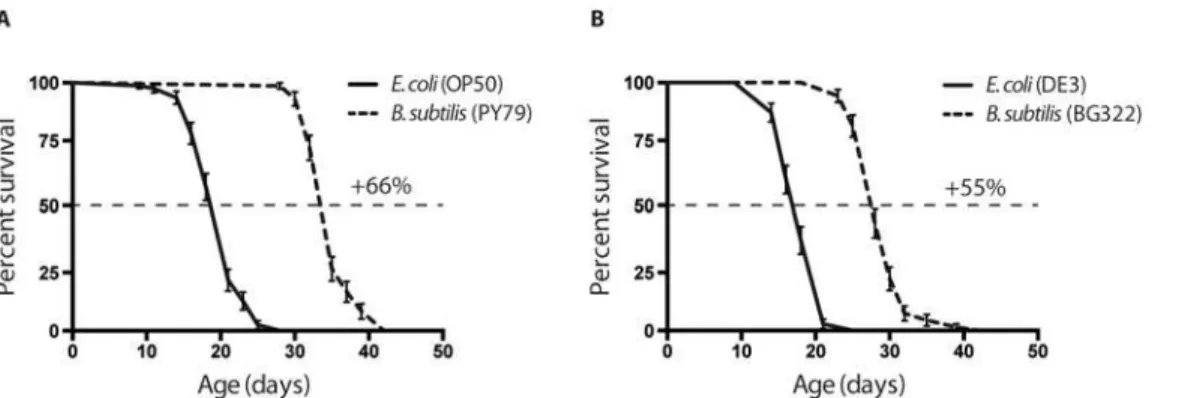
B.subtilisstrains, wild type (PY79) and BG322. Our results show that worms grown onB. sub-tilislive, on average, 50% longer than onE.coli(Fig 1). Both the worms fed on theE.coliand
B.subtilisRNAi strains live slightly shorter than their wild type counter parts, suggesting that the mutation in the RNase III gene might affect bacterial physiology and, as a consequence,
strains have on overall longevity. In addition we perform a wide variety of food switching ex-periments, and found that as soon asC.elegansgrow on non-pathogenic bacteria in adulthood (post-development), they would live longer (S1 FigandS1 Table). Worms grown onE.coli
fromL1to adulthood before being transferred toB.subtilislive as long as worms that lived on
B.subtilisfromL1larvae stage. It implies that developing worms either are not affected by pathogenicity ofE.coli, or manage to efficiently fight the infection.
Second, we designed a vector, pBSR (Bacillus subtilisRNAi), for bidirectional transcription of the desired dsRNAs. This vector has the following properties: (i) It carries two origins of rep-lication and two selection markers: the ColE1 reprep-lication sequence and aβ-lactamase gene (AmpR) for amplification and selection inE.coli; the pUB110 origin and kanamycin resistance gene, for amplification and selection inB.subtilis[26]. (ii) The vector has two bidirectional iso-propyl-β-D-thiogalactopyranoside (IPTG)-inducible Pspacpromoters. The Pspacpromoter is a hybrid of the phage SPO1 promoter and one of the three lac operators, designated“O1”
[27,28]. Separating these two promoters there is a 500 bp spacer, which is flanked by aHindIII and aSalI restriction sites to facilitate the cloning steps. (iii) The vector carries thelacI gene, encoding theE.coli Lacrepressor under control of the penicillinase promoter (Ppen) ofBacillus licheniformisthat promptslacIto be constitutively expressedin B.subtilis[27]. The Pspac pro-moter is repressed bylacIand consequently can only be induced when IPTG is present [27] (Fig 2A).
B. subtilis-induced RNAi is effective in inhibiting a GFP transgene
Next, we evaluated the effectiveness of dsRNA expressed from the pBSR vector to interfere with green fluorescent protein (GFP) transgene expression. For these experiments, we used two GFP-expressing strains: the SD1084, expressing the nuclear localized SUR-5::GFP transla-tional fusion, and the TJ356 strain, expressing the predominantly cytoplasmic DAF-16::GFP translational fusion. Thesur-5gene encodes a protein with a high similarity to theHomo sapi-ensAcetoacetyl-coenzyme A synthetase, and it is broadly expressed inC.elegans[29].daf-16encodes the soleC.elegansforkhead box O (FOXO) homologue that acts in the insulin/IGF-1-mediated signaling (IIS) pathway which regulates development, dauer formation, longevity, fat metabolism, stress response, and innate immunity [30]. When these animals expressing the GFP transgene were fedB.subtilisbacteria expressing dsRNA corresponding to the GFP re-porters [3,31], a decrease in GFP fluorescence was observed in about 90% of the worms in the Fig 1. Effects of variousE.coliandB.subtilisstrains on longevity.(A) Worms fed on wild typeB.subtilisstrain (PY79) live 65% longer compared to wild type, standard laboratory foodE.colistrain (OP50). (B) Worms fed on RNase III-nullB.subtilis(BG322) strain live 55% longer compared to DE3,E.colistrain used for expressing stable dsRNA for RNA interference. Median survival on: OP50 = 21 days, PY79 = 35 days DE3 = 18 days, BG322 = 28 days. Age refers to days of adulthood. Three biological replicates were observed for each experiment (n = 70–80 worms per experiment); error bars indicate Standard Error. In both graphs p<0.0001.
population (Fig2B–2D, andS2A Fig). GFP expression levels were reduced to 3% of the original expression level in the DAF-16::GFP worms, and were lowered almost 6 times in SUR-5::GFP animals. As onE.coli,B.subtilisexpressing dsRNA was not able to affect neuronal GFP expres-sion (Fig 2). These results suggest that dsRNA against GFP is produced byB.subtilisand is ca-pable of an effective gene-specific knock-down of GFP expression inC.elegansupon ingestion.
Effect of
B. subtilis
induced RNAi against known regulators of longevity
on DAF-16 nuclear localization
To further validate the effectiveness ofB.subtilisinduced RNAi, we created plasmids designed to produce dsRNAs corresponding to three endogenousC.elegansgenes that were found to be involved in the modulation of longevity.
Our first choice was thedaf-2gene, which encodes the insulin/IGF-1 signaling (IIS) receptor ortholog inC.elegans. Mutant worms with a defectivedaf-2function have been shown to live twice as long when compared to wild type animals [19] and this phenotype largely depends on the function of thedaf-16/FOXO transcription factor: loss-of-function mutations indaf-16
suppress the longevity phenotype ofdaf-2mutants, showing thatdaf-2acts upstream of the
daf-16FOXO transcription factor repressing its activity [32].
The second gene chosen wasglp-1which encodes a Notch family receptor, essential for germ line development and longevity [33]. RNAi inactivation of theglp-1gene leads to a signif-icant lifespan extension, and also this effect is dependent on thedaf-16/FOXO transcription factor [34].
Finally, we chose theunc-62gene.unc-62is an important developmental regulator, and an ortholog of the Drosophila Homothorax gene.unc-62directly binds to the promoter of many Fig 2. Genetic interference following ingestion of anti-GFP dsRNA-expressingB.subtilisbyC.elegans.(A) Physical map of the pBSR vector. The DNA sequence corresponding to dsRNA of interest was cloned between flanking copies of the Pspacpromoter to replace the spacer.B.subtilisstrain BG322 was used as a host. GFP-expressingC.elegansstrains TJ356 (B) and SD1084 (C) were fed on BG322 strains transformed with original pBSR vector and on bacteria expressing dsRNA corresponding to thegfpcoding region. Under these conditions, 95% of the animals showed dramatic decrease in GFP expression after 24 hours of feeding. DAF-16::GFP andSUR-5::GFP expression is significantly decreased in the GFP (RNAi) treated animals. RNAi was induced starting at L4 larvae stage by feeding wormsB.subtilisbacteria expressing dsRNA against GFP. GFP expression was measured at day 2 of adulthood. The y-axis denotes GFP expression (arbitrary units). Average expression and Standard Error from 20 animals are shown.*-p-value<0.001 (t-test p-values). Scale bar = 100μm.
age-regulated genes and in this way modulates lifespan [35]. Althoughunc-62is expressed in diverse tissues, its functions in the intestine play a particularly important role in modulating lifespan, as an intestine-specific knockdown ofunc-62by RNAi increases lifespan by 30% when compared to wild type worms. Even in this case, it has been shown thatunc-62(RNAi) leads to an activation ofdaf-16, which is required for the extended lifespan in the treated worms [35].
Although all of these three genes play a different role in the worm physiology, and are pressed in different tissues, mutation analysis of each of these showed a significant lifespan ex-tension when worms are grown onE.coli; and the long life of these mutants depends on the activity ofdaf-16/FOXO transcription factor. Therefore, we set out to test whether dsRNAs corresponding to each of these genes expressed inB.subtiliscan influencedaf-16and hence
C.eleganslifespan.
Fragments corresponding todaf-2,glp-1, orunc-62were cloned into the pBSR vector (see Materials and Methods). Clones that tested positive by PCR analysis and restriction digestion were transformed into theB.subtilisBG322 strain for further analysis. The efficiency of RNAi-mediated down-regulation of these genes was tested by qRT-PCR and by scoring previously de-scribed phenotypes. First, thedaf-2(RNAi) andglp-1(RNAi) treatments in wild type worms lead to approximately 20 to 40% decrease in the corresponding mRNA levels (S2B Fig). The
daf-2(RNAi) andglp-1(RNAi) treatments in RNAi-sensitive (NL2099) worms lead to approxi-mately 40 to 50% decrease in the corresponding mRNA levels (data not shown). Unfortunately we were unable to detect a significant reduction inunc-62level of expression. Second, we were able to score several known phenotypic effects on the treated animals (S2C Fig)[21–23]. For ex-ample, similar toE.colitreatments,daf-2(RNAi) treatment usingB.subtilisin combination with increased temperature (27°C) forces wild-type worms to enter into the dauer stage and the
glp-1(RNAi) treatment caused mild sterility, whereas theunc-62(RNAi) treatment led to em-bryonic and larval lethal phenotypes. Furthermore, these effects proved to be more prominent when the assays were carried out using the NL2099 strain.
To test the effectiveness of dsRNA againstdaf-2,glp-1, andunc-62in regulation ofdaf-16 lo-calization, we used the reporter strain TJ356, a DAF-16::GFP translational fusion strain. When the IIS pathway is active, DAF-16 is inactive and sequestered in the cytoplasm. However, upon inactivation of insulin signaling, DAF-16 becomes active and translocates to the nucleus. Mul-tiple publications have shown that when worms are fed onE.coli, a down-regulation ofdaf-2
orglp-1leads to DAF-16/FOXO nuclear translocation in various tissues, whereasunc-62
(RNAi) treatment didn’t lead to any nuclear localization of DAF-16/FOXO (Fig3Aand3Band [34–36]). Interestingly, 90% of worms grown on thedaf-2(RNAi)B.subtilisor 83% of worms grown on theglp-1(RNAi)B.subtilisexhibited DAF-16 nuclear localization, but mainly in cells located in the head region (Fig3C–3E,S2D Fig). In contrast, 95.5% of worms grown on thedaf-2(RNAi)E.coliexhibit strong nuclear localization in the head and intestinal cells. When the worms were grown onB.subtilis unc-62(RNAi), we could not see any DAF-16 nu-clear localization, neither in the head, nor in the intestine region (Fig 3F,S2D Fig). One possible explanation for this difference can be that the dsRNA is not optimally ingested by
C.elegansgrown onB.subtilisdsRNA expressing cells because the worm cannot efficiently grind and digest theB.subtilisvegetative cells due to the thick cell wall. Alternatively, the
B.subtilisstrain we used might form spores in our experimental conditions, preventing the worms from digesting these [37].
worms’intestine when they were fedE.coliorB.subtilisvegetative cells, suggesting the com-plete destruction of these bacteria by the worm’s grinder (data not shown; previously shown in [37,38]). Next, we tested theB.subtilisstrain BG322 ability to sporulate on NGM plates, but no spores could be found. In addition to circumvent this possible problem, we transferred the worms every 2–3 days onto fresh dsRNA expressing BG322 strains. However, the questions still remains, whydaf-2(RNAi) orglp-1(RNAi) inB.subtilisstrain is less effective to activate DAF-16 nuclear translocation, then the same clone expressed inE.coli? Based on our observa-tions, we can suggest, thatB.subtilisinduced RNAi causes milder effect on down-regulation of target mRNA levels thanE.coliinduced RNAi treatment. Another possibility is thatB.subtilis
is less pathogenic thanE.coli. Therefore, the knock-down of eitherdaf-2orglp-1gene is not sufficient to drive strong nuclear localization and activation of DAF-16/FOXO. More detailed studies are needed to validate or refute these alternatives.
DAF-16 activity in worms exposed to
B. subtilis
induced RNAi against
longevity regulators
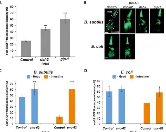
We next analyzed the activity of DAF-16 by measuring the expression ofsod-3gene, a well-known DAF-16 direct target, which encodes one of theC.elegansmanganese-superoxide dis-mutases involved in oxidative stress response and longevity [36,39,40]. To this purpose, we Fig 3. DAF-16/FOXO activated indaf-2(RNAi) andglp-1(RNAi) mutants when grown onE.coliorB.subtilis.Green fluorescent protein images of adult TJ356 (Isdaf-16::GFP) transgenic animals shown at 40X. (A) Images of an animal raised onE.coliexpressing empty vector for RNA interference. (B) Images of an animal grown onE.coliand exposed toE.coliexpressingdaf-2dsRNA for 48 hours post development. Note strong nuclear localization in most tissues, including the intestine. (C) Image of an animal grown onB.subtilisexpressing the empty vector for RNA interference. Images of animals fedB.subtilis expressing either (D)daf-2dsRNA, (E)glp-1dsRNA, or (F)unc-62dsRNA for 48 hours post development. Note weak activation of DAF-16/FOXO in the head area upon treatment with daf-2 and glp-1 dsRNA. Scale bar = 100μm
used the CF1553 strain expressing asod-3::GFP transcriptional fusion. We chose this gene be-cause our studies as well as recent publications have demonstrated that its expression highly correlates withC.eleganslifespan and can be used as a predictor of remaining life in the worm [25,35,41].
WhenC.elegansgrows on eitherB.subtilisorE.colias a food source,sod-3expression de-creases with age (S3 Fig). When young adults expressing thesod-3::GFP transgene were fed
B.subtilisbacteria expressing dsRNA corresponding to eitherdaf-2orglp-1genes, an increase in GFP fluorescence was observed mainly in the head region (pharynx, head hypodermis, and neurons) (Fig4Aand4B). These results are consistent with the DAF-16::GFP nuclear localiza-tion observed in the same region of the worm (Fig3Dand3E).
OnB.subtilis unc-62(RNAi) however, the expression ofsod-3significantly increased in the head and in the intestine region of the worm (Fig 4C). These expression levels remained high throughout theC.eleganslife (S3B Fig), whereas onE.coli unc-62(RNAi), the worms showed an increase insod-3::GFP expression mainly in the intestine (Fig 4D). This is a very puzzling phenotype since we were unable to see any nuclear localization of the DAF-16/FOXO inB. sub-tilis unc-62(RNAi) (Fig 3F). These data suggest that this increased expression ofsod-3may not entirely be dependent on DAF-16 activity. There are two other alternative explanations for this Fig 4. Consequences ofdaf-2(RNAi),glp-1(RNAi), andunc-62(RNAi) onsod-3expression.(A)sod-3::GFP expression is increased indaf-2(RNAi)
andglp-1(RNAi) treated animals. RNAi was induced at L4 larval stage by feeding wormsB.subtilisexpressing dsRNA.sod-3::GFP expression was
measured at day 2 of adulthood. The y-axis denotes GFP expression (arbitrary units). Average expression and Standard Error from 20 animals are shown. (B) Representative pictures of expression ofsod-3::GFP at day 2 of adulthood inunc-62(RNAi),daf-2(RNAi) andglp-1(RNAi) treated animals. The dashed lines indicate areas where fluorescence intensity was quantified to measure GFP intensity in the head or intestine (dashed lines in representative pictures (B) indicate the areas used in quantifications) upon RNAi treatment.sod-3::GFP expression is increased in the intestine ofunc-62(RNAi) treated animals. RNAi was induced starting at L4 larvae stage by feeding worms (C)B.subtilisor "(D)E.colibacteria expressing dsRNA.*-p-value<0.05,**- p-value><0.01, and **-p-value><0.001 (t-test p-values). Scale bar = 50μm.
observation. First, expression of theunc-62in worms grown onB.subtilisis much lower then in worms grown onE.coli. In this case using theB.subtilisas a food source gives as the same benefits asunc-62down-regulation. The second,unc-62is expressed at regular level, but be-causeB.subtilisis non-pathogenic, and does not cause any damage to intestine (see next sec-tion) down regulation of this gene does not provide additional benefit on worm life span. The further analysis ofunc-62expression behavior on different food sources is beyond the scope of this paper. More work is needed to investigate this observation further.
Worms grown on
B. subtilis
accumulate less lipofuscin
Lipofuscin is a fluorescent aggregate of lipids and damaged proteins which accumulates with age and it is often used as a biomarker for aging [42–45]. It was previously demonstrated that accumulation of lipofuscin is accelerated, as the worms grow older [9]. Interestingly, in long-lived mutants lipofuscin accumulates at low rates and has been associated more with physio-logical rather than chronophysio-logical life span [46]. Here, we would like to report a quite intriguing phenotype of lipofuscin accumulation when worms were grown onunc-62(RNAi) expressed in eitherE.coliorB.subtilis.
First, we observed very little lipofuscin accumulation in the gut of the worms grown on
B.subtiliscompared to worms grown onE.coli(S4 Fig). This result corroborates the previous observation thatB.subtiliscould be a healthier diet forC.elegansgrown under laboratory con-ditions. As the intestine is the primary site for the response to a pathogenic infection [8], one possible explanation for this effect could be thatB.subtilisis less pathogenic in this organ and therefore causes less damage to proteins and lipids residing in the worm’s intestine.
Second, we observed a significant reduction in intestinally accumulated lipofuscin inunc-62
(RNAi) mutant worms grown onE.coli(S4 Fig). It has been previously shown, that UNC-62 regulates many intestine specific age-regulated genes, including all six vitellogenin encoding genes [35] that are prone to damage and aggregation with age. In long-livedunc-62(RNAi) mutants grown onE.coli, expression of vitellogenin is reduced, and this might contribute to low overall gut/lipofuscin auto-fluorescence and longer life. OnB.subtilishowever, we ob-served quite the opposite effects of theunc-62(RNAi) knockdown on the lipofuscin accumula-tion in the gut of the worms compare to empty vector control bacteria. These mutant worms show almost seven fold increase in accumulation of gut autofluorescence (S4 Fig). These results show thatunc-62is expressed in worms grown onB.subtilis, and its inactivation leads to in-crease in accumulations of autofluourscent proteins in the gut. Several interesting questions were raised by these observations. Is the lipofuscin composition different when worms are usingB.subtilisas a food source? What kind of gene expression and metabolic changes accom-panyB.subtilispromoted health span extension? More experiments are needed to address these issues. It is entirely possible that we can useB.subtilisandC.elegansto identify a novel mechanism of longevity in worms by examining the beneficial effects of food on organ (intes-tine in this case) physiology.
In summary, we designed a second system that would allow continuous feeding ofC.elegans
In the aging research, this opens up the exiting opportunity to study this process in a less harmful and less pathogenic environmental condition; potentially allowing the discovery of new aging mechanisms went unnoticed before.
Supporting Information
S1 Fig. Analysis ofC.eleganslife span when grown on different bacteria as food source.
Lifespan curves of animals continuously grown on one food source (A-B), or transferred from a food source to another as L4 (C-F). In all the graphs: D = Development (from L1 to L4) A = Adulthood (from L4 on). Age refers to days of adulthood. N = 70–80 worms per experi-ment; error bars indicate Standard Error; all experiments performed at 15°C. For further infor-mation refer also toS1 Table.
(TIF)
S2 Fig. Quantification of RNAi treatment efficiency and induced phenotypes.(A) Absolute
quantification ofgfptranscript in SD1084 animals exposed to RNAi treatment vector for 48h (from L1 to L4/young adults stage at 20°C). (B) Relative quantification ofdaf-2andglp-1
mRNA levels, in N2 animals exposed to RNAi treatment for 48h. Fold change is calculated after normalization to the expression oftba-1gene. Error bars indicate Standard Error of the Mean.= p<0.01;= p<0.05 (t-test p-values). The experiment was conducted on three
bi-ological repeats. (C) Summary table of the RNAi-induced phenotypes.= Assay performed using the NL2099 strain (rrf-3 (pk1426) II), characterized by increased sensitivity to RNAi treatment compered to wt N2 strain. (D) The quantification of DAF-16::GFP nuclear localiza-tion was performed by calculating a percentage of the worms displaying nuclear localizalocaliza-tion of DAF-16::GFP. 0—non nuclear localization in the worm; 1–5—DAF-16::GFP was observed in just a few nuclei (1–5);>10—DAF-16::GFP was observed in more then 10 nuclei per worm.
(TIF)
S3 Fig. Regulation ofsod-3expression byunc-62.Quantification of levels ofsod-3::GFP ex-pression from 20 worms in control andunc-62(RNAi) mutants at three time points during aging. For direct comparison in this time course experiment worms were grown and treated with RNAi expressed in (A)E.coliand (B)B.subtilis. Expression levels were determined in the head and in the intestinal area of the worm by measuring pixel intensity from GFP images. Error bars represent Standard Error of the Mean pixel intensities.-p-value<0.05 and
-p-value<0.001 (t-test p-values).
(TIF)
S4 Fig.unc-62RNAi treatment has a different effect on lipofuscin formation in worms
grown onE.colivs.B.subtilis.(A) Representative photographs of lipofuscin in wild type and
unc-62(RNAi) animals intestines grown onE.coliorB.subtilisat day 3 of adulthood (excita-tion filter 360nm, emission 420nm [47]).unc-62(RNAi) results in decreased autofluorescence when worms are grown onE.colias a food source, whereasunc-62dsRNA treatment of ani-mals grown onB.subtiliscauses induction of gut autofluorescence. (B) Quantification of levels of gut autofluorescence from 20 worms in wild type andunc-62(RNAi) mutants at day 3 of adulthood. Expression levels were determined in the intestinal area of the worm by measuring pixel intensity from CFP images. Error bars represent the Standard Error of the Mean pixel in-tensities.-p-value<0.01 and-p-value<0.001 (t-test p-values). Scale bar = 50μm.
(TIF)
S1 Table. Summary of all the lifespan experiments performed in this study.Median and
information refer also toS1 Fig. # = log-rank (Mantel-Cox) p-values; ^ = RNAi host bacterial strain;= p<0.0001;= p<0.001; ns = not significant.
(DOCX)
Acknowledgments
We thank David Bechhofer for providing BG322 strain ofB.subtilis, without which this project would not be possible. We thank all members of Stanley Brul lab for discussions and comments on the manuscript; the wholeC.eleganscommunity andCaenorhabditisGenetic Center (CGC) funded by NIH office of Research Infrastructure Programs (P40 OD010440) for most of the worm strains used in this study.
Author Contributions
Conceived and designed the experiments: YB ML SB. Performed the experiments: KVDV MV ML. Analyzed the data: MV ML YB. Wrote the paper: ML YB SB.
References
1. Tissenbaum HA, Guarente L (2002) Model organisms as a guide to mammalian aging. Dev Cell 2: 9–19. PMID:11782310
2. Sulston JE, White JG (1980) Regulation and cell autonomy during postembryonic development of Cae-norhabditis elegans. Dev Biol 78: 577–597. Available: PMID:7190941
3. Fire A, Xu S, Montgomery MK, Kostas SA, Driver SE, Mello CC. (1998) Potent and specific genetic in-terference by double-stranded RNA in Caenorhabditis elegans. Nature 391: 806–811. PMID:9486653
4. Kamath RS, Ahringer J (2003) Genome-wide RNAi screening in Caenorhabditis elegans. Methods 30: 313–321. PMID:12828945
5. Timmons L, Fire A (1998) Specific interference by ingested dsRNA. Nature 395: 854. PMID:9804418
6. Smith ED, Tsuchiya M, Fox LA, Dang N, Hu D, Kerr EO, et al. (2008) Quantitative evidence for con-served longevity pathways between divergent eukaryotic species. Genome Res 18: 564–570. doi:10. 1101/gr.074724.107PMID:18340043
7. Brenner S (1974) The genetics of Caenorhabditis elegans. Genetics 77: 71–94. PMID:4366476
8. Garigan D, Hsu A-LL, Fraser AG, Kamath RS, Ahringer J, Kenyon C. (2002) Genetic analysis of tissue aging in Caenorhabditis elegans: a role for heat-shock factor and bacterial proliferation. Genetics 161: 1101–1112. PMID:12136014
9. Herndon LA, Schmeissner PJ, Dudaronek JM, Brown PA, Listner KM, Sakano Y, et al. (2002) Stochas-tic and geneStochas-tic factors influence tissue-specific decline in ageing C. elegans. Nature 419: 808–814. PMID:12397350
10. McGee MD, Weber D, Day N, Vitelli C, Crippen D, Herdon LA, et al. (2011) Loss of intestinal nuclei and intestinal integrity in aging C. elegans. Aging Cell 10: 699–710. doi:10.1111/j.1474-9726.2011.00713. xPMID:21501374
11. MacNeil LT, Watson E, Arda HE, Zhu LJ, Walhout AJM (2013) Diet-induced developmental accelera-tion independent of TOR and insulin in C. elegans. Cell 153: 240–252. doi:10.1016/j.cell.2013.02.049
PMID:23540701
12. Montalvo-Katz S, Huang H, Appel MD, Berg M, Shapira M (2013) Association with soil bacteria en-hances p38-dependent infection resistance in Caenorhabditis elegans. Infect Immun 81: 514–520. doi:
10.1128/IAI.00653-12PMID:23230286
13. Félix M-A, Duveau F (2012) Population dynamics and habitat sharing of natural populations of Caenor-habditis elegans and C. briggsae. BMC Biol 10: 59. doi:10.1186/1741-7007-10-59PMID:22731941
14. Garsin DA, Villanueva JM, Begun J, Kim DH, Sifri CD, Calderwood SB, et al. (2003) Long-lived C. ele-gans daf-2 mutants are resistant to bacterial pathogens. Science (80-) 300: 2003.
15. Casadaban MJ, Cohen SN (1980) Analysis of gene control signals by DNA fusion and cloning in Escherichia coli. J Mol Biol 138: 179–207. PMID:6997493
17. Miller JH (1972) Experiments in Molecular Genetics. Cold Spring Harbor: Cold Spring Harbor Labora-tory Press.
18. Chung CT, Niemela SL, Miller RH (1989) One-step preparation of competent Escherichia coli: transfor-mation and storage of bacterial cells in the same solution. Proc Natl Acad Sci U S A 86: 2172–2175. PMID:2648393
19. Kenyon C, Chang J, Gensch E, Rudner A, Tabtiang R (1993) A C. elegans mutant that lives twice as long as wild type. Nature 366: 461–464. PMID:8247153
20. Lawless JF (1982) Models and Methods for Lifetime Data. Wiley, New York.
21. Van Auken K, Weaver D, Robertson B, Sundaram M, Saldi T, Edgar L, et al. (2002) Roles of the Homothorax/Meis/Prep homolog UNC-62 and the Exd/Pbx homologs CEH-20 and CEH-40 in C. ele-gans embryogenesis. Development 129: 5255–5268. PMID:12399316
22. Dillin A, Crawford DK, Kenyon C (2002) Timing requirements for insulin/IGF-1 signaling in C. elegans. Science (80-) 298: 830–834. PMID:12399591
23. Lehner B, Calixto A, Crombie C, Tischler J, Fortunato A, Chalfie M, et al. (2006) Loss of LIN-35, the Caenorhabditis elegans ortholog of the tumor suppressor p105Rb, results in enhanced RNA interfer-ence. Genome Biol 7: R4. PMID:16507136
24. Timmons L, Court DL, Fire a (2001) Ingestion of bacterially expressed dsRNAs can produce specific and potent genetic interference in Caenorhabditis elegans. Gene 263: 103–112. PMID:11223248
25. Sánchez-Blanco A, Kim SK (2011) Variable Pathogenicity Determines Individual Lifespan in Caenor-habditis elegans. PLoS Genet 7: 14.
26. Joseph P, Fantino JR, Herbaud ML, Denizot F (2001) Rapid orientated cloning in a shuttle vector allow-ing modulated gene expression in Bacillus subtilis. FEMS Microbiol Lett 205: 91–97. PMID:11728721
27. Yansura DG, Henner DJ (1984) Use of the Escherichia coli lac repressor and operator to control gene expression in Bacillus subtilis. Proc Natl Acad Sci U S A 81: 439–443. PMID:6420789
28. Quisel JD, Burkholder WF, Grossman AD (2001) In vivo effects of sporulation kinases on mutant Spo0A proteins in Bacillus subtilis. J Bacteriol 183: 6573–6578. PMID:11673427
29. Gu T, Orita S, Han M (1998) Caenorhabditis elegans SUR-5, a novel but conserved protein, negatively regulates LET-60 Ras activity during vulval induction. Mol Cell Biol 18: 4556–4564. PMID:9671465
30. Gems D, Riddle DL (2000) Genetic, behavioral and environmental determinants of male longevity in Caenorhabditis elegans. Genetics 154: 1597–1610. PMID:10747056
31. Chalfie M, Tu Y, Euskirchen G, Ward W, Prasher D (1994) Green fluorescent protein as a marker for gene expression. Science (80-) 263: 802–805. PMID:8303295
32. Paradis S, Ruvkun G (1998) Caenorhabditis elegans Akt/PKB transduces insulin receptor-like signals from AGE-1 PI3 kinase to the DAF-16 transcription factor. Genes Dev 12: 2488–2498. PMID:9716402
33. Arantes-Oliveira N, Apfeld J, Dillin A, Kenyon C (2002) Regulation of life-span by germ-line stem cells in Caenorhabditis elegans. Science (80-) 295: 502–505. PMID:11799246
34. Curran SP, Ruvkun G (2007) Lifespan regulation by evolutionarily conserved genes essential for viabili-ty. PLoS Genet 3: e56. PMID:17411345
35. Van Nostrand EL, Sánchez-Blanco A, Wu B, Nguyen A, Kim SK (2013) Roles of the developmental reg-ulator unc-62/Homothorax in limiting longevity in Caenorhabditis elegans. PLoS Genet 9: e1003325. doi:10.1371/journal.pgen.1003325PMID:23468654
36. McCormick M, Chen K, Ramaswamy P, Kenyon C (2012) New genes that extend Caenorhabditis ele-gans’lifespan in response to reproductive signals. Aging Cell 11: 192–202. doi:10.1111/j.1474-9726. 2011.00768.xPMID:22081913
37. Laaberki M- H, Dworkin J (2008) Role of spore coat proteins in the resistance of Bacillus subtilis spores to Caenorhabditis elegans predation. J Bacteriol 190: 6197–6203. doi:10.1128/JB.00623-08PMID:
18586932
38. Laaberki M, Dworkin J (2008) Death and survival of spore-forming bacteria in the Caenorhabditis ele-gans intestine. SYMBIOSIS 46: 95–100.
39. Honda Y, Honda S (1999) The daf-2 gene network for longevity regulates oxidative stress resistance and Mn-superoxide dismutase gene expression in Caenorhabditis elegans. Faseb J 13: 1385–1393. PMID:10428762
40. Murphy CT, McCarroll SA, Bargmann CI, Fraser A, Kamath RS, Ahringer J, et al. (2003) Genes that act downstream of DAF-16 to influence the lifespan of Caenorhabditis elegans. Nature 424: 277–283. PMID:12845331
42. Davis BO, Anderson GL, Dusenbery DB (1982) Total luminescence spectroscopy of fluorescence changes during aging in Caenorhabditis elegans. Biochemistry 21: 4089–4095. PMID:7126532
43. Klass MR (1977) Aging in the nematode Caenorhabditis elegans: major biological and environmental factors influencing life span. Mech Ageing Dev 6: 413–429. PMID:926867
44. Brunk UT, Terman A (2002) Lipofuscin: mechanisms of age-related accumulation and influence on cell function. Free Radic Biol Med 33: 611–619. PMID:12208347
45. Jung T, Bader N, Grune T (2007) Lipofuscin: Formation, distribution, and metabolic consequences. An-nals of the New York Academy of Sciences. Vol. 1119. pp. 97–111. PMID:18056959
46. Iwasa H, Yu S, Xue J, Driscoll M (2010) Novel EGF pathway regulators modulate C. elegans health-span and lifehealth-span via EGF receptor, PLC-gamma, and IP3R activation. Aging Cell 9: 490–505. doi:10. 1111/j.1474-9726.2010.00575.xPMID:20497132