Salmonella
Sabrina S. Ali., Jeremy Soo., Chitong Rao, Andrea S. Leung, David Hon-Man Ngai, Alexander W. Ensminger, William Wiley Navarre*
Department of Molecular Genetics, University of Toronto, Toronto, Ontario, Canada
Abstract
The bacterial H-NS protein silences expression from sequences with higher AT-content than the host genome and is believed to buffer the fitness consequences associated with foreign gene acquisition. Loss of H-NS results in severe growth defects inSalmonella, but the underlying reasons were unclear. An experimental evolution approach was employed to determine which secondary mutations could compensate for the loss of H-NS inSalmonella. Six independently derivedS.
Typhimuriumhnsmutant strains were serially passaged for 300 generations prior to whole genome sequencing. Growth rates of all lineages dramatically improved during the course of the experiment. Each of thehnsmutant lineages acquired missense mutations in the gene encoding the H-NS paralog StpA encoding a poorly understood H-NS paralog, while 5 of the mutant lineages acquired deletions in the genes encoding theSalmonellaPathogenicity Island-1 (SPI-1) Type 3 secretion system critical to invoke inflammation. We further demonstrate that SPI-1 misregulation is a primary contributor to the decreased fitness inSalmonella hnsmutants. Three of the lineages acquired additional loss of function mutations in the PhoPQ virulence regulatory system. Similarly passaged wild typeSalmonellalineages did not acquire these mutations. The
stpAmissense mutations arose in the oligomerization domain and generated proteins that could compensate for the loss of H-NS to varying degrees. StpA variants most able to functionally substitute for H-NS displayed altered DNA binding and oligomerization properties that resembled those of H-NS. These findings indicate that H-NS was central to the evolution of the Salmonellae by buffering the negative fitness consequences caused by the secretion system that is the defining characteristic of the species.
Citation:Ali SS, Soo J, Rao C, Leung AS, Ngai DH-M, et al. (2014) Silencing by H-NS Potentiated the Evolution ofSalmonella. PLoS Pathog 10(11): e1004500. doi:10. 1371/journal.ppat.1004500
Editor:Brian K. Coombes, McMaster University, Canada
ReceivedMay 2, 2014;AcceptedOctober 2, 2014;PublishedNovember 6, 2014
Copyright:ß2014 Ali et al. This is an open-access article distributed under the terms of the Creative Commons Attribution License, which permits unrestricted use, distribution, and reproduction in any medium, provided the original author and source are credited.
Data Availability:The authors confirm that all data underlying the findings are fully available without restriction. All relevant data are within the paper and its Supporting Information files.
Funding:WWN is supported by an Operating Grant from the Canada Institutes for Health Research (MOP-86683). SSA was supported by a graduate training scholarship from the Natural Sciences and Engineering Research Council of Canada (NSERC). The funders had no role in study design, data collection and analysis, decision to publish, or preparation of the manuscript.
Competing Interests:The authors have declared that no competing interests exist.
* Email: [email protected]
.These authors contributed equally to this work.
Introduction
Horizontal gene transfer (HGT) has profoundly shaped the course of bacterial speciation and diversification. The uptake of ‘pre-assembled’ genetic loci involved in antibiotic resistance, virulence, phage resistance or novel modes of metabolism can instantly confer beneficial phenotypes to the recipient cell. HGT events have been critical in the evolution of almost all bacterial pathogens from their non-pathogenic progenitors [1–5]. Two of the critical events when the Salmonellae diverged from their last common ancestor with E. coli were the acquisition of the
Salmonella Pathogenicity Island-1 (SPI-1) and the tetrathionate reductasettrgene clusters [6–8]. SPI-1 is a 40 kb genomic island encoding a Type 3 Secretion System (TTSS) required for triggering inflammation and for invasion of cells lining the intestinal mucosa [9–12]. Together these systems enable Salmo-nellato outcompete other microbes in the mammalian gut where SPI-1 induces a potent oxidative inflammation that generates tetrathionate, which then serves as a terminal electron acceptor for
anaerobic respiration that is available solely toSalmonellabut not other gut microbes [7].
Despite its overall importance to bacterial evolution, any individual HGT event is more likely to reduce bacterial fitness than to improve it. Even potentially beneficial genes can disrupt regulatory networks or drain metabolic resources away from the production of energy or biomass if they are not properly regulated [13]. Indeed, studies examining the barriers to new gene acquisition found that genes expressed at high levels are much more likely to be selected against in the new host [14,15]. Virulence-associated genes, including those that encode secretion systems like the TTSS, can be particularly costly and are often lost in the absence of purifying selection (e.g. virulence attenuation by laboratory passage) [16–19]. For example, triggering TTSS activation from the Shigella virulence plasmid in liquid media causes the destabilization and eventual loss of the plasmid from the population [20].
%GC content significantly lower than the host genome average and are therefore likely to have been acquired from a foreign source [21–25]. H-NS confers this benefit both by counteracting transcription at standard promoters and by preventing spurious transcription within an adenine and thymine-rich (AT-rich) open reading frame at sequences that can adventitiously resemble a bacterial promoter [26]. H-NS exhibits low sequence specificity and targets DNA by recognizing specific structural features in the minor grove of AT-rich DNA [27,28]. H-NS polymerizes along target AT-rich sequences by virtue of two independent dimeriza-tion domains, leading to the formadimeriza-tion of extended nucleoprotein filaments [29–32]. As a result of its activity, H-NS regulates the majority of horizontally acquired sequences in species such asE. coli,Yersinia,ShigellaandSalmonella[1,33–35].
Members of the H-NS protein family are distributed between the alpha, beta and gamma proteobacteria. Functional analogues that bear minimal sequence or structural resemblance to H-NS have been identified inPseudomonas sp.(MvaT and MvaU) and
Mycobacteria sp.(Lsr2) [36,37]. While global gene expression data sets from Escherchia coli (E. coli), Yersinia enterolitica (Y. enterolitica),Salmonella entericaSv. Typhimurium (S. Typhimur-ium),Pseudomonas aeruginosa (P. aeruginosa) andMycobacteria smegmatis(M. smegmatis) point to a common role for the H-NS/ MvaT/Lsr2 proteins as silencers of foreign AT-rich sequences, the fitness consequences of mutating the xenogeneic silencers among these species differs significantly [21–24,38–40]. InP. aeruginosa, MvaT and MvaU together are essential and depletion of both of these proteins results in the activation of the Pf4 prophage, which kills the bacterial cell [41]. In most strains ofE. coli, mutations in
hns mildly impede growth rates whereas failed attempts at constructinghnsmutants inY. enteroliticaandY. pseudotubercu-losisstrains strongly suggesthnsis an essential gene inYersinia sp.
[42,43]. S. Typhimurium strain 14028s hns mutants are only viable if additional mutations are present in either the PhoP-PhoQ two component signaling system or the stationary phase sigma factor RpoS [22]. What remains unclear is why global H-NS mediated gene silencing is so critical for the fitness of S.
Typhimurium and Y. enterolitica, but is largely dispensable to other closely related species such asE. coli.
Several members of theEnterobacteriaceaeincludingE. coli,S.
Typhimurium andShigella flexneri(S. flexneri) encode a second H-NS-like protein, StpA. StpA shares 53% sequence identity with H-NS as well as several functional properties, such as the ability to self-associate and bind AT-rich DNA [44–48]. H-NS and StpA also share a similar domain architecture exemplified by the detection of StpA/H-NS heterodimersin vivoandin vitro [49– 52]. Global transcript analysis and ChIP-on-chip data sets indicate StpA and H-NS co-localize inE. coliand S.Typhimurium, but the loss ofstpAonly affects the transcript levels of a subset of these loci [47,48]. In fact, loss of StpA alone does not generate observable phenotypes but will further impair the fitness of strains lacking H-NS [45,53,54]. The mild effects ofstpAdepletion may be attributed to low intracellular StpA concentrations [46,55]. StpA is a substrate of the Lon protease and a StpA point mutation, F21C, that imparts resistance to proteolytic cleavage also restored stationary phase viability to an E. coli hns mutant strain [56]. Other reports, however, suggest H-NS and StpA exhibit similar expression levels with the StpA protein reaching 25 000 copies per cell at mid-exponential phase and H-NS reaching 20 000 copies [57]. Despite significant sequence homology between H-NS and StpA, the basis for their functional dissimilarities remains unknown. In this study, we employed an experimental evolution strategy to select for mutations that compensate for the strong fitness defects of
S.Typhimuriumhnsmutants. Using whole genome sequencing we identified parallel adaptations in many of thehnsmutant lineages including genomic deletions in the pathogenicity locus SPI-1 and non-synonomous changes in the gene encoding StpA. ThestpA
mutations altered residues in the oligomerization domain and several enhanced the ability of StpA to silencehnsregulated genes without having an effect on StpA expression levels. Much of the fitness defect in thehnsmutants could be attributed to overexpres-sion of SPI-1. This work provides compelling evidence that H-NS potentiates bacterial speciation by improving bacterial tolerance for horizontally acquired sequences. These findings also suggest that fitness-cost buffering by xenogeneic silencing proteins contributes to the observed tendency for genomic islands to be AT-rich.
Results
Passaging ofS.Typhimurium hnsmutants for 30 days leads to strains with improved fitness
Disruption of the hns gene in the wild type S.Typhimurium 14028s strain background severely restricts its growth rate to the point where cultivation is difficult [22]. However, we previously demonstrated thathnsmutations can be achieved in strains that harbor additional mutations in the gene encoding the alternative sigma factor RpoS (sS
or s38
). Alleles that reduce sS
activity frequently arise during laboratory passage and are present in another commonly usedSalmonellalaboratory strain, LT2. The alleviating effect ofrpoSmutations in thehnsmutants may be due to the fact that loss of H-NS dramatically improves the stability of RpoS [58], which may cause the inappropriate overexpression of stationary-phase genes and interfere with the expression of housekeeping genes controlled by RpoD. To facilitate this study thehnsgene fromS. Typhimurium 14028s was replaced with a kanamycin resistance cassette in a background harboring a 5 amino acid in frame deletion within the coding region ofrpoSthat reduces RpoS activity (referred to asrpoS*) [22]. Although this additional mutation improved the tolerance of 14028s for hns
mutations,Dhns/rpoS*strains continue to display severe growth defects including dramatically reduced colony size.
Author Summary
H-NS is an abundant DNA-binding protein found in enteric bacteria including the important pathogens Escherichia,
Salmonella,Vibrio, andYersinia, that plays a primary role in defending the bacterial genome by silencing AT-rich foreign genes. H-NS has been hypothesized to facilitate the evolution of bacterial species by acting as a buffer against the negative consequences that can occur when new genes are incorporated into pre-existing genetic landscapes. Here experimental evolution and whole-genome sequencing were employed to determine the factors underlying the severe growth defects displayed by
Salmonella strains lacking H-NS. Through tracking the evolution of several independently derived mutant line-ages, we find that compensatory mutations arise quickly and that they occur in loci related to virulence. A frequent outcome was loss of theSalmonellaPathogenicity Island-1, the defining genetic island of the genus Salmonella. Among other things these findings demonstrate that H-NS has enabled the birth of a new and important bacterial pathogen by buffering the fitness consequences caused by overexpression of SPI-1. These findings are likely generalizable to pathogens such as E. coli, Yersinia,
Shigella, andVibrio cholerae, all of which maintain a pool of ‘‘expensive’’ AT-rich virulence genes that are repressed by H-NS.
In the course of an earlier microarray study of a S.
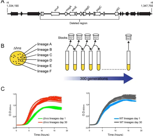
TyphimuriumDhns/rpoS* strain we noted one isolate appeared to lose a large cluster of genes at some point during laboratory passage [22]. To identify the nature of this deletion the isolate was further analyzed by whole genome sequencing where reads were assembled against the S. Typhimurium 14028s reference genome (Genbank ID CP001363.1) using Geneious Pro 5.5.6 software. This analysis revealed that the isolate incurred a 10 kb genomic deletion spanning nucleotides 1,334,560 to 1,344,664 (Figure 1A). The deleted region is highly AT-rich (GC% = 40% as compared to the genome average of %GC = 52) and encodes several putative envelope proteins including the PhoP activated genes pagC, pagD, pliC, envE, envFand msgA [59]. Multiple studies have shown that expression ofpagCis strongly repressed by H-NS, and the spontaneous loss of these genes from theDhns
isolate suggested that hnsmutants are genetically unstable and may shed horizontally acquired sequences during passage [21,22,60].
We sought to experimentally determine if the loss of horizon-tally acquired sequences is a reproducible outcome of deletinghns
fromS.Typhimurium, as well as to identify novel compensatory mutations that may alleviate the fitness defects associated with the loss of H-NS. Toward this end anin vitro evolution screen was performed where six independently derived freshly constructed
Dhns/rpoS*mutant lineages were serially passaged alongside six lineages of the isogenicrpoS* background (the ‘‘wild type’’ strain) in Luria-Bertani broth for 30 days, or approximately 300 generations (Figure 1B). The lineages were designated WT or
Dhns, ‘‘A’’ through to ‘‘F’’. Each day during the experiment, aliquots from the cultures were stocked and stored at280uC to enable the retrospective analysis of genomic changes in each lineage over time. At the end of the evolution period, the growth rates of the passaged wild type and the passaged Dhns lineages were monitored alongside their unpassaged (day 0) counterparts (Figure 1C). All six lineages lacking H-NS displayed significant increases in their growth rates compared to their respective day 0
Figure 1. Experimental evolution ofS.Typhimuriumhnsmutants.(A) Anhnsmutant isolate prepared for microarray analysis incurred a spontaneous 10 kb deletion after minimal laboratory passage. The white arrows represent the deleted open reading frames (ORFs) and the black arrows represent the ORFs adjacent to the deleted region. (B) Schematic diagram illustrating the experimental design of the serial passaging experiment. Six independently derivedS.Typhimuriumhnsmutant colonies were selected to initiate cultures of LB media, namedDhnslineages A–F. TheDhnslineages were serially passaged in parallel with six wild type cultures for 30 days (approx. 300 generations) and samples from each lineage were stocked daily. Genomic DNA from each population at Day 1 and Day 30 of the evolution period was prepared for Illumina sequencing. (C) Left hand side: growth curves of thehnsmutant populations at day 1 (green curves) and day 30 (red curves) of the evolution period. Right hand side: growth curves of the wild type populations at day 1 (blue curves) and day 30 (grey curves) of the evolution period. Liquid growth assays were performed in a 96-well plate reader.
clone, while the wild type lineages displayed modest improvements in growth (Figure 1C). Notably, by day 30 the Dhnslineages all exhibited growth rates similar to that of the wild type strains at day 30.
Thehns mutant lineages evolved parallel genetic changes
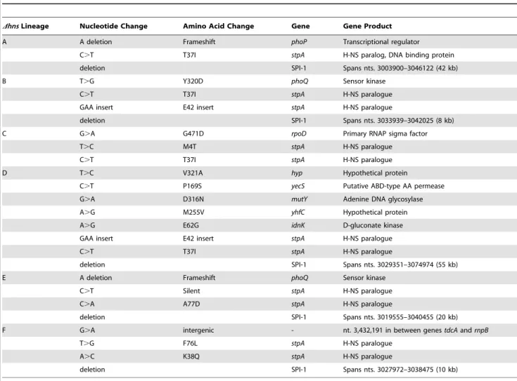
To identify mutations that arose throughout the evolution period, genomic DNA from the passaged WT andDhnslineages and their progenitor lines was analyzed by Illumina whole genome sequencing. In total, the sixDhns lineages acquired 15 missense mutations, 2 small deletions, 2 small insertions and 5 chromosomal deletions larger than 10 kb (Table 1). Most striking was the high degree of similarity in these mutations. Five of sixDhnslineages incurred unique 10–50 kb deletions within the Salmonella
Pathogenicity Island 1 (SPI-1) and all sixDhnslineages accumu-lated missense mutations within thestpAgene encoding the H-NS paralogue StpA (Figure 2, Table 1).
In agreement with our earlier observations [22], three Dhns
lineages acquired mutations in the genes encoding the PhoP/ PhoQ two component system that activates many H-NS repressed genes involved in virulence, acid stress, resistance to antimicrobial peptides and intramacrophage survival [59,61–64]. Specifically, lineages A and E acquired frameshift mutations in PhoP and PhoQ respectively while Dhns lineage B acquired a missense
mutation (Y320D) in the cytoplasmic sensor kinase domain of PhoQ.
Throughout the experiment eachDhnslineage acquired a total of three to four mutations with the exception ofDhnslineage D, which acquired eight. It is notable thatDhnslineage D incurred the largest chromosomal deletion that extended beyond SPI-1 into the locus encodingmutSand mutL, essential components of the methyl-directed mismatch repair pathway [65]. The loss of either
mutSormutLfromE. colihas been shown to result in a mutator phenotype and may explain the accumulation of other missense mutations specific to the Dhns lineage D, namely idnK(E62G),
mutY(D316N), yecS(P169S), yhfC(M255V) and stm1881(V321A) [66]. Analysis of the SPI-1 deletion junction regions revealed that 3 of the 5 deletions occurred without any homology in the sequences flanking the deleted segment. The other 2 SPI-1 deletions occurred between segments homologous in only 4 nucleotides. This suggests that RecA mediated recombination did not play a role in the loss of this island in theDhns mutants (Figure S1).
Analysis of the wild type lineages revealed that comparatively fewer genetic changes arose during the course of the experiment. 3 of the 6 wild type lineages acquired large chromosomal deletions that extended from 10 kb to 58 kb downstream of theuvrClocus (Table 2). Common to all three deleted fragments were compo-nents of theuvrABCnucleotide excision pathway and constituents
Table 1.Summary of the genetic changes specific to thehnsmutant lineages.
DhnsLineage Nucleotide Change Amino Acid Change Gene Gene Product
A A deletion Frameshift phoP Transcriptional regulator
C.T T37I stpA H-NS paralog, DNA binding protein
deletion SPI-1 Spans nts. 3003900–3046122 (42 kb)
B T.G Y320D phoQ Sensor kinase
C.T T37I stpA H-NS paralogue
GAA insert E42 insert stpA H-NS paralogue
deletion SPI-1 Spans nts. 3033939–3042025 (8 kb)
C G.A G471D rpoD Primary RNAP sigma factor
T.C M4T stpA H-NS paralogue
C.T T37I stpA H-NS paralogue
D T.C V321A hyp Hypothetical protein
C.T P169S yecS Putative ABD-type AA permease
G.A D316N mutY Adenine DNA glycosylase
A.G M255V yhfC Hypothetical protein
A.G E62G idnK D-gluconate kinase
GAA insert E42 insert stpA H-NS paralogue
C.T T37I stpA H-NS paralogue
deletion SPI-1 Spans nts. 3029351–3074974 (55 kb)
E A deletion Frameshift phoQ Sensor kinase
C.T Silent stpA H-NS paralogue
C.A A77D stpA H-NS paralogue
deletion SPI-1 Spans nts. 3019555–3040455 (20 kb)
F G.A intergenic - nt. 3,432,191 in between genestdcAandrnpB
T.G F76L stpA H-NS paralogue
A.C K38Q stpA H-NS paralogue
deletion SPI-1 Spans nts. 3027972–3038475 (10 kb)
doi:10.1371/journal.ppat.1004500.t001
of the flagellar apparatus. Under the laboratory growth conditions used in this study, expression of theuvrABCand flagellar genes likely resulted in a disadvantageous use of cellular resources. Apart from these deletions no mutations common among the wild type lineages were observed.
The temporal emergence of the mutations
To determine the timeline of the genetic changes that took place, genes of interest were PCR amplified from the frozen daily stocks of the hns mutant lineages and the PCR products were submitted for Sanger sequencing. This assay enabled the detection of mutant alleles soon after they arose in a given lineage and the relative proportion of the wild type and mutant alleles in the population at each day could be estimated from the sequencing chromatograms by analyzing the dual fluorescence peaks at a particular nucleotide. The relative signal strength of wild type vs. mutated nucleotides was used to approximate the emergence and dominance of each mutation in each population over time.
To determine when the large chromosomal SPI-1 deletions arose a PCR assay was employed; amplifying a region bridging the deleted segment. This detection method did not enable us to estimate the relative proportion of SPI-1 deletion strains in the population.
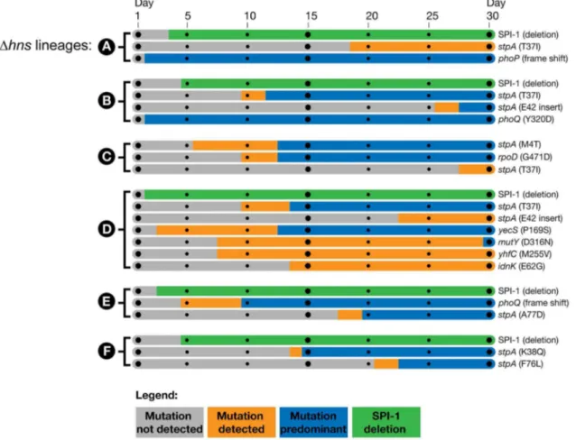
We found the mutations in the PhoP/PhoQ regulatory system and the SPI-1 deletions were acquired by thehnsmutant lineages in the early stages of the passaging period, prior to the missense mutations in stpA (Figure 3). The PhoP/PhoQ and SPI-1 mutations were detected as early as day 2 of the evolution period inDhnslineages A, B and D, suggesting these mutations confer the greatest growth advantages and/or are most easily acquired.
Of particular interest is the Dhns lineage C, which did not obtain inactivating mutations in either the PhoP/PhoQ or SPI-1 but displayed a comparable increase in fitness asDhnslineages A, B, D, E and F in liquid growth assays (Figure 1C).Dhnslineage C acquired astpAmissense mutation (M4T) by day 5 that persisted at low frequency until it also acquired a second mutation in the housekeeping sigma factor RpoD (G471D), at which point the
Dhns/stpA/rpoDmutant rapidly outcompeted both theDhns and
Dhns/stpAmutant strains in the population by day 13. To address the concern that lineage C acquired SPI-1 inactivating mutations that were not detected with the Geneious Pro software we performed a reference alignment of the rawDhnslineage C paired end reads to theS.Typhimurium 14028s reference genome using the Bowtie software package and also preformed ade novogenomic assembly of the evolvedDhnslineage C with Velvet [67,68]. A list of variants from both the Bowtie and Velvet assemblies was generated with Samtools and no other mutations besides for the StpAM4Tand
RpoDG471Dvariants were identified [69].
Inactivation of Salmonella Pathogenicity Island 1 improves growth ofhns mutants
The fact that five out of six Dhns lineages rapidly and independently incurred deletions within the SPI-1 locus suggested that SPI-1 misregulation is a major contributor to fitness defects in
S.TyphimuriumDhnsmutants. SPI-1 expression is repressed by
hnsand activated by a complex positive feedback loop where the production of the HilD regulatory protein induces the expression of HilA, a transcription factor that directly activates expression of the TTSS and effector proteins [70]. To determine the degree to which SPI-1 impairs growth of theS.TyphimuriumDhnsmutant,
Table 2.Summary of the genetic changes specific to the wild-type lineages.
WT Lineage Nucleotide Change Gene Gene Product
A deletion uvrC-fliB Spans nts. 2048083–2058350 (10 kb)
C deletion uvrC-fliG Spans nts. 2049159–2070524 (21 kb)
D deletion uvrC-stm2443 Spans nts. 2049052–2107464 (58 kb)
E deletion stm3185 Hypothetical protein, frameshift deletion spans nts. 3086531–3086545
doi:10.1371/journal.ppat.1004500.t002
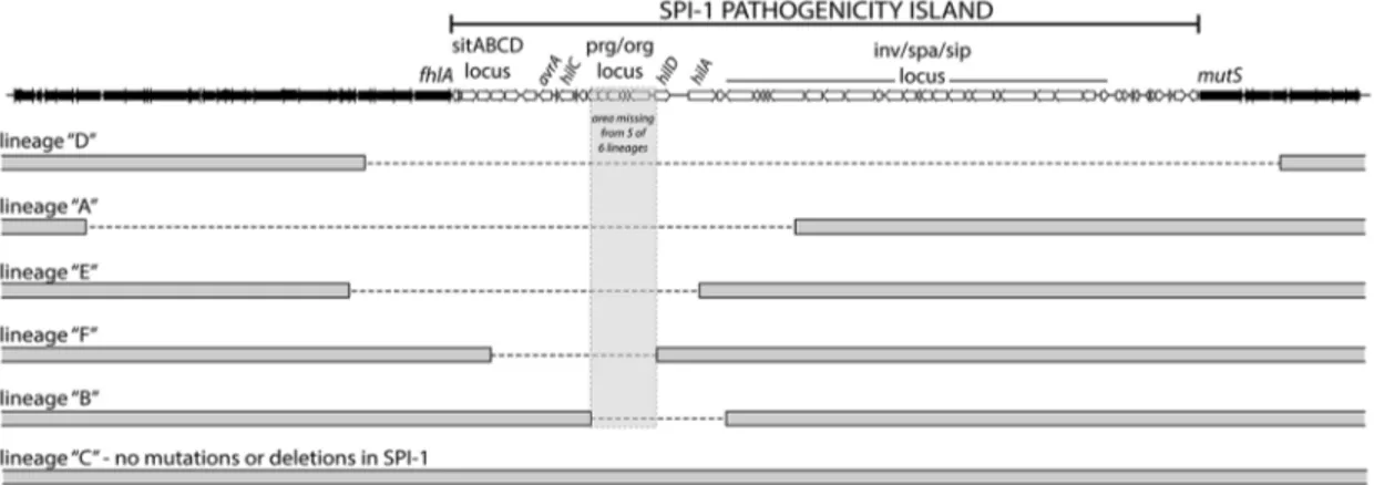
Figure 2.S.Typhimuriumhnsmutant lineages acquired large chromosomal deletions in the SPI-1 invasion locus.Throughout the 30-day evolution period, 5 out 6Dhnslineages incurred 10–50 kb deletions in SPI-1. The lineages are arranged according to the size of the deletion they incurred. The deleted regions are marked with a dashed line. The grey box encompasses a region common to all five deletions that encoded theprg/ orglocus and the promoter and start codon ofhilD. Lineage C did not acquire a deletion or any other mutations in the SPI-1 region.
we deleted the 40 kb genomic island from a wild type strain prior to introducing the hns deletion by transduction. The SPI-1 deletion significantly improved the growth of theDhns strain and also provided a mild improvement in growth of the wild type strain (Figure 4A). The region of SPI-1 lost in allDhnslineages included the promoter upstream ofhilD. Introduction of ahilD mutation into theDhns background conferred a growth benefit similar to that of the 40 kb SPI-1 deletion (Figure 4B). These results indicate that in the absence of H-NS, SPI-1 is activated through ahilD
dependent pathway and that the uncontrolled expression of SPI-1 encoded virulence determinants significantly impairs Salmonella
growth.
Salmonella entericaharbors a second pathogenicity island, SPI-2, that encodes a type-3 secretion system distinct from the one encoded on SPI-1. Lucchini et al., previously reported that construction of aSalmonellaDssrA/Dhns double mutant unable to express the genes encoded in SPI-2 significantly increased the growth rate of theDhnsstrain (grown in LB media and using strain LT2) [21]. To determine if inactivation of SPI-2 encoded TTSS would offer the same fitness benefit as deletion of SPI-1 from a
Dhnsbackground, we introduced a 25 kb SPI-2 genomic deletion into Dhns and Dhns/DSPI-1 strains (Figure S2). Inactivation of SPI-2 did not significantly improve growth of either theDhnsor
Dhns/DSPI-1 14028s strain to the same extent as loss of SPI-1. A similar experiment was conducted in LPM (low pH, low Mg2+
and low phosphate) media known to activate SPI-2 to determine if fitness of theDhns mutant would be adversely affected in a manner dependent on SPI-2. TheDhns mutant failed to grow in this media but this growth defect was not alleviated in theDssrA/Dhns double mutant indicating that other factors, not SPI-2, impact fitness in our strain under these particular conditions.
Mutations in the H-NS paralog, StpA, provide partial complementation for the impaired growth and motility phenotypes ofhns mutants
The only gene that acquired mutations in all six passagedDhns
lineages encodes the H-NS paralogue StpA. All of the acquired
stpAmutations resulted in single amino acid substitutions or in-frame insertions that map to the predicted N-terminal and central dimerization domains of the protein. Because disruption ofstpA
in a Dhns background is known to exacerbate hns mutant phenotypes, we found it unlikely that these substitutions impaired
stpAfunction. Intriguingly, the StpA mutations arise exclusively at sites where the unchanged amino acid is not conserved with H-NS, and the residue changes appear to render StpA more ‘‘H-NS-like’’
Figure 3. Timeline of the genetic changes in thehnsmutant lineages.The mutations present in thehnsmutant lineages at day 30 of the passaging period were detected by whole genome sequencing of the mixed populations. The identified genes of interest were PCR amplified directly from samples of the frozen daily culture stocks. The PCR products were sequenced and the relative signal intensities of the wild type vs. mutant alleles from each daily stock were used to estimate when the mutations emerged and became dominant in each population. Bars shaded in grey indicate the mutation was not detected via PCR sequencing, the orange bars indicate the mutation was detected in a fraction of the sequencing reads (,50%) and the blue bars indicate the mutant allele was predominant in the population (.50%). The large SPI-1 chromosomal deletions were detected by the appearance of a PCR product from the frozen stocks that spanned the deleted region. The green bars indicate detection of the SPI-1 deletions, the method employed did enable approximation of their abundance in the population.
doi:10.1371/journal.ppat.1004500.g003
(Figure 5A). We hypothesized that thestpAmutations impart H-NS-like silencing properties to StpA and therefore partially compensated for the loss of hns at loci outside of SPI-1 in the serial passaging experiment.
To test the ability of the StpA variants to complement hns
mutant phenotypes, we cloned thestpAlocus from each passaged
Dhnslineage and wild typestpAinto a low copy vector with the native stpA promoter. The resulting plasmids were pStpAWT,
pStpAT37I cloned from Dhns lineage A, pStpAT37I/E42ins from Dhnslineages B and D which both acquired the T37I substitution and an E42 insertion, pStpAM4TfromDhnslineage C, pStpAA77D
from Dhns lineage E and pStpAK38Q/F76Lfrom Dhns lineage F.
The StpA plasmids were transformed into a Dhns/DstpA S.
Typhiumurim background in order to determine whether or not the isolated StpA variants could ameliorate bacterial fitness in the absence ofhns. Introducing either pStpAWTor StpAT37Idid not
significantly improve growth of the Dhns/DstpA mutant (Fig-ure 5B). On the other hand expression of the StpAM4T mutant
significantly improved bacterial fitness in the liquid growth assay. Likewise, the StpAT37I/E42ins variant also offered an observable
growth advantage. The strains expressing StpAA77D and
StpAK38Q/F76L initially displayed a slight growth advantage and
then plateaued at a similar final optical density as the StpAWT
expressing strain.
Given that expression of the StpA variants identified in the serial passaging experiment enhanced bacterial fitness to varying degrees, we next tested the ability of the modified StpA proteins to complement the impaired motility phenotype ofhnsmutants. H-NS is required for both the expression and assembly of a functional flagellum [71–73]. H-NS indirectly stimulates flagellar gene expression by repressing hdfR, a known repressor of the flhDC
regulatory locus and, in addition, H-NS directly binds to the
Figure 4. Disruption of SPI-1 expression improves fitness of anhnsmutant.(A) Growth of a wild typeS.Typhimurium (black curve) and a Dhnsstrain (green curve) was monitored in liquid media alongside wild type andDhnsstrains harboring a 40 kb SPI-1 deletion (blue and red curves respectively). Elimination of the SPI-1 locus significantly increased growth of theDhnsstrain and also provided a slight growth advantage in the wild type background. (B) Targeted disruption ofhilD, the master SPI-1 activator produces a similar growth outcome as the SPI-1 deletion. Growth curves of theDhilDandDhilD/Dhnsstrains are represented in orange and purple respectively. The corresponding wild type (black) andhnsmutant (green) growth curves are the same as panel A. Plotted is the average of three biological replicates and standard error (panels A and B).
flagellar protein FliG and helps organize rotor subunit assembly [22,74]. StpA has also been shown to bind FliG, but does not promote motility in the absence of H-NS unless cellular StpA levels are artificially elevated [74]. To determine if the StpA variants stimulate motility to a greater extent than wild type StpA, we employed the same strains used in the liquid growth assays and
measured their radial swarming diameters on soft agar motility plates. After a 16 hr incubation period, wild typeS.Typhimurium displayed a swarming diameter of 62 mm (Figure 5C). Similar to the hns mutant strain, the S. Typhimurium Dhns/DstpA strains harboring pStpAWT and pStpAT37Idid not migrate beyond the
original inoculation zone. Remarkably, the StpA variants
Figure 5. Missense mutations in the H-NS paralogue StpA partially restore the impaired growth and motility phenotypes ofhns
mutants.(A) Sequence alignment of H-NS and StpA fromS.Typhimurium, conserved residues are highlighted in purple. The StpA amino acid substitutions acquired throughout thehnsmutant passaging are indicated below the alignment in orange. (B) Plasmids harboring the StpA variants isolated from the passagedDhnslineages were introduced into aDhns/DstpAbackground and growth of the resulting strains was monitored in a 96-well plate reader. The wild type (black) andDhns/DstpAstrain expressing StpAWT(green) growth curves are the same in each panel. (C) Swarming
motility of the same strains used in the growth assays were measured on soft agar plates following a 12 hour incubation period at 37uC. Expression of StpAT37I/E42, StpAM4T, StpAA77Dand StpAK38Q/F76Lrestored motility by 16%, 30%, 44% and 34% respectively. Bars represent the average swarm
diameter of three independent experiments and error bars represent the standard error. doi:10.1371/journal.ppat.1004500.g005
StpAM4T, StpAA77D and StpAK38Q/F76L restored motility to the Dhns/DstpAstrain by 30%, 44% and 34% that of the wild type strain respectively (Figure 5C). StpAT37I/E42ins provided a small
yet significant increase in swarming diameter to 16% the wild type diameter.
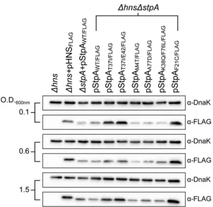
One possibility by which the StpA variants could restore motility to the Dhns mutant would be if the single amino acid substitutions increase StpA protein stability. Intracellular StpA pools are reportedly subject to proteolysis by the Lon protease in strains lackinghns[56]. In this study a mutation in the N-terminal dimerization domain of StpA, F21C, was shown to impart resistance to proteolysis and increase intracellular StpA concen-trations. To determine if any of the StpA mutations identified in our laboratory passage screen influenced protein levels, the amount of intracellular StpA was quantified by western blot analysis.Dhnsstrains harboring epitope tagged StpA or its variants was probed with ana-FLAG antibody. DnaK levels were analyzed on the same blot as a loading control. Similar to the StpAF21C
variant, StpAT37I and StpAT37I/E42 accumulated to higher
intracellular levels than StpAWT(Figure 6).In contrast the variants
StpAM4T, StpAA77Dand StpAK38Q/F76L were detected at similar
levels to that of StpAWT. This suggests that the StpA variants
identified in this study fall into one of two categories, mutations that increase intracellular StpA levels similar to the previously identified StpAF21Cvariant, and a novel class of mutations that do
not significantly alter intracellular StpA levels. Notably, it was the latter class of variants that provided partial complementation for the loss ofhnsin the growth and motility assays suggesting that the amino acid substitutions M4T, A77D and K38Q/F76L alter the functional properties of StpA and not its stability.
Amino acid substitutions M4T and A77D alter StpA’s silencing properties
Much like H-NS, StpA has also been implicated in silencing AT-rich regions of the genome. Although the set of genes under control of StpA shares significant overlap with the set of genes regulated by H-NS, in the absence of H-NS, the silencing activity of StpA alone does not provide sufficient repression of H-NS regulated loci [46,48,75]. To determine if the missense mutations acquired throughout the evolution of theDhnslineages enhanced StpA’s silencing activity, we measured the steady state transcript levels of four model H-NS and StpA regulated loci from aDhns/ DstpA strain harboring pStpAWT, pStpAM4T, pStpAA77D and
pStpAF21C. The StpAM4Tand StpAA77Dvariants were chosen for
transcript analysis because they provided the greatest restoration of theDhns growth and motility defects without altering protein stability, while the StpAF21Cvariant was included to determine the
regulatory consequences of increased intracellular StpA levels. Also included in the analysis were a Dhns complemented strain (Dhns+pHNS) and aDhnsstrain, which served as reference points for repressed and derepressed transcript levels. cDNA from mid-log cultures was analyzed by Q-PCR with primers specific toproV,
hilA, ssrAand yciG.proV is a well studied H-NS regulated gene target that resides outside the Salmonella pathogenicity islands, whilehilAandssrAare transcriptional activators encoded within SPI-1 and SPI-2 respectively.yciGis part of therpoSregulon and was previously shown to be highly induced in a Salmonella
SL1344 strain lackingstpA[48].
Relative to theDhnscomplemented strain, the transcript levels ofproV,hilA,ssrAandyciGincreased by 20-fold or greater in the
Dhnsstrain (Figure 7). The expression ofyciGis highly repressed
Figure 6. StpA missense mutations do not affect intracellular protein concentrations.Expression levels of FLAG-tagged StpA variants were monitored by western blot analysis at early exponential phase (O.D. 600 nm 0.1) mid exponential phase (O.D. 600 nm 0.6) and early stationary phase (O.D. 600 nm 1.5).a-DnaK served as a loading control.
in theDhns+pHNS strain, its transcript levels were lower than the detection limit of the Q-PCR cycler and could not be reported with confidence. The Dhns/DstpA strain harboring pStpAWT
displayed a greater increase in the transcripts levels ofproV,ssrA
andyciGcompared to theDhnsstrain, whilehilAtranscript levels were reduced by 4.5-fold in the presence of pStpAWT. Substituting
StpAWTwith StpAM4Tsignificantly reduced the expression levels
ofproVandssrAby approximately 2-fold and 10-fold respectively. The StpAA77Dvariant provided even greater repression ofproV
andssrAby reducing their transcript levels by 4-fold and 20-fold relative to StpAWT. Similar to the Dhns+pHNS strain, both
StpAM4Tand StpAA77DmaintainedyciGexpression levels close to
the detection limit of the sensor. In contrast, the StpAF21Cvariant
that accumulates to higher intracellular levels than StpAWTdid
not maintain significantly lower expression levels of any of the four genes tested relative to the pStpAWTstrain. This further establishes
that the StpAM4T and StpAA77D variants as a novel set of
mutations that enhance StpA silencing activity without affecting protein stability.
While the two single point mutations, M4T and A77D, significantly enhanced StpA’s silencing activity at the proV, ssrA
and yciG promoters regions these substitutions did not provide increased repression of hilA, encoding the SPI-1 transcriptional activator HilA.hilAexpression is induced by three transcriptional activators, HilC, HilD and RtsA [70]. In the absence of H-NS it is possible that silencing complexes generated by StpAM4T and
StpAA77D, although more effective than StpAWT, were unable to
impede the combined HilC and HilD-mediated activation ofhilA.
Mutations instpAarise reproducibly as a consequence of mutations in hns
We repeated ourin vitroevolution on an expanded number of freshly constructedhnsdeletion mutants to determine if loss ofhns
invariably led to mutations instpAand, if so, to use this technique as a novel method of mapping functional residues instpA. Toward this endhnsdeletion mutations were introduced by transduction into therpoS-low strain to generate 12 independent lineages. To assess the impact SPI-1 may have on the evolution ofstpAanother 12 linages were generated by introducing thehnsmutation into a strain already lacking SPI-1. Each of the 24 lineages were serially
passaged in LB media over the course of 21 days and thestpA
genes of each lineage were amplified by PCR and sequenced. Sequencing of thestpA genes revealed missense mutations in 10/12 of thehnsmutants in therpoSbackground and 12/12 of the rpoS*/SPI-1 mutant background (Table 3). Remarkably the twohnsmutant strains that did not acquire misssense mutations in
stpAdid acquire silent mutations, suggesting that either thatstpAis prone to mutation in the absence ofhnsor that the presumably silent mutations actually affect StpA levels or function by increasing mRNA stability or by altering codon usage. As before all missense mutations mapped to the oligomerization domain between residues 2 and 80 of thestpAprotein. Furthermore some lineages acquired as many as 4 different nucleotide substitutions. The fact that 30 independent lineages (24 in this experiment and 6 in the initial experiment) acquired mutations instpAand that none of these were nonsense mutations confirms that there is strong selective pressure to acquire mutations instpAin the absence of H-NS.
Notably there were some differences observed in the specific mutations acquired between the two lineages (those with or without SPI-1). In the presence of SPI-1 the StpA protein was altered at several different residues but a cluster of mutations occurred at or near codon 38 (nucleotides 112–114) encoding lysine including a silent mutation at nucleotide 111. Strains that evolved in the absence of SPI-1 acquired a notably different set of mutations where all but one lineage acquired a mutation at nucleotide 110 resulting in the StpA(T37I) variant. Additional mutations changed the asparagine at positions 2 or 7 to an aspartic acid (N2D or N7D). This suggests that the pressures that select for mutations in StpA may differ in the absence of SPI-1.
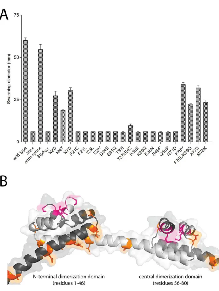
The results of the evolution experiment provided an opportu-nity to map what single or double residue changes in StpA would be sufficient to engender it with H-NS-like functionality. This functionality of each StpA variant was assessed by their ability to restore motility (Figure 8A) when expressed in the hns mutant background. This assay was chosen because our data with the earlier StpA variants indicated motility restoration correlates closely with their to silence H-NS regulated loci. These assays uncovered functional changes in single amino acids that cluster to two discrete regions of the StpA protein (Figure 8). The functional variants StpAN2D, StpAM4T, and StpAN7Dmap to the short helix 1
Figure 7. Point mutations A77D and M4T enhance StpA repression of selecthnsregulated loci.Transcript levels of four H-NS regulated genes,proV,ssrA,yciGandhilA, were measured by reverse transcriptase QPCR in aDhns/DstpAstrain expressing StpAWTor the variants StpAM4T,
StpAA77Dand StpAF21C. Transcript levels were also assayed inDhnsandDhnscomplemented backgrounds (Dhns+pHNS).
doi:10.1371/journal.ppat.1004500.g007
that lies within the N-terminal dimerization domain while the variants StpAF76V, StpAF76L, StpAA77Dand StpAM78Kall map to
helix 4 which is contained in the central dimerization domain. Other single residue StpA variants, where changes mapped to helix 3 or the short linker segments that connect helix 3 to the other helices, failed to restore significant motility to the hns
mutant. Modeling these changes on the previously published H-NS oligomer structure show that the individual changes that confer H-NS-like function to StpA are buried within the dimerization interfaces or present on the outer, convex, surface of the H-NS filament while the residues that do not lie predominantly on the concave surface of the filament, and are largely predicted to have surface exposed side chains (Figure 8B). It is important to note the StpA residues were mostly assessed individually (only two double-mutants were assessed) and that some residues that appear to have no gain of function in our assays may have a more dramatic impact in combination with other changes.
The M4T and A77D StpA variants, but not the T37I StpA variant, have DNA binding properties that are more similar to H-NS than to that of StpA
Electrophoretic mobility shift assays were used to determine if changes in the StpA variants that led to increased ‘‘H-NS-like’’ function manifest as differences in their ability to form nucleo-protein complexes on DNA. Like H-NS, StpAWT displays
cooperative binding to a model 289 bp AT-rich sequence
(%GC = 34) but forms nucleoprotein complexes are consistently observed to have significantly lower mobility than those formed by H-NS on the same DNA target (Figure 9). Remarkably the nucleoprotein complexes formed by the StpAM4T and StpAA77D
variants formed complexes with motility more similar to H-NS than wild type StpA. StpAM4T formed two complexes on DNA,
one that migrated with the top band of the DNA ladder like StpA and one that migrated further into the gel at the same position as the H-NS complex. StpAA77Dalmost exclusively formed a single
H-NS like complex. StpAT37I,which had enhanced protein levels in vivo, but failed to complement for H-NS for either motility or silencing, formed a lower mobility nucleoprotein complex identical to that of the wild type StpA protein. Notably, there were no differences in overall affinity for DNA between the different variants.
This data indicates that subtle changes in the dimerization domains of StpA can generate large and quantifiable differences in properties of the nucleoprotein complex and that the functional differences observed between StpA variants manifest as differences in their effects on nucleoprotein structure. At high protein concentrations both StpA and H-NS have the ability to spontaneously oligomerize into higher order structures in the absence of DNA, a phenomenon that can be measured by changes by analytical gel filtration chromatography. We assessed the gel filtration profiles of StpA and its variants (Figure 10) to determine if any changes in their oligomerization states could be observed. StpAWTand the StpAT37I, which do not effectively substitute for Table 3.Summary ofstpAchanges in second evolution experiment.
Lineage Nucleotide Change Amino Acid Change
A* (WT) G.C 146, T.G 226 R49P, F76V
B* (WT) T.A 111 Silent
C* (WT) T.C 64, C.G 72 S22P, D24E
D* (WT) A.C 114 K38N
E* (WT) A.C 112, A.C 149 K38Q, Q50P
F* (WT) A.C 67 I23L
G (WT) A.C 114 K38E
H (WT) A.C 112 K38Q
I (WT) C.A 63, T.G 226 F21L, F76V
J (WT) A.G 112 K38E
K (WT) C.T 63 Silent
L (WT) A.G 19, T.C 64, G.C 91 N7D, S22P, E31Q
M (DSPI-1) A.G 19, C.T 110 N7D, T37I
N (DSPI-1) T.C 61, T.A 228 F21L, F76L
O (DSPI-1) A.G 19, T.C 61, C.T 110, G.C 146 N7D, F21L, T37I, R49P
P (DSPI-1) A.G 19, C.T 110 N7D, T37I
Q (DSPI-1) A.G 4, C.T 110, T.A 233 N2D, T37I, M78K
R (DSPI-1) A.G 4, C.T 110 N2D, T37I
S (DSPI-1) A.G 4, C.T 110, A.G 112 N2D, T37I
T (DSPI-1) A.G 4, A.G 19, C.T 110 N2D, N7D, T37I
U (DSPI-1) A.G 4, C.T110 N7D, T37I
V (DSPI-1) A.G 4, C.T 110 N7D, T37I
W (DSPI-1) A.G 67, C.T 110, T.G 226 I23V, T37I F76V
X (DSPI-1) A.G 19, C.T 110, A.G 211 N7D, T37I, N71D
Figure 8. StpA mutants that partially restore the motility phenotype inhnsmutants cluster in regions.(A) Swarming motility of the
strains expressing either StpAWTor individual StpA variants were measured on soft agar plates following a 12 hour incubation period at 37uC. Bars
represent the average swarm diameter of three independent experiments and error bars represent the standard error. (B) Location of the StpA mutations mapped on the structure of the H-NS oliomerization domain. Orange residues indicate individual changes that could not impart H-NS functionality upon StpA.
doi:10.1371/journal.ppat.1004500.g008
H-NS, displayed two prominent peaks with calculated molecular weights of approximately 450 and 150 kDa (StpA monomer is
,15 kDa). The chromatographic profiles of the StpAM4T and
StpAA77Dproteins indicate that these proteins have a dramatically
reduced propensity to form the oligomeric species that elutes early during chromatography. We note that the asymmetrical rod-like structure of the StpA and H-NS oligomers prevent an accurate determination of molecular weight based on mobility through the column when compared to a set of globular standards. Differences in shape or flexibility would also manifest as different elution profiles by gel filtration. Nevertheless these findings when taken as a whole indicate that the functional differences between the StpA
variants (and also the functional differences between H-NS and StpA) are primarily due to differences in manner of their oligomerization and not in the specificity of their DNA binding domains.
Discussion
The xenogeneic silencing model predicts that the selective silencing of foreign DNA accelerates bacterial evolution by reducing the fitness cost associated with HGT. While multiple studies have established a role for the H-NS, MvaT and Lsr2 protein families in regulating newly acquired sequences, the
Figure 9. StpAM4Tand StpAA77Dform DNA-protein complexes more similar to H-NS than StpAWT.Electrophoretic mobility shift assays
were conducted using purified H-NS, StpAWT, StpAT37I, StpAM4T, and StpAA77D. As indicated, the purified proteins were added to 289 bp AT-rich
(%GC = 34) or 204 bp GC-rich (%GC = 74) model DNA fragments in increasing protein concentrations of 150 nM, 200 nM, 300 nM, 400 nM, and 500 nM. DNA/protein complexes were separated by 6% native polyacrylamide gel and stained with SYBR green. StpA complexes (x) migrated alongside the top band of a nucleotide standard, while H-NS complexes (c) migrated further into the gel as indicated.
evolutionary advantage of foreign gene repression by H-NS and its impact on genome content had not been assessed by experimental evolution [21,22,39,40]. The genetic adaptations we identify that improve growth in strains lacking H-NS indicate that xenogeneic silencing played a major role in the evolution of the Salmonellae by buffering the fitness consequences caused by the SPI-1 encoded TTSS, a defining characteristic of the species. Indeed a recent study on the evolution of Salmonella revealed that, while many sequences acquired by HGT will adopt the %GC of their host over time, the major pathogenicity islands have selectively retained their AT-richness, presumably to maintain their silencing by H-NS [6]. The fact that we observed large deletions in SPI-1, rather than inactivating point mutations or small indels, is somewhat surprising and suggests that this region may be naturally unstable and prone to gene loss. The 5 deletions independently occurred at different sequences, each with limited no flanking homology, suggesting that replication errors and not homologous or site-specific recombination likely caused the loss of these regions from the genome.
Multiple lines of evidence suggest that maintaining a TTSS represents a costly investment of cellular resources. Induction of the Yersinia TTSS by low calcium essentially halts bacterial growth and the plasmid-encoded Shigella TTSS is readily lost during laboratory passage [20]. An association between impaired bacterial growth and SPI-1 expression in wild type cells was recently reported in a study conducted by Sturmet al[76]. This study tracked the spontaneous induction of the SPI-1 encoded TTSS at a single cell level using time-lapse microscopy imaging. Sturmet al.correlated the expression of the TTSS with retarded growth rates that were alleviated by mutations in the SPI-1 activator hilA. Under the conditions employed in this study the
hnsmutant strains only incurred genomic deletions within SPI-1. However, a targeted disruption of SPI-2 was previously shown to partly improve growth in an hns mutant background [21]. We believe the loss of SPI-1 and not SPI-2 from our hns mutant lineages likely reflects that the conditions we employed in this study favored SPI-1 expression. It is important to note that the conditions employed during the experimental evolution experi-ment were arbitrarily chosen and it is entirely likely that subtle changes in environment will significantly impact which loci will
impinge on fitness in the absence of H-NS. While the wild type lineages passaged in parallel to ourhnsmutants did not acquire mutations in the SPI-1 locus, genomic deletions encompassing components of the flagellar apparatus were noted in 3 out of 6 of the wild type lineages. Flagella and TTSS are evolutionarily related and highly homologous in both primary sequence and structure [77]. The fact that the flagellar loci of thehnsmutant lineages did not acquire mutations is consistent with the fact that
hnsmutants fail to express flagella to begin with.
Pathogens likeSalmonella spend a substantial amount of time outside of the host environment and our studies suggest that H-NS is essential for enteric bacteria to retain virulence in the absence of selective pressures. Naturally occurring SPI-I deletions have occasionally been identified among environmental Salmonella
isolates that have consequentially lost the ability to invade host cells [78]. Spontaneous SPI-1 mutations are thought to arise throughout host infection generating a subpopulation of ‘‘avirulent defectors’’ that propagate much faster than their TTSS-positive predecessors [79]. Diard et al demonstrated that Salmonella
infection with a constitutively active SPI-1 TTSS strain resulted in a sharp rise of the genetically avirulent subpopulation and consequently premature clearing of the infection [79].
The onlyhnsmutant lineage that did not incur a SPI-1 deletion,
Dhns lineage C, acquired a missense mutation in rpoD
(RpoDG471D). The recent crystal structure of theE. coliRNAP/
sD
holoenzyme shows RpoD residue G471 is located in an exposed loop region, enriched in highly conserved aromatic and positively charged residues [80]. An alignment of the E. coli
RNAP/sDstructure with the RNAP/sDinitiation complex from
Thermus thermophilusreveals the conserved loop region harboring residue G471 is in close proximity the template strand during transcriptional initiation [81]. It is possible that introduction of the negatively charged aspartic acid residue at position 471 could hinder transcriptional initiation and result in reduced expression of the SPI-1 locus, however it is currently unclear how this mutation would affect SPI-1 but not impede the expression of many other importantsD
targets.
Another central and important outcome of this study was the identification StpA oligomerization variants that partially com-pensate for several H-NS dependent phenotypes. Many of these
Figure 10. Analytical gel filtration of StpA and the StpAM4T, StpAA77D, and StpAT37Ivariants.Purified StpAWT, StpAT37I, StpAM4T, StpAA77D
proteins at a concentration of 100mM were applied to a Superdex 200 10/300 GL column pre-equilibrated with 20 mM Tris pH 8, 1 M NaCl, 1 mM
EDTA and 5% glycerol. The column was calibrated using globular protein standards ranging from 12.4 kDa to 443 kDa. doi:10.1371/journal.ppat.1004500.g010
mutations do not increase cellular StpA protein concentrations, as has been observed previously [46,54]. Notably a recent study on a spontaneous mutant that improved fitness of an E. coli strain lacking Hha and YdgT, two molecules that collaborate with H-NS to facilitate gene silencing, identified a promoter mutation that dramatically enhanced H-NS levels [82]. StpA inS.Typhimurium was recently proposed to repress the rpoS regulon during exponential growth and the major caveat of our study is that we started with strain encoding a defective RpoS, which would alleviate the selective pressure to maintain a wild type copy of StpA [48]. H-NS also represses numerous genes activated byrpoS
in response to cellular stress [83–85]. A study of the hdeAB
promoter region suggested H-NS repression was overcome by the RNAPNsS complex, while RNAP associated with the house
keeping sigma factor sD was more effectively inhibited by the presence of H-NS [85]. A similar finding was also reported for the
dpspromoter [86]. One model that could be extrapolated from these observations is that StpA restricts transcription of the RNAP complexed withsSwhile H-NS more efficiently represses RNAP bound tosD
. Complicating this model is the fact that H-NS and StpA can heterodimerize and that each may individually regulate cellularsS
concentrations [48–50,87,88].
The story that is emerging from this and other recent studies is that subtle changes in local nucleoid architecture, directed by the structure of the oligomerized protein, underlies the diverse functions ascribed to the H-NS like molecules. Several findings indicate that changes in DNA shape and tension are the relevant outputs of this class of transcriptional modulators; a mode of gene regulation that is particularly challenging to study using conven-tional assays like EMSA and footprinting. Our results indicate that StpA and H-NS differ primarily not in their ability to bind AT-rich DNAper se, in fact StpA binds DNA with an apparent affinity higher than that of H-NS, but by some physical property that manifests as a change in promoter architecture once bound by the protein. Due to its apparent higher affinity for DNA and elevated propensity to form higher-order oligomers in conventional assays one would predict that StpA would be a more effective silencer than H-NS in most situations. We note that there are significant qualitative differences in the shifted DNA complexes between the StpA variants that can complement for the loss of H-NS and those variants that cannot. This supposition is further supported by recent studies on the H-NS-like transcriptional activator Ler and H-NS paralogs encoded on plasmids demonstrating that the central linking domain, not the DNA binding domain, is the primary determinant in how these molecules functionally differ from H-NS [89,90].
Evidence that H-NS, StpA, and the ‘‘H-NS-like’’ Ler proteins each form characteristically distinct higher order protein/DNA complexes has been more directly provided by recent atomic force microscopy imaging studies and single molecule ‘‘DNA stretching’’ experiments [91–94]. Lim et al reported that StpA-induced DNA/protein filaments were significantly more rigid than those produced by H-NS, and that the StpA filaments were insensitive to changes in pH, temperature, and osmolarity; conditions known to disrupt H-NS-DNA binding [92]. Another observation that might support divergent oligomerization prop-erties of StpA and H-NS is that StpA can silence theE.coli bglG
operon, but only in the presence of H-NS molecules deficient in DNA binding [50,95]. This observation was used to suggest that the H-NS proteins can heterodimerize with StpA to facilitate silencing of bglG. However, based on our new findings, we cannot exclude the possibility that thehnsmutant strain used in that study acquired mutation(s) in stpA during routine lab passaging that enabled it to act like H-NS.
The fact that compensatory stpAmutations arise rapidly and reliably in the absence of H-NS is a worrying outcome of this study. Complicating matters further is the apparent functional heterogeneity in the variousstpAmutations we uncovered, i.e. the different compensatory mutations do not share the exact same properties. It is unclear how much care has been taken in the maintenance of the varioushnsmutant strains employed in many prior studies and in all but one case it is clear that thestpAlocus was not sequenced to check for mutations. Regrettably this leaves some doubt regarding the validity of earlier studies on the phenotypes of strains lacking H-NS. Given their genetic instability, all future work on hns mutants in either E. coli or Salmonella
should be performed on multiple freshly constructed (transduced) isolates and laboratory passaging of such strains should be kept to a minimum. Whenever possible the genomes of hns mutants should be re-sequenced to verify that phenotypes ascribed to H-NS are not, in fact, due to a mutation in a different gene.
Materials and Methods
Plasmid and strain construction
The plasmids and strains employed in this study are listed in Table 4 and a complete list of oligonucleotides sequences is provided in Table 5. In a previous study, a FLAG-epitope tag was incorporated into the XhoI and BamHI sites of the low copy vector pHSG576 to generate pWN425 [22]. The stpA coding sequence and 206 bp upstream region (comprising nucleotides 2976460 to 2968067 in the S. Typhimurium 14028s genome Genbank ID CP001363.1) was PCR-amplified from S. Typhi-murium 14028s genomic DNA with primers ALO115 and ALO116. The amplified fragment was ligated into the PstI and BamHI sites of pHSG576 backbone for expression of StpA harboring a C-terminal FLAG epitope tag. The StpA coding sequence and promoter region were incorporated into pHSG576 in the opposite orientation of the lac promoter, such that stpA
expression levels were controlled by the nativestpApromoter. The resulting plasmid pStpAWTwas used for complementation studies.
Similarly, plasmids harboring the StpA variants identified in the experimental evolution screen were constructed using the pHSG576 backbone with a C-terminal FLAG epitope tag. The mutated StpA alleles were PCR amplified from the genomic DNA of their respectivehnsmutant lineages that had been passaged for 30 days. The same primer pair used to amplify the wild typestpA
coding sequence and 59 promoter region was employed. The mutantstpAallele PCR fragments were inserted into the PstI and BamHI sites of vector pHSG576 harboring the FLAG epitope sequence 39 of the BamHI site. The plasmids generated and the corresponding hns mutant lineage that the stpA alleles were cloned from were as follows: pStpAT37I from Dhns lineage A,
pStpAT37I/E42insertfromDhnslineages B and D, pStpAM4Tfrom Dhnslineage C, pStpAA77DfromDhnslineage E and pStpAK38Q/ F76L from Dhns lineage F. The sequences of all the plasmids
constructed in this study were confirmed by Sanger Sequencing at the TCAG Sequencing Facility (Centre for Applied Genomics, Hospital for Sick Children).
The Salmonella entericaserovar Typhimurium 14028s strains used in this study possess a mutantrpoSallele (calledrpoS*) that encodes a five residue in-frame deletion that significantly reduces RpoS (s32
Table 4.Plasmids and strains used in this study.
Plasmid or strain Description Reference
Plasmids Vector
pStpAWT Low copy vector encoding StpAWTwith C-terminal FLAG epitope, under control of thestpApromoter. Variants listed below are constructed identically.
pHSG576 This study
pStpAT37I FLAG-tagged StpAT37I pHSG576 This study
pStpAT37I/E42 FLAG-tagged StpAT37I/E42 pHSG576 This study
pStpAM4T FLAG-tagged StpAM4T pHSG576 This study
pStpAA77D FLAG-tagged StpAA77D pHSG576 This study
pStpAK38Q/F76L FLAG-tagged StpAK38Q/F26L pHSG576 This study
pStpAN2D FLAG-tagged pStpAN2D pHSG576 This study
pStpAN7D FLAG-tagged pStpAN7D pHSG576 This study
pStpAF21C FLAG-tagged pStpAF21C pHSG576 This study
pStpAF21L FLAG-tagged pStpAF21L pHSG576 This study
pStpAS22P FLAG-tagged pStpAS22P pHSG576 This study
pStpAI23L FLAG-tagged pStpAI23L pHSG576 This study
pStpAI23V FLAG-tagged pStpAI23V pHSG576 This study
pStpAD24E FLAG-tagged pStpAD24E pHSG576 This study
pStpAE31Q FLAG-tagged pStpAE31Q pHSG576 This study
pStpAK38N FLAG-tagged pStpAK38N pHSG576 This study
pStpAK38Q FLAG-tagged pStpAK38Q pHSG576 This study
pStpAK38E FLAG-tagged pStpAK38E pHSG576 This study
pStpAR49P FLAG-tagged pStpAR49P pHSG576 This study
pStpAQ50P FLAG-tagged pStpAQ50P pHSG576 This study
pStpAN71D FLAG-tagged pStpAN71D pHSG576 This study
pStpAF76V FLAG-tagged pStpAF76V pHSG576 This study
pStpAM78K FLAG-tagged pStpAM78K pHSG576 This study
pWN425 (pHNSWT) FLAG-tagged H-NS pHSG576 [22]
pET21b StpAwt Inducible expression of His-tag StpAwt This study
pET21b StpAT37I Inducible expression of His-tag StpAT37I This study
pET21b StpAM4T Inducible expression of His-tag StpAM4T This study
pET21b StpAA77D Inducible expression of His-tag StpAA77D This study
Strains
WN153 S.entericaserovar Typhimurium 14028srpoS*(five amino acid in frame deletion that reduces RpoS activity) [34]
SSA105 WN153Dhns::Km/pHSG576 vector only [22]
SSA162 WN153DstpADhns::Km/pStpAWT This study
SSA163 WN153DstpADhns::Km/pStpAT37I This study
SSA164 WN153DstpADhns::Km/pStpAT37I/E42insert This study
SSA165 WN153DstpADhns::Km/pStpAM4T This study
SSA166 WN153DstpADhns::Km/pStpAA77D This study
SSA167 WN153DstpADhns::Km/pStpAK38Q/F76L This study
SSA171 WN153DSPI-2::CmDhns::Km This study
SSA172 WN153DSPI-1DSPI-2::CmDhns::Km This study
JS008 BL21Dhns/pET21b StpAwt This study
JS009 BL21Dhns/pET21b StpAT37I This study
JS011 BL21Dhns/pET21b StpAM4T This study
JS012 BL21Dhns/pET21b StpAA77D This study
JS065 WN153DstpADhns::Km/pStpAF21C This study
JS076 WN153DstpADhns::Km/pStpAN7D This study
JS077 WN153DstpADhns::Km/pStpAF21L This study
JS078 WN153DstpADhns::Km/pStpAI23L This study
JS079 WN153DstpADhns::Km/pStpAD24E This study
cassette was subsequently flipped out of the chromosome by introducing the pCP20 plasmid expressing the FLP recombinase. This generated aS.TyphimuriumDstpAstrain without antibiotic resistance markers. To test the ability of the StpA variants to compensate for the loss of H-NS, each of the StpA complemen-tation plasmids were transformed into the S. Typhimurium 14028s DstpAmutant. Next, thehns null allele was moved into the DstpA mutant strains harboring the StpA complementation plasmids by P22 transduction. The resulting clones were selected for on Miller’s Luria Bertani (LB) 1% agar plates supplemented with 50mg/ml kanamycin (to select for thehnsnull mutation) and 20mg/ml chloramphenicol (to select to the StpA plasmids).
SPI-1 deletion mutants were constructed by deleting a 44.4 kb region spanning the SPI-1 region using the lambda red recombinase method described by Datsenko and Wanner [96]. In brief, the region between S. Typhimurium 14028s genome coordinates 3005740–3050161 (Genbank ID CP001363.1) in each parent strain was replaced by a chloramphenicol resistance cassette flanked by FRT recombinase sites from plasmid pKD3 using primers ALO76 and ALO77. Each knockout mutation was then transduced into a fresh strain background by P22 HT105/1
int-201 transduction. Similarly, the SPI-2 deletion mutants were generated using the lambda red recombinase method. A chloramphenicol resistance cassette was amplified from plasmid pKD3 with primers ALO83 and ALO84, which were designed with flanking sequences complementary to the SPI-2 region. Following lambda red recombinase with the amplified PCR product, a 25 kb SPI-2 deletion spanning nucleotides 1,486,143– 1,511,465 (Genbank ID CP001363.1) was introduced into theS.
Typhimurium 14028s genome. The SPI-2 mutation was then transduced into a fresh strain background by P22 HT105/1int -201 transduction. The doubleDSPI-1/DSPI-2 mutants strain were generated by first flipping theDSPI-1 chloramphenicol resistance cassette out of the chromosome by introducing the plasmid PCP20 expressing the FLP recombinase, and then introducing the SPI-2 deletion via P22 transduction.
Strains containing thehilD mutants were constructed by P22 transduction of a previously constructed mutation provided generously by the lab of Dr. Ferric Fang at the University of Washington [97].
Experimental evolution ofSalmonella hnsmutants
Anhnsgene knockout fromS.Typhimurium 14028s harboring a kanamycin resistance cassette in place ofhnswas moved into a freshS.Typhimurium 14028s background containing a mutated
rpoSallele (rpoS*) via P22 phage transduction. The transductants were selected on LB-agar plates supplemented with 50mg/ml kanamycin. Six independently derived colonies from the original transduction were streaked twice on LB-kanamycin plates to eliminate trace P22 phage lysate, with each passage on solid media corresponding to a 16 hour incubation period at 37uC. All six transductants harbored the kanamycin resistance cassette in place ofhnsand were free of contaminating P22 phage as determined by PCR. Thesehns mutant isolates were selected to inoculate 5 ml LB in conical 25 ml polypropylene culture tubes. The cultures were grown at 37uC with 200 rpm shaking and every 24 hr, 5ml from each culture was transferred to 5 ml of fresh media. The 1:1000 dilution corresponds to approximately 9.96 doublings a day for a total of,300 doublings over the course of the 30 day
evolution period. Daily samples from each lineage were taken and stored at280uC in culture media supplemented with 10% DMSO for later analysis.
Preparation of genomic libraries for Illumina sequencing
Samples from the frozen DMSO stocks representing day 1 and day 30 of the evolution period were scraped into LB media and grown at 37uC with shaking until mid-stationary phase (approx-imately 8 hours). The genomic DNA from approx(approx-imately 46109 cells from each culture was purified using the Qiagen DNeasy blood and tissue kit. 5mg of the purified DNA in 130ml water was sheared to a mean fragment size of 400 nt using a Covaris S2 focused ultrasonicator (Woburn, Massachusetts). The fragmented Table 4.Cont.
Plasmid or strain Description Reference
JS080 WN153DstpADhns::Km/pStpAK38E This study
JS081 WN153DstpADhns::Km/pStpAF76V This study
JS082 WN153DstpADhns::Km/pStpAM78K This study
JS083 WN153DstpADhns::Km/pStpAI23V This study
JS084 WN153DstpADhns::Km/pStpAR49P This study
JS085 WN153DstpADhns::Km/pStpAQ50P This study
JS086 WN153DstpADhns::Km/pStpAS22P This study
JS087 WN153DstpADhns::Km/pStpAK38Q This study
JS088 WN153DstpADhns::Km/pStpAK38N This study
JS089 WN153DstpADhns::Km/pStpAN2D This study
JS090 WN153DstpADhns::Km/pStpAE31Q This study
JS091 WN153DstpADhns::Km/pStpAN71D This study
AL089 WN153DSPI-1::Cm This study
AL108 WN153DhilD Laboratory of Dr. Fang
AL109 WN153DSPI-1::CmDhns::Km This study
AL113 WN153DSPI-2::Cm This study
AL118 WN153DhilDDhns::Km This study
DNA was concentrated in a centrifugal evaporator to less than 34ml and treated with the End-IT DNA repair kit from Epicenter to blunt-end the DNA. Following a 1 hr incubation period at
room temperature, 50ml H20 and 400ml buffer QG from the
Qiaquick Gel extraction kit were added to the blunted DNA fragments. The samples were purified with the QIAquick spin Table 5.Oligonucleotides used in this study.
Name Sequence
ALO76 59TATGAAGCGATTGGGTATTGATAAAGACGCGTTAGCGTAAGTGTAGGCTGGAGCTGCTTC 39
ALO77 59TACATTCTCATCATTCCTGGACTACAACAGGGGTGATACTCATATGAATATCCTCCTTAG 39
ALO83 59TTAACCTTCGCAGTGGCCTGAAGAAGCATACCAAAAGCATCATATGAATATCCTCCTTAG 39
ALO84 59CAAAATATGACCAATGCTTAATACCATCGGACGCCCCTGGGTGTAGGCTGGAGCTGCTTC 39
ALO115 59AAAACTGCAGTTAGATTAAGAAATCATCCAGAG 39
ALO116 59AAAAGGATCCAAACCTACAGATATACCGTAG 39
ALO117 59ATAGAGACAGGAAACGAAGCGCCA 39
ALO118 59CAATCGCATACAATACCGCCTGTAATAG 39
ALO122 59TCACGGTTTAAGCAGCGAAGAACG 39
ALO123 59AAATTCTGCAGGCCAAATAGGGCG 39
ALO127 59GTTGGCGACGTCGGTTTCCAG 39
ALO128 59GCAGCAATCTTACGCACCTTGTTGAC 39
ALO129 59GGTGCCCGGTTCAACTTTTGC 39
ALO130 59GTTTGCCGCGATAGAAAGTCTTTTGT 39
ALO131 59GGAAGCCGAATGCTGGTGTGAG 39
ALO132 59GGAGATCGGGATGGCGCTG 39
ALO133 59GCAACGCCACATCGCGAATC 39
ALO134 59CAGGTGATAACCTTTAACCCGAACTATCTC 39
ALO135 59GCAACAGTCAGTCAACAAGGCAC 39
ALO136 59CTTCCACTGTCTACTGGGTGACGAAG 39
ALO139 59CGTAATATTGTGTTGTGGAATATGACTCAGAGG 39
ALO140 59GACTTCTGACCGCCTGACTCAGC 39
ALO141 59GTGTTTGAGTTCTGAAAACGGGCATTATCC 39
ALO142 59GTTCTGCTCATAAGTCATCCTCTTCATCG 39
ALO143 59GGTATTCCATGTTAAGTATCCGCAGGCTG 39
ALO144 59CCAGTATCCGCACGATGTCATTACCAC 39
ALO145 59CCAGCGCCACTATGCCATATGC 39
ALO146 59GCACCATAATCAACGCTAGACTGTTC 39
ALO151 59CGTGCAGCACCGTTTGACCATG 39
ALO152 59CTGCGCTGTTTGCCTACCATCC 39
ALO153 59CCTCCACGCGCTTCACTTCTCTTC 39
GT49 59CCGCAGGTGGCTGAACA
GT50 59CGAATGCGGTGCGTTGATGG
GT68 59GGCATGATAATAGTGTATTCTCTT
GT77 59CTCTCTCTGCACCAGGATA
SSA198 59TCGGGAAGTTTAACCGTCACCTCA 39
SSA199 59ACATAAACGGCAAGGGATGGCACG 39
SSA200 59ATATGCCGTTCTGGTCATCCTGCT 39
SSA201 59TCCGACAGAATACGTCGTAAGGCA 39
SSA202 59TCTGGCCATTGAAGAAGGCGAGAT 39
SSA203 59TCAACGCCGTCAATCAGTACCTGT 39
SSA232 59CATCGTGGTGGTTCAGGAAAT 39
SSA233 59TGAAATTCCCGCCGCTATG 39
WNp233 59GATGGGTTTTCCAGCAGGTATTC 39
WNp234 59AGGTCTGATTGCGGTGGTTTC 39
doi:10.1371/journal.ppat.1004500.t005