Vesicles on the Development of Atopic Dermatitis
Induced by
Staphylococcus aureus
Sung-Wook Hong1, Eun-Byul Choi1, Taek-Ki Min2, Ji-Hyun Kim1, Min-Hye Kim3, Seong Gyu Jeon1, Byung-Jae Lee4, Yong Song Gho1, Young-Koo Jee5, Bok-Yang Pyun2*", Yoon-Keun Kim3
*"
1Department of Life Sciences, Pohang University of Science and Technology (POSTECH), Pohang, Republic of Korea,2Department of Pediatrics, Sooncheonhyang University College of Medicine, Seoul, Republic of Korea,3Department of Medicine and Institute of Convergence Medicine, Ewha Womans Medical Center, Seoul, Republic of Korea,4Department of Allergy and Clinical Immunology, Samsung Medical Center, Sungkyunkwan University School of Medicine, Seoul, Republic of Korea, 5Department of Internal Medicine, Dankook University College of Medicine, Cheonan, Republic of Korea
Abstract
Skin barrier disruption and dermal inflammation are key phenotypes of atopic dermatitis (AD). Staphylococcus aureus secretes extracellular vesicles (EVs), which are involved in AD pathogenesis. Here, we evaluated the role of EVs-associateda -hemolysin derived fromS. aureusin AD pathogenesis.a-hemolysin production fromS. aureuswas detected using western blot analyses. The cytotoxic activity ofa-hemolysin on HaCaT keratinocytes was evaluated by measuring cell viability after treating cells with soluble and EVs-associateda-hemolysin. To determine the type of cell death, HaCaT keratinocytes were stained with annexin V and 7-AAD. Thein vivoeffects ofa-hemolysin were evaluated by application of soluble and EV-associateda-hemolysin on the mouse skin. The present study showed that increased a-hemolysin was produced by S. aureuscolonized on AD patients compared to healthy subjects.a-hemolysin production was also related to AD severity. In addition, EV-associated a-hemolysin was more cytotoxic to HaCaT keratinocytes than soluble a-hemolysin, and a -hemolysin-negative EVs did not induce keratinocyte death. EV-associateda-hemolysin induced necrosis, but soluble a -hemolysin induced apoptosis of keratinocytes.In vivo,skin barrier disruption and epidermal hyperplasia were induced by soluble and EV-associated a-hemolysin. However, AD-like dermal inflammation was only caused by EV-associated a -hemolysin. Moreover, neither skin barrier disruption nor AD-like skin inflammation was induced bya-hemolysin-negative EVs. Taken together,a-Hemolysin secreted fromS. aureus, particularly the EV-associated form, induces both skin barrier disruption and AD-like skin inflammation, suggesting that EV-associateda-hemolysin is a novel diagnostic and therapeutic target for the control of AD.
Citation:Hong S-W, Choi E-B, Min T-K, Kim J-H, Kim M-H, et al. (2014) An Important Role ofa-Hemolysin in Extracellular Vesicles on the Development of Atopic Dermatitis Induced byStaphylococcus aureus. PLoS ONE 9(7): e100499. doi:10.1371/journal.pone.0100499
Editor:Malcolm James Horsburgh, University of Liverpool, United Kingdom
ReceivedFebruary 7, 2014;AcceptedMay 28, 2014;PublishedJuly 3, 2014
Copyright:ß2014 Hong et al. This is an open-access article distributed under the terms of the Creative Commons Attribution License, which permits unrestricted use, distribution, and reproduction in any medium, provided the original author and source are credited.
Funding:This study was supported by grants from the Korea Ministry of Health & Welfare, Republic of Korea (HI 13C 0040-010013). The funders had no role in study design, data collection and analysis, decision to publish, or preparation of the manuscript.
Competing Interests:The authors have declared that no competing interests exist.
* Email: [email protected] (BYP); [email protected] (YKK)
"Contributed equally to the supervision of the studies.
Introduction
Atopic dermatitis (AD) is a chronic inflammatory skin disease that is characterized by eczematous lesions with pruritus and xerosis [1]. AD skin lesions show distinct features, such as disrupted barrier function with epidermal hyperplasia and Staphylococcus aureus colonization. In particular, abnormal skin barrier function induced by the death of keratinocytes is one of the major causes in the etiology of AD [2,3,4]. Through the disrupted skin barrier, pathogen-associated antigens and allergens can penetrate the skin and subsequently affect host immune responses. Skin lesions of AD patients show increased keratinocyte cell death induced by immunologic mediators [5,6,7].
S. aureusis a gram-positive bacterium and notorious pathogen that can induce many human diseases [8]. Human skin and nasal anterior are well-known reservoirs of S. aureus, and it has been reported that carriage ofS. aureusis related to allergic diseases. In
particular,S. aureusis closely related to AD because it colonizes the skin lesions of most AD patients and augments disease severity [9,10]. S. aureus affects the host immune system by producing pathogenic molecules and toxins. For example,S. aureusproduces staphylococcal enterotoxins that can induce excessive T-cell responses, as well as hemolysins, that cause cell death by forming heptameric membrane pores [8,11]. Among these, a-hemolysin (also called as a-toxin) is an important toxin, which kills many types of cells [8,12,13]. It has also been reported thata-hemolysin can target keratinocytes and is related to AD disease severity [14,15].
that EVs harbor pathogenic toxins, includinga-hemolysin [16]. Thus, we hypothesized that a-hemolysin, particularly the EV-associated form, is a key mediator of AD pathogenesis.
Results
Comparison ofa-hemolysin production between AD patients and healthy subjects
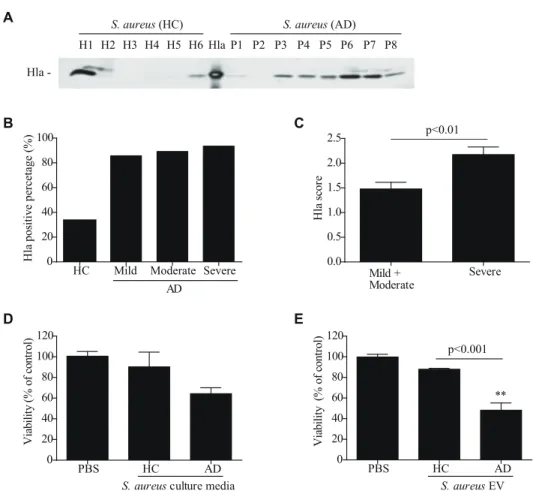
To evaluate the relationship betweena-hemolysin production from S. aureus and AD, a-hemolysin levels were measured in culture media ofS. aureusfrom AD patients and healthy controls. All bacteria were cultured under the same conditions and a -hemolysin levels in the culture media were measured by western blot. Although two of six samples ofS. aureusfrom healthy controls produceda-hemolysin, mostS. aureusfrom AD patients (seven of eight) produced it (Fig. 1, A). Furthermore, whena-hemolysin production was measured inS. aureusculture media from 90 AD patients, 91% of S. aureusfrom AD patients were positive fora -hemolysin compared to 33% of healthy controls (Fig. 1, B and Table 1). In terms of a-hemolysin production according to AD severity,a-hemolysin production was significantly higher fromS. aureusfrom the severe group compared toS. aureusfrom the mild and moderate groups (Fig. 1, C and Table 1). These findings suggest that a-hemolysin fromS. aureus is related to AD disease development and/or progression.
The effect ofa-hemolysin on keratinocyte cell death
Numerous reports have shown thata-hemolysin can kill many types of cells, including epidermal keratinocytes [12,13,14]. Becausea-hemolysin-producing S. aureuswere more frequent in AD patients compared to healthy controls, we evaluated the effect ofa-hemolysin on keratinocyte death by measuring cell viability after treatment with bacterial culture media. We found that HaCaT keratinocyte viability was decreased upon treatment with culture media ofS. aureusfrom AD patients compared to healthy controls (Fig. 1, D). In addition, we evaluated the effect ofS. aureus EVs on HaCaT keratinocyte death. The results indicate that keratinocyte viability was significantly decreased upon treatment withS. aureusEVs from AD patients (Fig. 1, E). These findings suggest that a-hemolysin in S. aureus EVs may be an important etiologic agent in AD pathogenesis.
The role ofa-hemolysin inS. aureusEVs on keratinocyte cell death
Based on these data, we sought to determine whetherS. aureus EVs harbora-hemolysin, which induces keratinocyte cell death. For these experiments, we used the S. aureus 14458 strain, a reference strain used previously [16,17]. Our data show thata -hemolysin was present in both culture media and EVs from theS. aureus14458 strain (Fig. 2, A). In terms ofa-hemolysin hemolytic activity, we found that hemolysis was induced byS. aureusEVs in a dose-dependent manner, as well as by a soluble form of a
-Figure 1.S. aureuson atopic dermatitis skin producesa-hemolysin. A,Detection ofa-hemolysin in culture media ofS. aureusisolated from
the skin of healthy controls (HC) and atopic dermatitis patients (AD).B,The percentage ofa-hemolysin-producingS. aureusfrom 90 AD patients.C, The amount ofa-hemolysin in culture media was evaluated by scoring western blot band sizes from 0 to 3.DandE,Human keratinocyte viability after treatment with 10mg/mlS. aureusculture media (D) and 25mg/ml EVs (E) for 24 hr. ** P,0.01 versus the PBS group.
hemolysin (sHla) and theS. aureusculture media (Fig. 2, B and Fig. S1, A). Next, to evaluate the cytotoxic effect ofa-hemolysin on keratinocytes, S. aureusEVs were added to HaCaT keratino-cytes. Keratinocyte viability was significantly decreased upon treatment with S. aureus EVs, S. aureusculture media, and sHla compared to PBS alone (Fig. 2, C).S. aureusEVs and sHla also killed primary human keratinocytes (data not shown). To elucidate the role of a-hemolysin in EVs on keratinocyte cell death, we performed experiments usingS. aureusstrains that produce various amounts of a-hemolysin. We found that the amounts of a -hemolysin in S. aureus EVs were positively associated with keratinocyte death (Fig. S1, B). Next, to evaluate the effect of a-hemolysin deficiency on keratinocyte death, we isolated EVs froma-hemolysin-positive WT (Newman strain) anda -hemolysin-deficient mutant strains. Cell viability of HaCaT keratinocytes was significantly higher after treatment withS. aureusEVs from thea -hemolysin-positive strain compared to the a-hemolysin-negative strain. In addition, keratinocyte death was reversed by treatment with EVs isolated from the a-hemolysin-negative strain that complemented a-hemolysin using a plasmid (Fig. 2, D). Furthermore, the results indicate that EVs from a -hemolysin-negative S. aureus strains from either AD patients or healthy controls did not induce HaCaT keratinocyte death, whereas death was induced by a-hemolysin-positive S. aureus strains from AD patients (Fig. 2, E). Taken together, these findings suggest thata -hemolysin inS. aureusEVs are a key player in AD pathogenesis via keratinocyte death.
Comparison between soluble and EV-associateda -hemolysin on keratinocyte cell death
Although sHla was reported to act on the plasma membrane of keratinocytes [14], the working mechanism of S. aureus EVs on keratinocyte death remains unknown. First, we evaluated the cellular localization of EVs. When fluorescence-labeled EVs were added to HaCaT keratinocytes, EVs were internalized into the cytoplasm (Fig. 3, A). In our measurements, EVs contained approximately 0.6mg ofa-hemolysin after treatment with 10mg of EVs (quantified by total protein amount) (Fig. S2). When HaCaT keratinocytes were treated with equal amounts of soluble and EVs forms of a-hemolysin, a-hemolysin in EVs was more effectively delivered into the keratinocytes (Fig. 3, B). Furthermore, to compare the cytotoxic effects of EV-associateda-hemolysin and
sHla, HaCaT keratinocytes were treated with equal amounts ofa -hemolysin in the soluble and EV forms. This study showed that cytotoxicity was enhanced after treatment with EV-associateda -hemolysin compared to sHla (Fig. 3, C). Keratinocyte death was induced faster by EV-associateda-hemolysin versus sHla (Fig. 3, D). Together, these findings suggest that compared to sHla, EV-associateda-hemolysin potently induces keratinocyte death.
a-hemolysin localization inS. aureusEVs
Generally, secreted soluble toxins are neutralized and lose their activity by engaging the host immune system [18,19,20]. Compared to soluble toxins, EVs may protect toxins by enveloping them with cell membrane. Our data show that EV-associateda -hemolysin remained intact after proteinase K treatment; however, after EVs were disrupted by lysis buffer, EV-associated a -hemolysin was degraded by proteinase K (Fig. 3, E). This finding suggests thata-hemolysin is localized in the EV lumen, not on the EV surface. Moreover, when both soluble and EV-associateda -hemolysin were treated with the anti-a-hemolysin antibody, keratinocyte cell death induced by EV-associated a-hemolysin was unaffected, whereas the cytotoxicity induced by sHla was reversed (Fig. 3, F). Collectively, these findings suggest that a -hemolysin in the EV lumen enhances killing of keratinocytes and evasion of host immune defenses.
Comparison of cell death mechanisms between EV-associated and solublea-hemolysin
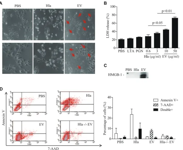
Although several reports have suggested that sHla induces host cell apoptosis [12,21,22], the exact mechanism of EV-associateda -hemolysin in keratinocyte death is unknown. We found that the morphology of cell death differed between soluble and EV-associateda-hemolysin; HaCaT keratinocytes were rounded and many cells were floating upon sHla treatment, which is indicative of apoptotic cell death; whereas cells underwent cell rupture upon EVs treatment, suggestive of necrosis (Fig. 4, A). In accordance with the observed cell morphology, LDH release, a marker of necrosis or cell rupture, was significantly increased after EVs treatment (Fig. 4, B). In addition, high-mobility group box (HMGB)-1, another marker for necrosis [23] was detected in culture media of EV-treated HaCaT keratinocytes, but not from that of sHla-treated cells (Fig. 4, C). Flow cytometry also showed that the number of 7-AAD-positive cells (necrotic cells) increased Table 1.Demographic and clinical characteristics of atopic dermatitis patients.
Mild Moderate Severe Total
n = 7 n = 37 n = 46 n = 90
Age, months 66.43 51 62 55
(range) (36–145) (4–163) (2–510) (2–510)
Sex (male: female) 01:02.5 01:01.2 1:01 01:01.1
MRSA (%) 42.8 43.2 28.6 38.9
a-hemolysin (%)"
0 14.3 10.8 6.5 8.9
1 28.6 48.7 26.1 35.5
2 42.8 24.3 10.9 18.9
3 14.3 16.2 56.5 36.7
Values shown are age in months (median), ratio of males versus females, percentage of methicillin-resistantStaphylococcus aureus, and percentage of patients colonized witha-hemolysin-producingS. aureus.
"
The percentage was determined by the amount ofa-hemolysin produced byS. aureusisolated from the patients. The amount ofa-hemolysin was measured by band intensity using Multi Gauge V3.1. Scores are as follows: 0: zero; 1: up to 6000 arbitrary units (AU); 2: from 6001 to 15,000 AU; and 3: over 15,001 AU.
after EVs treatment, whereas annexin V-positive cells (apoptotic cells) increased after sHla treatment (Fig. 4, D). To sum up, these findings suggest that EV-associateda-hemolysin induces necrotic cell death by carrying a-hemolysin into the cytoplasm of keratinocytes, whereas sHla induces keratinocyte death via apoptosis.
In vivoeffects of EV-associated and solublea-hemolysin on skin barrier disruption
Because keratinocytes are major constituents of the skin barrier, keratinocyte death is a key contributor of skin barrier disruption in AD pathogenesis [2,5]. To examine the in vivo effects of EV-associated a-hemolysin on skin barrier disruption, EVs froma -hemolysin-positive and -negativeS. aureusstrains, as well as sHla, were applied to the back skin of mice. Evans blue dye penetration was enhanced by a-hemolysin-positive EVs or sHla treatment compared to PBS ora-hemolysin-negative EVs treatment (Fig. 5, A). Next, we aimed to evaluate thein vivo effects of skin barrier disruption on the penetration of high-molecular-weight allergens. Therefore, fluorescein-labeled ovalbumin (OVA) was administered to EV- and sHla-treated skin, and OVA levels were measured in
excised skins by quantification of fluorescein. We found that OVA-fluorescein levels were increased in mice treated witha -hemolysin-positive EVs or sHla compared toa-hemolysin-negative EVs or PBS treatment (Fig. 5, B). Together, these findings suggest that both EV-associated and solublea-hemolysin induce skin barrier disruption via keratinocyte cell death and consequently enhance penetration of high-molecular-weight allergens.
Effects of EV-associated and solublea-hemolysin on pro-inflammatory mediator production by keratinocytes
Our previous reports showed that S. aureus EVs induced pro-inflammatory cytokine production by dermal fibroblasts and airway epithelial cells [17,24]. To evaluate the effect of EV-associated and soluble a-hemolysin on the production of pro-inflammatory mediators from keratinocytes, we measured the production of pro-inflammatory cytokines from HaCaT keratino-cytes after treatment with equal amounts ofa-hemolysin in the EV and soluble forms. We found that the cytokine production profile differed between EV-associated and solublea-hemolysin. IL-6 was enhanced by both EV-associated and solublea-hemolysin, IL-1b was enhanced only by EV-associated a-hemolysin, and TNF-a Figure 2.a-Hemolysin inS. aureusEVs is a key factor for EVs cytotoxicity. A,The presence ofa-hemolysin in culture media, EVs-removed
culture media (media-EVs), and EVs from theS. aureusATCC14458 strain.B,Hemolytic function of solublea-hemolysin and EVs.C,Viability of human keratinocytes after treatment with soluble hemolysin (5mg/ml),S. aureusculture media (10mg/ml), and EVs (20mg/ml).D,a-Hemolysin in EVs from the Newman strain,a-hemolysin-deficient mutant strain, anda-hemolysin complemented strain (pHla). Human keratinocyte viability after treatment with each EVs (40mg/ml).E,a-Hemolysin in EVs from randomly selectedS. aureusfrom healthy controls (HC) and atopic dermatitis (AD) patients. Viability of human keratinocytes after treatment with EVs (25mg/ml). * P,0.05; ** P,0.01 versus the PBS group.
was enhanced only by sHla (Fig. 6, A). Moreover, IL-1band IL-6 production by keratinocytes was decreased after treatment with EVs derived from the a-hemolysin-negative S. aureus strain compared to the a-hemolysin-positive strain, whereas TNF-a production was enhanced in the former group versus the latter group (Fig. 6, B). Collectively, these findings suggest that EV-associated a-hemolysin induces IL-1band IL-6 production from keratinocytes but may inhibit TNF-a production induced by S. aureusEVs or other signals.
Effects of EV-associated and solublea-hemolysin on the development of AD-like skin inflammation
Finally, we evaluatedin vivoeffects of EV-associated and soluble a-hemolysin on the development of skin inflammation. To do this, the same amounts ofa-hemolysin in the EV-associated and soluble forms were administered epicutaneously into the mouse skin and
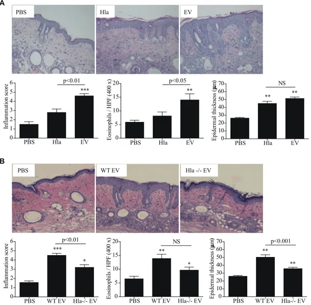
histological alterations were evaluated. Dermal infiltration of inflammatory cells, particularly eosinophils, was enhanced by EV-associateda-hemolysin but not by sHla. However, both forms of a-hemolysin increased epidermal cell hyperplasia (Fig. 7, A). Furthermore, dermal infiltration of inflammatory cells, including eosinophils, and epidermal thickening were reduced in skin treated with repeated applications ofa-hemolysin-negative EVs (Fig. 7, B). Taken together, our data show that a-hemolysin inS. aureus EVs are a key player for the development of AD phenotypes, including epidermal thickening and eosinophilic inflammation in the dermis.
Discussion
If the skin barrier is disrupted, pathogen-associated antigens and allergens can penetrate into the human body. Skin barrier disruption is considered one of the major causes of AD Figure 3. EVs are more potent mediators of keratinocyte death compared to solublea-hemolysin. A,Confocal microscopy of human
keratinocytes with DiI-labeledS. aureusEVs (red:S. aureusEVs, blue: nucleus).S. aureusEVs and nucleus are shown merged on DIC image (lower panel). Scale bar, 20mm.B,a-Hemolysin in keratinocytes after treatment with identical amounts of solublea-hemolysin and EVs.C,Viability of keratinocytes after treatment with each reagent.D,Time dependence of cell death.E,a-Hemolysin on intact and disrupted EVs after treatment with proteinase K.F,Keratinocyte viability after treatment with solublea-hemolysin (3mg/ml) and EVs (10mg/ml) with the anti-a-hemolysin antibody (5% of culture media volume). * P,0.05; ** P,0.01 versus the PBS group; NS, not significant.
exacerbation, but many studies have focused on intrinsic factors, such as host molecules that maintain the skin barrier [4,25]. Though it is known that staphylococcal toxins can affect skin barrier integrity for a long time, the role ofa-hemolysin in skin barrier disruption via killing keratinocytes have been reported recently [26,27]. In the present study, we elucidated the role of extrinsic factor (a-hemolysin and EVs) on the development of skin barrier disruption and AD-like inflammation. The present data show that a-hemolysin-producing S. aureus had colonized AD patients, and that soluble and EV-associateda-hemolysin induce keratinocyte cell death, consequently enhancing skin penetration of high-molecular-weight allergens. However, EV-associated a -hemolysin was found to induce keratinocyte necrosis, whereas sHla induced keratinocyte apoptosis. Additionally, EV-associated a-hemolysin induced epidermal thickening and eosinophilic inflammation in the dermis, whereas the soluble form induced only epidermal thickening. This is the first report thata-hemolysin in EVs derived fromS. aureusinduces skin barrier disruption and AD-like skin inflammation, predominantly via keratinocyte necrosis and the production of pro-inflammatory mediators by keratinocytes.
S. aureussecretes toxins, includinga-hemolysin, as both soluble and EV-associated forms. EV-associated toxins have some advantages in intercellular communication compared to soluble forms. Toxins in the EV lumen, such as EV-associated a -hemolysin, can be protected from clearance by host defense
systems, including antibody-mediated neutralization and protease-mediated destruction, enabling toxins to retain their function and travel long distances without interference [28]. In addition, because EVs are membrane-enveloped complexes, they can deliver their contents easily into the cytoplasm by fusing with the host cell membrane or by endocytosis after interaction between ligands on EVs and receptors on host cells [18,29]. Indeed, the present data show thatS. aureus EVs were internalized into the cytoplasm of keratinocytes and that EVs efficiently delivered a -hemolysin to the cytoplasm. Collectively, these findings suggest that EVs-associated toxins are key molecules in disease pathogen-esis.
EVs derived from several bacteria kill host cells by transferring cytotoxic factors [29,30,31]. It was reported thatS. aureusEVs can induce death of human epidermoid cancer cells [32]. Additionally, recent data indicate thata-hemolysin associated withS. aureusEVs can induce death of cervical cancer cells [33]. In addition, the present data showed that EV-associated a-hemolysin can also induce keratinocyte necrosis, whereas the soluble form induces apoptosis. This difference can be partly attributed to the differences in delivery efficacy between EV-associated and soluble a-hemolysin. We can speculate thata-hemolysin can be efficiently delivered by EVs and that a higha-hemolysin concentration in the cytoplasm attacks cell membranes and/or cytoplasm organelles, thereby initiating necrosis pathways.
Figure 4.S. aureusEVs induces necrotic cell death, in contrast to solublea-hemolysin. A,Micrographs of cell death by solublea-hemolysin
(3mg/ml) and EVs (25mg/ml). Pictures were taken under microscope at x 100 (upper panel) and x 200 (lower panel) magnification. Red arrows indicate ruptured cells.B,LDH levels in culture media after treatment with solublea-hemolysin and EVs.C,HMGB-1 in culture media after treatment with solublea-hemolysin and EVs.D,Flow cytometry analyses of annexin V and 7-AAD staining.
Figure 5.a-Hemolysin fromS. aureusinduces skin barrier disruption. A,Evans blue dye penetration into mouse skin after treatment with
solublea-hemolysin, EVs (5mg), EVs from the Newman strain (10mg), and EVs from thea-hemolysin-deficient strain (10mg) (n = 2 mice per group).B, Level of penetrated fluorescein-labeled OVA in skin treated with solublea-hemolysin (1.5mg), Newman EVs (10mg), anda-hemolysin-deficient EVs (10mg) (n = 3 mice per group).
doi:10.1371/journal.pone.0100499.g005
Figure 6.S. aureusEVs and solublea-hemolysin induce production of different cytokines. AandB,Pro-inflammatory cytokines in human
keratinocyte culture media after treatment with solublea-hemolysin and EVs (A) and 40mg/ml of EVs from each strain (B). * P,0.05; ** P,0.01 versus the PBS group.
Cell death via necrosis or apoptosis can result in immunolog-ically distinct consequences [22]. The present data showed that EV-associateda-hemolysin up-regulateed the production of IL-6, a key mediator of Th17 polarization, which is associated with keratinocyte necrosis. In contrast, sHla enhanced the production of TNF-a, but not of IL-6. Our previous data demonstrated that skin exposure to S. aureusEVs induced a Th17-cell response in regional lymph nodes and ultimately resulted in AD-like skin inflammation [17]. Together, these findings suggest thatS. aureus EVs induce skin barrier disruption and Th17-associated inflam-mation in the dermis via effective delivery of a-hemolysin into keratinocytes.
The expression of toxins from S. aureus can be regulated by environmental stress, and also host factors, such as filaggrin production [11,34,35,36]. In the present study, most of S. aureus isolated from the skin of AD patients was found to produce a -hemolysin, as soluble and EV-associated forms, which can induce skin barrier disruption. In contrast, we can hardly detect the production of a-hemolysin from S. aureus isolated from healthy control subjects. Moreover, the levels of a-hemolysin in lavage
fluids from the AD patient skin were found to be positively correlated with AD severity. Because EVs are produced via shedding of bacterial membrane and contain many pathogenic molecules [16,18], it is hard to define the effect of EVs by deleting or adding specific molecules. We can assume that many proteins interact with other proteins and the relationship between various proteins can affect overall characteristics of EVs. Nevertheless, out present findings suggest thata-hemolysin, especially EV-associated form, is a good biomarker for the diagnosis and therapy of AD.
In summary, the present study showed that a-hemolysin, present in the EV lumen, induces skin barrier disruption and AD-like skin inflammation via keratinocyte necrosis and/or up-regulation of pro-inflammatory mediator production from kerati-nocytes. Moreover,S. aureuscolonized on AD patient skin secretes a-hemolysin, which is significantly related to AD severity. These findings indicate thata-hemolysin, particularly the EV-associated form, is a novel target for diagnosis and treatment of AD. Figure 7.a-Hemolysin-positiveS. aureusEVs induces atopic dermatitis-like skin inflammation. AandB,Skin alterations after treatment
with 5mg of solublea-hemolysin and EVs (A) and 10mg of EVs from the Newman wild-type anda-hemolysin-deficient strains (B) (n = 5 mice per group). * P,0.05; ** P,0.01; *** P,0.001 versus the PBS group; NS, not significant.
Materials and Methods
Ethics statement
This study was carried out in strict accordance with the recommendations in the Guide for the Care and Use of Laboratory Animals of the National Institute of Health. The experimental protocols were approved by the Institutional Animal Care and Use Committee at POSTECH, Pohang, Republic of Korea (Permit Number: 2011-01-0027). All animal experiments were planned in order to minimize mice suffering. The study protocol for human samples was approved by the Ethics Committee of Seoul Suncheonhyang Hospital (Permit Number: 2010-01-0016). Participants provided their written informed consent to participate in the present study.
Mice
SKH-HR1 hairless mice were purchased from Charles River Laboratories Japan, Inc. (Yokohama, Japan) and were bred in a specific-pathogen-free facility at Pohang University of Science and Technology (POSTECH; Pohang, Republic of Korea).
Keratinocytes and bacteria
Immortalized human epidermal keratinocytes (HaCaT cells) were kindly donated by Jeung-Hoon Lee (Chungnam National University, Daejeon, Korea). Primary human epidermal keratino-cytes (HEK cells) were purchased from ScienCell (Carlsbad, CA). HaCaT cells were maintained in DMEM (Hyclone Laboratories, South Logan, UT) containing fetal bovine serum (Gibco) and antibiotics (Hyclone Laboratories). ATCC14458 S. aureus strain was purchased from ATCC. The Newman strain, a-hemolysin deficient strain, anda-hemolysin complemented strain were kindly provided by Juliane Bubeck Wardenburg (University of Chicago, IL).
S. aureusisolation from human samples
S. aureuswas collected from the skin lesions of 90 AD patients visiting the Pediatric Clinic of Seoul Suncheonhyang Hospital (Seoul, Republic of Korea). S. aureus from healthy controls was isolated from the skin of the upper limbs and subungual spaces from 36 volunteers who had no AD symptoms.
Isolation ofS. aureusEVs
S. aureusEVs were obtained as described previously [17]. Briefly, S. aureuswas cultured in nutrient broth or tryptic soybean broth (Difco, Sparks, MD) at 37uC to an optical density (OD) of 1.5 at 600 nm. Bacteria were removed by centrifugation and filtration. The filtrate was concentrated and the resulting concentrate was filtered and ultracentrifuged at 150,0006g for 3 h. The pellet was resuspended in PBS.S. aureus-derived EVs protein concentrations were measured using BCA assays (Thermo Scientific, Rockford, IL). Hereafter, the dose ofS. aureusEVs refers to the quantity ofS. aureus-derived EVs proteins.
Cytotoxicity measurements
Keratinocyte viability was measured at 24 h after treatment using thiazolyl blue tetrazolium bromide (MTT) purchased from Sigma Aldrich (St. Louis, MO). The PBS control group was used as 100% viability. Lactate dehydrogenase (LDH) activity in the culture supernatant was measured using the LDH cytotoxicity detection kit purchased from Takara Bio Inc. (Otsu, Japan) according to the manufacturer’s instructions.
Hemolysis measurements
Red blood cells (RBCs) were isolated from mouse whole blood. a-Hemolysin (Sigma-Aldrich, USA) andS. aureusEVs were added to RBCs and incubated at 37uC. After 1 h, the remaining RBCs were removed by centrifugation and the optical density at 540 nm of the supernatant was measured. RBC lysis buffer was used as a positive control.
Flow cytometry analyses of cell death
Annexin V (BD biosciences) and 7-AAD (Biolegend) were used detect cell death using flow cytometry. Cells were treated withS. aureusEVs ora-hemolysin, and cells in the culture supernatant and remaining cells were collected. Cells were processed according to the manufacturer’s instructions. Processed cells were analyzed using FACSCalibur (Becton Dickinson, USA).
In vivoassays
For Evans blue dye assays and fluorescein-labeled OVA penetration, gauze soaked with 100-ml PBS containing S. aureus EVs or a-hemolysin was placed and secured on mildly tape-stripped skin. Mice were treated five times in 1 week withS. aureus EVs anda-hemolysin. Dorsal skin was serially fixed with 30, 50, 70, and 100% methanol. After fixation, 0.1% Evans blue was added for 10 min, followed by washing with PBS. Skin was excised, immersed in formamide, and incubated at 60uC. After 6 h, the optical density at 620 nm was measured. For OVA-fluorescein penetration, 50mg of fluorescein-conjugated OVA
were added twice toS. aureus EVs- or a-hemolysin-treated skin. Next, the skin was excised and homogenized. Fluorescence was measured using a Wallac 1420 Victor luminometer (American Instrument Exchange, Inc., Harverville, MA). Skin alterations were evaluated after S. aureus EVs and a-hemolysin treatment three times per week for 3 weeks, as reported previously [17].
Statistical analyses
For multiple comparisons, one-way analysis of variance (ANOVA) was used first. If significant differences were found, individual t-tests or Wilcoxon’s rank-sum tests were performed between pairs of groups. Differences were considered statistically significant if P,0.05.
Supporting Information
Figure S1 A,Hemolytic activity of EVs and culture media from the ATCC14458 strain and a-hemolysin-deficient EVs strain. Hemolysis mediated by RBC lysis buffer is used as 100%.B,The amount ofa-hemolysin on EVs from different strains (left panels) and cytotoxicity of EVs on keratinocytes (right panel). ** P,0.01 versus the PBS group.
(EPS)
Figure S2 Quantification of a-hemolysin in EVs. Western blotting showed that 0.6mg ofa-hemolysin was present in 10mg
of EVs protein from the ATCC14458 strain and 0.25mg of a
-hemolysin was present in 10mg of EVs protein from the Newman
strain. (EPS)
Acknowledgments
Author Contributions
Conceived and designed the experiments: SH EC BP YK. Performed the experiments: SH EC TM JK SJ BL. Analyzed the data: SH YG MK YJ BP
YK. Contributed reagents/materials/analysis tools: BL YG BP YK. Wrote the paper: SH TM BP YK. Obtained funding: BP YK.
References
1. Bieber T (2008) Atopic dermatitis. N Engl J Med 358: 1483–1494.
2. Cork MJ, Danby SG, Vasilopoulos Y, Hadgraft J, Lane ME, et al. (2009) Epidermal barrier dysfunction in atopic dermatitis. J Invest Dermatol 129: 1892–1908.
3. Elias PM, Hatano Y, Williams ML (2008) Basis for the barrier abnormality in atopic dermatitis: outside-inside-outside pathogenic mechanisms. J Allergy Clin Immunol 121: 1337–1343.
4. Boguniewicz M, Leung DY (2011) Atopic dermatitis: a disease of altered skin barrier and immune dysregulation. Immunol Rev 242: 233–246.
5. Trautmann A, Akdis M, Kleemann D, Altznauer F, Simon HU, et al. (2000) T cell-mediated Fas-induced keratinocyte apoptosis plays a key pathogenetic role in eczematous dermatitis. J Clin Invest 106: 25–35.
6. Rebane A, Zimmermann M, Aab A, Baurecht H, Koreck A, et al. (2012) Mechanisms of IFN-gamma-induced apoptosis of human skin keratinocytes in patients with atopic dermatitis. J Allergy Clin Immunol 129: 1297–1306. 7. Zimmermann M, Koreck A, Meyer N, Basinski T, Meiler F, et al. (2011)
TNF-like weak inducer of apoptosis (TWEAK) and TNF-alpha cooperate in the induction of keratinocyte apoptosis. J Allergy Clin Immunol 127: 200–207, 207 e201–210.
8. Otto M (2010) Basis of virulence in community-associated methicillin-resistant Staphylococcus aureus. Annu Rev Microbiol 64: 143–162.
9. Huang JT, Abrams M, Tlougan B, Rademaker A, Paller AS (2009) Treatment of Staphylococcus aureus colonization in atopic dermatitis decreases disease severity. Pediatrics 123: e808–814.
10. Semic-Jusufagic A, Bachert C, Gevaert P, Holtappels G, Lowe L, et al. (2007) Staphylococcus aureus sensitization and allergic disease in early childhood: population-based birth cohort study. J Allergy Clin Immunol 119: 930–936. 11. Schlievert PM, Strandberg KL, Lin YC, Peterson ML, Leung DY (2010)
Secreted virulence factor comparison between methicillin-resistant and methi-cillin-sensitive Staphylococcus aureus, and its relevance to atopic dermatitis. J Allergy Clin Immunol 125: 39–49.
12. Bantel H, Sinha B, Domschke W, Peters G, Schulze-Osthoff K, et al. (2001) alpha-Toxin is a mediator of Staphylococcus aureus-induced cell death and activates caspases via the intrinsic death pathway independently of death receptor signaling. J Cell Biol 155: 637–648.
13. Prince LR, Graham KJ, Connolly J, Anwar S, Ridley R, et al. (2012) Staphylococcus aureus induces eosinophil cell death mediated by alpha-hemolysin. PLoS One 7: e31506.
14. Walev I, Martin E, Jonas D, Mohamadzadeh M, Muller-Klieser W, et al. (1993) Staphylococcal alpha-toxin kills human keratinocytes by permeabilizing the plasma membrane for monovalent ions. Infect Immun 61: 4972–4979. 15. Wichmann K, Uter W, Weiss J, Breuer K, Heratizadeh A, et al. (2009) Isolation
of alpha-toxin-producing Staphylococcus aureus from the skin of highly sensitized adult patients with severe atopic dermatitis. Br J Dermatol 161: 300–305.
16. Lee EY, Choi DY, Kim DK, Kim JW, Park JO, et al. (2009) Gram-positive bacteria produce membrane vesicles: proteomics-based characterization of Staphylococcus aureus-derived membrane vesicles. Proteomics 9: 5425–5436. 17. Hong SW, Kim MR, Lee EY, Kim JH, Kim YS, et al. (2011) Extracellular
vesicles derived from Staphylococcus aureus induce atopic dermatitis-like skin inflammation. Allergy 66: 351–359.
18. Kulp A, Kuehn MJ (2010) Biological functions and biogenesis of secreted bacterial outer membrane vesicles. Annu Rev Microbiol 64: 163–184.
19. Radjainia M, Hyun JK, Leysath CE, Leppla SH, Mitra AK (2010) Anthrax toxin-neutralizing antibody reconfigures the protective antigen heptamer into a supercomplex. Proc Natl Acad Sci U S A 107: 14070–14074.
20. Sato H, Sato Y, Ito A, Ohishi I (1987) Effect of monoclonal antibody to pertussis toxin on toxin activity. Infect Immun 55: 909–915.
21. Bayles KW, Wesson CA, Liou LE, Fox LK, Bohach GA, et al. (1998) Intracellular Staphylococcus aureus escapes the endosome and induces apoptosis in epithelial cells. Infect Immun 66: 336–342.
22. Haslinger B, Strangfeld K, Peters G, Schulze-Osthoff K, Sinha B (2003) Staphylococcus aureus alpha-toxin induces apoptosis in peripheral blood mononuclear cells: role of endogenous tumour necrosis factor-alpha and the mitochondrial death pathway. Cell Microbiol 5: 729–741.
23. Scaffidi P, Misteli T, Bianchi ME (2002) Release of chromatin protein HMGB1 by necrotic cells triggers inflammation. Nature 418: 191–195.
24. Kim MR, Hong SW, Choi EB, Lee WH, Kim YS, et al. (2012) Staphylococcus aureus-derived extracellular vesicles induce neutrophilic pulmonary inflamma-tion via both Th1 and Th17 cell responses. Allergy 67: 1271–1281. 25. O’Regan GM, Sandilands A, McLean WH, Irvine AD (2009) Filaggrin in atopic
dermatitis. J Allergy Clin Immunol 124: R2–6.
26. Bin L, Kim BE, Brauweiler A, Goleva E, Streib J, et al. (2012) Staphylococcus aureus alpha-toxin modulates skin host response to viral infection. J Allergy Clin Immunol 130: 683–691 e682.
27. Brauweiler AM, Bin L, Kim BE, Oyoshi MK, Geha RS, et al. (2013) Filaggrin-dependent secretion of sphingomyelinase protects against staphylococcal alpha-toxin-induced keratinocyte death. J Allergy Clin Immunol 131: 421–427 e421– 422.
28. Bomberger JM, Maceachran DP, Coutermarsh BA, Ye S, O’Toole GA, et al. (2009) Long-distance delivery of bacterial virulence factors by Pseudomonas aeruginosa outer membrane vesicles. PLoS Pathog 5: e1000382.
29. Parker H, Chitcholtan K, Hampton MB, Keenan JI (2010) Uptake of Helicobacter pylori outer membrane vesicles by gastric epithelial cells. Infect Immun 78: 5054–5061.
30. Kim YR, Kim BU, Kim SY, Kim CM, Na HS, et al. (2010) Outer membrane vesicles of Vibrio vulnificus deliver cytolysin-hemolysin VvhA into epithelial cells to induce cytotoxicity. Biochem Biophys Res Commun 399: 607–612. 31. Jin JS, Kwon SO, Moon DC, Gurung M, Lee JH, et al. (2011) Acinetobacter
baumannii secretes cytotoxic outer membrane protein A via outer membrane vesicles. PLoS One 6: e17027.
32. Gurung M, Moon DC, Choi CW, Lee JH, Bae YC, et al. (2011) Staphylococcus aureus produces membrane-derived vesicles that induce host cell death. PLoS One 6: e27958.
33. Thay B, Wai SN, Oscarsson J (2013) Staphylococcus aureus alpha-toxin-dependent induction of host cell death by membrane-derived vesicles. PLoS One 8: e54661.
34. Morfeldt E, Taylor D, von Gabain A, Arvidson S (1995) Activation of alpha-toxin translation in Staphylococcus aureus by the trans-encoded antisense RNA, RNAIII. EMBO J 14: 4569–4577.
35. Ohlsen K, Koller KP, Hacker J (1997) Analysis of expression of the alpha-toxin gene (hla) of Staphylococcus aureus by using a chromosomally encoded hla::lacZ gene fusion. Infect Immun 65: 3606–3614.