Dependent
Tao Xu, William Brown, Martin G. Marinus*
Department of Biochemistry and Molecular Pharmacology, University of Massachusetts Medical School, Worcester, Massachusetts, United States of America
Abstract
Bleomycin (BLM) is a glycopeptide antibiotic and anti-tumor agent that targets primarily the furanose rings of DNA and in the presence of ferrous ions produces oxidative damage and DNA strand breaks.Escherichia colicells growing in broth medium and exposed to low concentrations of BLM contain double-strand breaks and require homologous recombination to survive. To a lesser extent, the cells also require the abasic (AP) endonucleases associated with base excision repair, presumably to repair oxidative damage. As expected, there is strong induction of the SOS system in treated cells. In contrast,E. colicells growing in glucose or glycerol minimal medium are resistant to the lethal action of BLM and do not require either homologous recombination functions or AP-endonucleases for survival. DNA ligase activity, however, is needed for cells growing in minimal medium to resist the lethal effects of BLM. There is weak SOS induction in such treated cells.
Citation: Xu T, Brown W, Marinus MG (2012) Bleomycin Sensitivity in Escherichia coli is Medium-Dependent. PLoS ONE 7(3): e33256. doi:10.1371/ journal.pone.0033256
Editor:Michael R. Volkert, University of Massachusetts Medical School, United States of America
ReceivedDecember 6, 2011;AcceptedFebruary 6, 2012;PublishedMarch 15, 2012
Copyright:ß2012 Xu et al. This is an open-access article distributed under the terms of the Creative Commons Attribution License, which permits unrestricted use, distribution, and reproduction in any medium, provided the original author and source are credited.
Funding:This work was supported by grant R01 GM0637900-08 from the National Institutes of Health (www.nih.gov). The grant paid for all supplies and wages and salaries connected with this work. The funder had no role in study design, data collection and analysis, decision to publish, or preparation of the manuscript.
Competing Interests:Co-author MG Marinus is a PLoS ONE Editorial Board member. This does not alter the authors’ adherence to all PLoS ONE policies on sharing data and materials.
* E-mail: [email protected]
Introduction
Bleomycin (BLM) is a glycopeptide antibiotic and anti-tumor agent isolated fromStreptomyces verticillis[1,2] that targets primarily the furanose rings of DNA. Degradation by BLM is initiated by generating a free radical, in the presence of ferrous ion, in the deoxyribose resulting in two different types of DNA damage [3,4]. At low oxygen tension, oxidized abasic (AP) sites are favored while at high oxygen tension single-and double-strand breaks (DSBs) predominate. These alternative pathways lead to a mix of abasic sites and strand breaks which occur at a 1:1 ratio [5]. The DSBs are suspected to be the major cause of cell death. Up to one-third of BLM-induced lesions are double-strand breaks which consist of either two identical breaks in opposite strands or arise from an abasic site with a closely opposed strand break [6].
In order to more fully understand the mechanism of BLM toxicity in Escherichia coli, previous studies [7,8] have shown an increased BLM sensitivity of lexA and recA mutant strains indicating that the SOS response is an important mechanism cells use to resist the toxic effects of the drug. The increased sensitivity ofrecAandrecBCmutant strains to BLM indicates a role for homologous recombination in cell survival [8,9]. This result is consistent with a requirement for recombinational repair as a consequence of DSBs in DNA. Surprisingly,recNandrecGmutant strains, in an otherwise wildtype background, were also found to be sensitive to BLM exposure, a feature not shared with other common DNA damaging agents [8]. The role of RecN in recombination/repair is unknown while RecG is a helicase that translocates Holliday junction intermediates in recombination and repair [10].
We have confirmed and extended the above observations to additional mutant strains and show that at the low concentrations of BLM used here, homologous recombination is the principal pathway of repair in cells growing in broth. We also show that there is differential sensitivity ofE. coliin glucose minimal medium versus rich medium after exposure to BLM.
Results
Strain construction
A previous study [8] used mutations affecting repair/recombi-nation in an AB1157 background. These mutations are either transposon insertions or point mutations, many of which have not been characterized at the DNA sequence level. We have constructed a panel of mutant strains in a different genetic background (GM7330) using deletion alleles wherever possible and have used them in this study. These strains have been used previously to study the toxic effects of cisplatin and MNNG [11,12].
DNA repair-deficient mutant strains
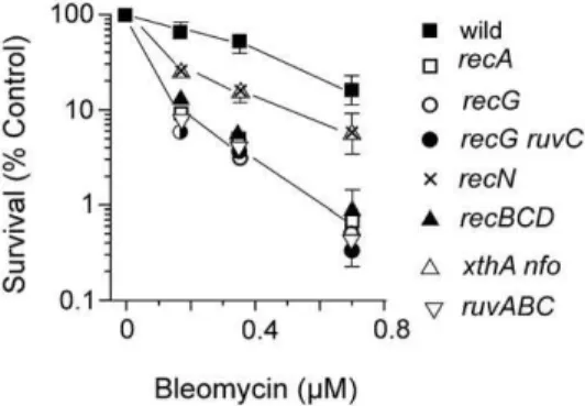
An xthA nfo strain, defective for AP endonucleases, was more sensitive than wildtype after BLM treatment (Fig. 1). This result is consistent with the known base damage inflicted by BLM requiring base excision repair enzymes. The Fpg and MutY glycosylases are active on oxidative base lesions butfpg(mutM) and
and mutS strains affected for mismatch repair (data not shown). Two temperature-sensitive mutant strains defective in DNA ligase (lig-4 and lig-7) were more sensitive at the higher temperature when exposed to BLM (see below).
Mutant strains affecting homologous recombination We confirmed the previous observations [8] that recA and
recBCDstrains growing in L broth were more sensitive after BLM exposure than the wild type strain (Fig. 1). We also confirmed the increased sensitivity ofrecNandrecGmutants relative to wildtype [8]. Bacteria deleted for the ruvABC genes (Holliday junction translocation and resolution) were among the most sensitive to BLM action (Fig. 1). However,recF,recO,recR,sbcDC,recJandrecQ
mutant alleles did not affect survival to BLM relative to wildtype (data not shown). Double mutants bearingrecBCDwithrecForrecG
orrecQwere no more sensitive to BLM than therecBCDstrain (data not shown). Similarly, therecG ruvCmutant had the same survival to BLM as therecGmutant (Fig. 1).
Survival in minimal versus rich medium
The survival experiments described above utilized wildtype and mutant cells grown in L broth. However, when wildtype cells were grown and exposed to BLM in glucose or glycerol minimal medium there was greater survival compared to the same doses used with wildtype cells in L broth (Fig. 2). The increased survival in glucose minimal versus L medium, was seen with all the mutant
strains involved in recombination that were tested above (see Fig. 2 for the recA strain survival) and the xthA nfo AP-endonuclease-deficient strain (data not shown). At high BLM concentrations, there was increased killing of the wildtype cells (Fig. 3); however, even at these high concentrations the survival of therecAmutant strain paralleled that of the wildtype (Fig. 3). This result suggests that homologous recombination and SOS induction are not required to resist the toxic effects of BLM at these high concentrations.
Only the temperature sensitive DNA ligase-deficient strains showed sensitivity to BLM when cultivated in glucose minimal medium (Fig. 4). In L broth, reduced survival of thelig-7strain was apparent even at the permissive temperature and was decreased further at the non-permissive temperature. In glucose minimal medium, survival at both temperatures was higher compared to L broth. The survival at the permissive temperature was identical to a wildtype strain but there was a sharp decrease in survival at the non-permissive temperature (Fig. 4). The lig-4 strain was also tested and gave qualitatively the same result as thelig-7strain (data not shown).
BLM requires ferrous ions to generate radicals but supplement-ing the glucose minimal medium with 1mg/ml Fe2SO4 did not
alter survival of wildtype or recA bacteria. L broth addition to glucose minimal medium in a 1:1 ratio only partially reduced survival of wildtype cells (data not shown). Supplementing the glucose minimal medium with casamino acids (0.2%) did not change the survival compared to glucose minimal medium (data not shown). Supplementing L broth with individual components of glucose minimal medium did not produce an increase in survival (data not shown). Replacement of glucose with glycerol in the minimal medium did not alter survival (data not shown).
Survival of cells growing in minimal medium to MNNG The surprising result that E. coli cells growing in glucose minimal medium were resistant to the lethal effects of BLM, prompted us to test another cytotoxic chemical – N-methyl-N9 -nitro-N-nitrosoguanidine (MNNG). MNNG was chosen because its action results in the formation of methylated bases that are subject to base excision repair and that the lability of methylated bases also leads to abasic sites. Furthermore, the modified bases are blocks to replicative polymerases and MNNG exposure leads to the formation of DSBs in DNA of cells exposed to it. Widtype and recA strains were exposed to MNNG in broth and glucose minimal medium and the results are shown in Fig. 5. The strains are sensitive in both media and MNNG appears more toxic in glucose minimal versus broth medium. It appears, therefore, that Figure 1. Survival ofE. colistrains after exposure to BLM.Cells in
the logarithmic phase of growth in L broth at 37uC were exposed to various concentrations of BLM for 30 min, and then diluted and plated on L medium to determine survival.
doi:10.1371/journal.pone.0033256.g001
Figure 2. Survival of wildtype andrecAcells to BLM in L broth and glucose minimal medium.Logarithmic phase cells growing at 37uC in L broth or glucose minimal medium were exposed to various concentrations of BLM for 30 min, and then diluted and plated on L medium to determine survival.
doi:10.1371/journal.pone.0033256.g002
Figure 3. Survival of wildtype andrecAcells to high concen-trations of BLM.Logarithmic phase cells growing at 37uC in glucose minimal medium were exposed to various concentrations of BLM for 30 min, and then diluted and plated on L medium to determine survival. The BLM concentrations used in the Figure are ten-times greater than those used in Figures 1 and 2.
drug-resistance in minimal medium is associated only with BLM and is not a general phenomenon of DNA damaging agents.
Detection of DNA breaks
We used pulsed field gel electrophoresis to detect DNA DSBs in cells after BLM exposure. The RecBCD exonuclease acts on DNA ends generated by DSBs [10] and so we used arecBCDstrain to stabilize such ends. Wildtype orrecBCDcells were grown in either L broth or glucose or minimal medium and exposed to various concentrations of BLM for 30 min, centrifuged and washed once to remove BLM, and then incubated for a further 30 and 60 min in respective growth medium. Samples of the washed and incubated cell population were removed, embedded in agarose and subjected to pulse field gel electrophoresis. The results are shown in Fig. 6. The wildtype strain, GM7330, growing in L broth, shows a dose-dependent increase in low molecular weight DNA after BLM exposure (Fig. 6A, lanes 1–4). Compared to the wildtype strain, therecBCDstrain growing in L broth shows a dose-dependent increased accumulation of low molecular weight DNA after BLM exposure (Fig. 6A, lanes 5–8). In glucose minimal medium, however, there is no dose-dependent accumulation of low molecular weight DNA in either the wildtype orrecBCDcells (Fig. 6B). These results indicate that, after BLM exposure, DSBs are detectable in cells growing in broth but not in cells growing in glucose minimal medium.
Microarray analysis of treated cells
To gain insight on the mechanism of resistance of cells to BLM, total RNA was isolated from bacteria growing in either L broth or glucose minimal medium and challenged or not with 0.7mM
BLM. The microarray data from exponentially growing BLM treated cells was compared to that from untreated cells. The abbreviated results are shown in Table 1 and the complete data are in Tables S1 and S2.
Wildtype bacteria exposed to BLM in L broth show a robust SOS regulon induction with the recNgene showing the greatest increase (Table 1). As expected, there is no induction of SOS genes in therecAstrain in either media. In therecAstrain growing in L broth and exposed to BLM, there is increased induction of the initiator of chromosome replication protein, DnaA; the ribonu-cleotide reductase (nrd) genes, and its associated cofactor ferridoxin gene,yfaE. These results suggest that there is increased initiation at
oriCand increased levels of deoxyribonucleoside triphosphates in BLM-stressedrecAcells growing in L broth.
In glucose minimal medium, SOS induction occurs in wildtype cells exposed to BLM but the fold induction and the number of expressed SOS genes is less than in L broth (Table 1). ThesokC
gene encodes a small regulatory RNA that indirectly blocks translation of the HokC toxic membrane polypeptide. The mltA
gene encodes a membrane-bound murein hydrolase and thenapA
gene encodes a nitrate reductase. TheproVgene produces a high affinity glycine transporter and thefdnHgene encodes a subunit for formate hydrogenase N, an integral inner membrane protein. It is not clear how increased expression of any of these non-SOS genes can produce BLM resistance.
In therecAbacteria in glucose minimal medium and exposed to BLM, the gene with the largest fold increase isrygA(omrA), which encodes a small non-coding regulatory RNA (Table 1). Together with OmrB, another small RNA, OmrA may regulate outer membrane composition in response to environmental stress. The
hycDgene product is part of the formate hydrogenlyase complex; the tnaL gene encodes tryptophanase; the cryptic bglB gene encodes phospho-beta-glucosidase; b1011 (rutB) is required for utilization of pyrimidines; b0941 encodes a fimbrin subunit; b2209 (eco) encodes a serine protease inhibitor; thespoTgene product is a ppGpp pyrophosphohydrolase; and sgcC encodes a predicted phosphotransferase . The functions of theyjeT, ygeW, ydfD, yibK
genes are not known. Figure 5. Survival of wildtype andrecAcells to MNNG in L broth
and glucose minimal medium.Logarithmic phase wildtype (filled symbols) or recA (open symbols) cells growing at 37uC in L broth (squares) or glucose minimal medium (circles) were exposed to various concentrations of MNNG for 30 min, and then diluted and plated on L medium to determine survival.
doi:10.1371/journal.pone.0033256.g005
Figure 6. Pulse field gel electrophoresis of DNA from cells exposed, or not, to BLM.(A). Wildtype (lanes 1–4) orrecBCDstrains (lanes 5–8) growing in L broth were exposed to 0, 0.175, 0.35, and 0.70mM BLM (lanes 1–4, 5–8) for 30 min before harvesting and processing. (B). The same as in A except that cells were cultivated in glycerol minimal medium.
doi:10.1371/journal.pone.0033256.g006 Figure 4. Survival of lig-7 (Ts) cells to BLM in L broth and
glucose minimal medium. Logarithmic phaselig-7 (Ts) cells in L broth or glucose minimal medium at 30uC (permissive) or 42uC (non-permissive) were exposed to various concentrations of BLM for 30 min, and then diluted and plated on L medium to determine survival. The plates were incubated at 30uC.
There was no overlap (other than tryptophanase) between wildtype and therecAstrains in genes showing increased expression after BLM treatment in glucose minimal medium. Genes showing greater than a two-fold decrease in transcription were few and not informative. The microarray analysis did not, therefore, reveal a transcriptional mechanism responsible for BLM resistance. It should be noted that the array data represent only a single set of mRNA samples.
Discussion
DNA breaks in BLM-exposed cells growing in broth ForE. colicells growing in broth, exposure to BLM results in the formation of DSBs. We base this conclusion on the detection of such breaks by pulse field gel electrophoresis; by the induction of the SOS regulon; and by the requirement for homologous recombination for cells to survive BLM challenge. The require-ment for the RecBCD pathway of homologous recombination, and not the RecF pathway, is consistent with the preferential ability of this pathway to repair DSBs [10]. Single-strand breaks are also formed based on the sensitivity of DNA ligase mutant strains. These results for cells growing in broth were expected based on previous studies with other cytotoxic agents [13].
While this work was in progress, Nichols et al. [14] published the results of a high throughput method to determine E. coli
phenotypes using the response of the Keio collection strains to various drugs including BLM. These studies were conducted in cells growing in rich (L) media. In general, the results of their study are in agreement with those reported here in terms of the requirement for recombination gene products (recA,recC,recN,recG) and DNA ligase. In addition, the Nichols et al. study identifiedhfq,
fis,rusAandsbcAmutant strains as BLM sensitive as well as a large number of mutant bacteria affecting the structure/function of the cell envelope. The RusA and SbcA proteins are part of a prophage recombination system. The requirement for Hfq likely reflects a requirement for one or more small regulatory non-coding RNA molecules and their study identified istR and gvcB although additional RNA molecules are possible. The Fis protein is important to maintain nucleoid integrity. In sum, both studies indicate the importance of recombination in the resistance ofE. colicells growing in broth to BLM.
The results of the Nichols et al. study [14] also confirmed the data from two previous investigations. Girgis et al. [15] mutagenized E. coli with a transposon and selected for mutant strains with greater or less resistance to a panel of antibiotics including BLM. Becket et al. [16] screened the Keio collection Table 1.Increase in gene expression after challenge with BLM.
GM7330 (wildtype) GM7661 (D(recA-srl)306::Tn10)
L broth MS L broth MS
Gene Fold increase Gene Fold increase Gene Fold increase Gene Fold increase
recN 15.0 recN 4.0 nrd 2.5 rygA 4.5
umuD 10.2 sokC 3.1 yfaE 2.4 hycD 3.1
umuC 6.2 mltA 3.0 b0423 2.1 yjeT 3.1
sulA 5.5 dinD 2.4 nrdA 2.1 tnaL 3.0
oraA 4.9 sulA 2.4 dnaA 2.1 bglB 2.9
dinI 4.1 dinI 2.4 gltB 2.1 b1011 2.5
dinD 4.0 umuC 2.2 yigB 2.0 ygeW 2.1
dinB 3.4 napA 2.1 b0941 2.1
sbmC 3.3 proV 2.0 b2209 2.1
yebF 3.3 fdnH 2.0 ydfD 2.1
polB 3.1 tnaL 2.0 spoT 2.0
dinF 3.0 mutL 2.0 yibK 2.0
yebG 2.8 sgcC 2.0
uvrA 2.7
ybiA 2.7
recA 2.6
yigN 2.4
yjgL 2.2
yafP 2.1
ybiB 2.0
uvrB 2.0
yeeA 2.0
ruvA 2.0
lexA 1.9
dinG 1.7
[17] for sensitivity to various antibiotics. These determinations were carried out in L broth and, in general, the results from all three studies are in agreement.
The Nichols study did not identify a requirement for gene products involved in repair of oxidative damage to DNA. In this study, however, we showed that xthA nfo mutant bacteria were sensitive to BLM. This discrepancy is most probably because the singlexthAandnfomutant cells are not as sensitive as the double mutant.
Sensitivity ofrecGandrecN mutants strains to BLM The sensitivity ofrecGandrecNmutant strains growing in broth to BLM was confirmed. These mutant strains are not sensitive to other commonly used DNA damaging agents such as ultra-violet light or the methylating agents, ethyl- and methyl methane sulfonate [10]. BLM must cause a specific DNA lesion(s) that requires RecG and RecN and which is not produced by other commonly used DNA damaging agents. In the absence of RecBCD,recN mutant strains are sensitive to ionizing radiation, but not ultra-violet light, suggesting a role in repair of DSBs and/ or oxidative damage through the RecF pathway [10,18,19]. Transcription of the recN gene is strongly stimulated during the SOS response [20,21]. However, the function of the recN gene product remains unknown although it is a member of the SMC (structural maintenance of chromosomes) family of proteins.
RecG is a helicase that can translocate Holliday junctions but in the opposite direction to RuvAB [10,22].recG mutants are only slightly sensitive to ultra-violet light or X-rays but show increased sensitivity in aruvABmutant background [23]. Recently, Rudolph et al. [24] proposed that RecG prevents PriA mediated over-replication of UV-irradiated chromosomal DNA. In the absence of RecG, extensive replication occurs at chromosomal sites where replication forks have been inactivated. New forks are initiated through the action of PriA, which can load the DnaC protein, which in turn can load the replicative DnaB helicase. It is possible that such a ‘‘pathological cascade’’ may also occur in recGcells exposed to BLM. Alternatively, RecG might unwind persistent recombination intermediates that would otherwise compromise chromosome replication.
DNA breaks in BLM-exposed cells growing in glucose minimal medium
In contrast to bacteria growing in broth, cells growing in glucose minimal medium were resistant to the cytotoxic effects of BLM. This result was not expected as other DNA-damaging agents are effective in both types of media. Single-strand breaks were formed inE. colicells growing in minimal medium based on the increased sensitivity to BLM of ligase-deficient strains (Fig. 4). Alternatively, the requirement for DNA ligase in both broth and minimal medium might reflect the increased need for this enzyme as a result of base excision repair of lesions. There is no requirement for homologous recombination functions and only a weak SOS response in wildtype cells exposed to BLM. The sensitivity of the ligase-deficient strains would minimize, but not exclude, the possibility of reduced transport of BLM into the cell as an explanation for resistance.
The lack of BLM sensitivity for cells growing in minimal medium could be due to the reduced number of replication forks in such cells as compared to those grown in broth. Fewer forks would reduce encounters with lesions to form DSBs. This explanation would account for the observation of reduced number of DSBs, and reduced SOS induction in minimal medium growing cells. However, when the BLM dose was increased to cause substantial cytotoxicity in the wildtype (Fig. 3), there was no
requirement for homologous recombination which is not consis-tent with replication fork breakdown and restart. Furthermore, cells growing in minimal medium were killed by MNNG, a chemical known to produce replication-blocking lesions, indicating that sufficient forks are present in bacteria growing in minimal medium.
BLM can produce DNA strand breaks in the test tube when oxygen and ferrous ion are supplied. It may not be necessary, therefore, for cells to metabolize BLM to an active form. Drug resistance through failure to activate BLM seems unlikely especially given the susceptibility of the DNA ligase-deficient mutants to BLM toxicity. Alternatively, cells can become resistant to BLM either by protein sequestration [25] or by having a cysteine peptidase to cleave BLM to an inactive form [2]. Either of these possibilities could explain BLM resistance of E. coli cells growing in glucose minimal medium but such proteins have not yet been identified. Alternatively, ferrous ions may be more available in fast growing cells than cells growing slowly; that is, there may be greater exchange of metal from stores into a cellular pool for use by active enzymes or BLM.
The control samples for the microarray analysis allowed us to compare the transcriptional profiles of cells growing in broth and glucose minimal medium. Transcription profiles of cells in the logarithmic phase of growth were compared to stationary phase cells. For cells growing in broth, 46 of the 100 most highly transcribed genes were those encoding flagella biosynthesis and chemotaxis. Others in the top 100 were tryptophanase, succinate dehydrogenase, and D-ribose binding and transport proteins. For cells growing in glucose minimal medium, the transcription profile was very different. There were only 19 genes encoding flagella biosynthesis and chemotaxis in the top 100. The most highly expressed genes (6 in the top 12) were for maltose binding and transport. There were 21 genes in the top 100 associated with Fe binding and transport. A putative zinc protease gene,pqqL, was the 93rd most highly transcribed gene. However, the pqqL mutant strain in the Keio collection [17] behaved like the GM7330 wildtype strain in its survival after BLM challenge (data not shown). Unfortunately, the array transcriptional data do not allow identification of the mechanism of BLM resistance.
Previous studies investigating the action of BLM onE. coliin L broth identified a putative protease that when inactivated increased toxicity [14–16]. We confirmed that the mutant strain (yfgC) encoding this protein was more sensitive than wildtype when cultivated in L broth but was as resistant as wildtype when tested in glucose minimal medium (data not shown).
Further studies could include a screen of the Keio collection for cells sensitive to BLM on glucoase minimal medium. Alternatively, or in addition, mutagenized cultures could be screened for this property.
Materials and Methods
Bacterial strains
The strains used are derived from GM7330 (except for thelig-7
(Ts) mutant) and the most important are described in Table 2. The full genotypes of other GM7330 mutant strains have been described elsewhere [12,26].
Doubling times for the wildtype strain was 36 min in L broth, 54 min in glucose minimal salts, and 84 min in glycerol minimal salts.
Media
NaOH per l, and solidified when required with 16 g agar (Difco). Minimal medium contained minimal salts as described by Davis and Mingioli [27] supplemented with 0.4% glucose or glycerol and 0.2mg thiamine per ml. Ampicillin and rifampicin were used at a
concentration of 100mg/ml, kanamycin at 20mg/ml, tetracycline
and chloramphenicol at 10mg/ml and carbenicillin at 50mg/ml.
BLM toxicity assay
Cells in L broth or minimal medium with glucose or glycerol as carbon source were grown to an OD600= 0.2–0.3 and exposed to
BLM (Bleomycin sulfate, Sigma-Aldrich) for 30 min after which they were diluted in minimal salts and plated on L media and incubated overnight at 37uC. There was no difference in viable counts from the cells exposed to BLM in minimal medium when plated on rich medium or minimal medium plates. For the temperature-sensitive (Ts)lig-7cells, cultures were grown at 30uC and then shifted to 42uC for 30 min before adding BLM. In the absence of BLM, there was no decrease in survival after a 60 min incubation at 42uC.
Preparation of plugs and Pulse field gel electrophoresis (PFGE) migration
We have used the procedure described by Seigneur et al. [28] with modifications [29]. Cells were cultivated in L broth or minimal medium with glycerol or glucose as carbon source and grown at 37uC to an OD600= 0.2–0.3 and exposed to BLM for
30 min after which the cells were harvested by centrifugation, washed once with minimal salts, twice in SE buffer (75 mM NaCl, 25 mM EDTA, pH 7.4) and resuspended in 160ml of distilled
water. The cells were mixed with an equal volume of 2% low melt agarose (in 0.56TBE buffer), distributed in 60ml portions in molds
and left for a few minutes at 4uC until set. The plugs were incubated overnight at 56uC in 1 ml LE buffer (1% N-laurylsarcosine, 0.5 M EDTA, pH 9.6) with proteinase K (0.5%) [30]. The plugs were then washed three times with TE buffer and stored in TE buffer at 4uC. Portions of each agarose plug were loaded into the wells of 1% agarose gels (Seakem Gold Agarose) and sealed with 1.0% low melt agarose and were subjected to electrophoresis at 14uC for 24 h at 6 V/cm in 0.56TBE, with an initial switching time 60 s and final switching time of 120 s in a BioRad CHEF-DR II apparatus. Plugs containing the chromo-somes of the yeast Saccharomyces cerevisiae were routinely used as molecular weight standards (New England Biolabs). For each of
the PFGE figures, the experiment was done at least three times and a representative figure is shown.
Gene Chip arrays
The procedure was that recommended for the Affymetrix Gene ChipE. coliGenome 2.0 Array. Cells were cultivated and exposed or not to 0.7mM BLM as described above and total
RNA was isolated using the MasterPure RNA Purification kit (Epicentre Technologies). Further steps in the procedure were carried out at the UMass Medical School Genomics Core. Random primers (Life Technologies) were annealed to 10mg of
the RNA and SuperScript II Reverse Transcriptase used for cDNA synthesis. Residual RNA was removed by alkaline hydrolysis and the cDNA was purified using a Qiagen MinElute PCR Purification Kit. The cDNA was fragmented with DNAse I (Amersham Biosciences) in One-Phor-All buffer (Amersham Biosciences) and end-labeled using Terminal Deoxynucleotidyl Transferase (Promega) and GeneChip DNA Labeling Reagent (Affymetrix). The end-labeled RNA was hybridized to an Affymetrix GeneChipE. coliGenome 2.0 Array in a GeneChip Hybridization Oven 320 at 45uC for 16 hours, then washed in the Affymetrix Fluidics Station 450 and scanned in the Affymetrix GeneChip Scanner 3000. Full details of this procedure are available at the Affymetrix website (www.affymetrix.com). The results from the hybridization were exported as Microsoft Pivot files and imported into Microsoft Excel for sorting. The arrays were done only once as the results were not informative as to the mechanism of BLM resistance (see Results).
Supporting Information
Table S1 Gene Chip 2.0 values from GM7330 cells exposed or not to BLM in L broth and minimal medium.
(XLSX)
Table S2 Gene Chip 2.0 values from GM7661 cells exposed or
not to BLM in L broth and minimal medium. (XLSX)
Author Contributions
Conceived and designed the experiments: MGM. Performed the experiments: TX WB. Analyzed the data: TX WB MGM. Wrote the paper: TX WB MGM.
Table 2.Some of theE. coliK-12 strains used in this study.
Strain Description Source of mutation/strain
GM7330 F2D(lacY-lacZ)286(Q80dIIDlacZ9)ara thi(?) KS418
GM7332 GM7330DrecG263::Kan Met2 R.G. Lloyd
GM7338 GM7330ruvC53 eda51::Tn10DrecG263::Kan Met2 GM7332
GM7346 GM7330DrecBCD::Kan K.C. Murphy
GM7390 GM7330DruvABC::Cam R.G. Lloyd
GM7661 GM7330D(recA-srl)306::Tn10 A.J. Clarke
GM7663 GM7330DrecN::Tet A. Poteete
GM8492 GM7330DxthA::KanDnfo::Cam R. Cunningham
KS418 F2D(lacY-lacZ)286(Q80dIIDlacZ9)ara thi(?) Met2 B. Konrad
N2668 lig-7(Ts)rpsL M. Gottesman
References
1. Umezawa H, Suhara Y, Takita T, Maeda K (1966) Purification of bleomycins. J Antibiot (Tokyo) 19: 210–215.
2. Umezawa H, Maeda K, Takeuchi T, Okami Y (1966) New antibiotics, bleomycin A and B. J Antibiot (Tokyo) 19: 200–209.
3. Burger RM, Peisach J, Horwitz SB (1981) Activated bleomycin. A transient complex of drug, iron, and oxygen that degrades DNA. J Biol Chem 256: 11636–11644.
4. Kane SA, Hecht SM (1994) Polynucleotide recognition and degradation by bleomycin. Prog Nucleic Acid Res Mol Biol 49: 313–352.
5. Povirk LF, Wubter W, Kohnlein W, Hutchinson F (1977) DNA double-strand breaks and alkali-labile bonds produced by bleomycin. Nucleic Acids Res 4: 3573–3580.
6. Steighner RJ, Povirk LF (1990) Bleomycin-induced DNA lesions at mutational hot spots: implications for the mechanism of double-strand cleavage. Proc Natl Acad Sci U S A 87: 8350–8354.
7. Yamamoto K, Hutchinson F (1984) The effect of bleomycin on DNA in Escherichia coli K12 cells. Chem Biol Interact 51: 233–246.
8. Kosa JL, Zdraveski ZZ, Currier S, Marinus MG, Essigmann JM (2004) RecN and RecG are required forEscherichia colisurvival of Bleomycin-induced damage. Mutat Res 554: 149–157.
9. Knezevic-Vukcevic J, Simic D (1991) RecBC promoted repair of bleomycin damage inEscherichia coli. Biochimie 73: 497–500.
10. Kuzminov A (1999) Recombinational repair of DNA damage inEscherichia coli
and bacteriophage lambda. Microbiol Mol Biol Rev 63: 751–813.
11. Zdraveski ZZ, Mello JA, Marinus MG, Essigmann JM (2000) Multiple pathways of recombination define cellular responses to cisplatin. Chem Biol 7: 39–50. 12. Nowosielska A, Smith SA, Engelward BP, Marinus MG (2006) Homologous
recombination prevents methylation-induced toxicity inEscherichia coli. Nucleic Acids Res 34: 2258–2268.
13. Friedberg EC, Walker GC, Siede W, Wood RD, Schultz RA, et al. (2006) DNA Repair and Mutagenesis. Washington DC: ASM Press.
14. Nichols RJ, Sen S, Choo YJ, Beltrao P, Zietek M, et al. (2011) Phenotypic landscape of a bacterial cell. Cell 144: 143–156.
15. Girgis HS, Hottes AK, Tavazoie S (2009) Genetic architecture of intrinsic antibiotic susceptibility. PLoS ONE 4: e5629.
16. Becket E, Chen F, Tamae C, Miller JH (2010) Determination of hypersensitivity to genotoxic agents amongEscherichia colisingle gene knockout mutants. DNA Repair (Amst) 9: 949–957.
17. Baba T, Ara T, Hasegawa M, Takai Y, Okumura Y, et al. (2006) Construction of Escherichia coli K-12 in-frame, single-gene knockout mutants: the Keio collection. Mol Syst Biol 2: 2006.
18. Picksley SM, Morton SJ, Lloyd RG (1985) The recN locus of Escherichia coli K12: molecular analysis and identification of the gene product. Mol Gen Genet 201: 301–307.
19. Sargentini NJ, Smith KC (1986) Quantitation of the involvement of therecA,
recB,recC,recF,recJ,recN,lexA,radA,radB,uvrD, andumuCgenes in the repair of X-ray-induced DNA double-strand breaks in Escherichia coli. Radiat Res 107: 58–72.
20. Finch PW, Chambers P, Emmerson PT (1985) Identification of theEscherichia coli recNgene product as a major SOS protein. J Bacteriol 164: 653–658. 21. Courcelle J, Khodursky A, Peter B, Brown PO, Hanawalt PC (2001)
Comparative gene expression profiles following UV exposure in wild-type and SOS-deficientEscherichia coli. Genetics 158: 41–64.
22. Lloyd RG (1991) Conjugal recombination in resolvase-deficientruvCmutants of
Escherichia coliK-12 depends onrecG. J Bacteriol 173: 5414–5418.
23. Lloyd RG, Buckman C (1991) Genetic analysis of therecGlocus ofEscherichia coli
K-12 and of its role in recombination and DNA repair. J Bacteriol 173: 1004–1011.
24. Rudolph CJ, Upton AL, Lloyd RG (2009) Replication fork collisions cause pathological chromosomal amplification in cells lacking RecG DNA translocase. Mol Microbiol 74: 940–955.
25. Gatignol A, Durand H, Tiraby G (1988) Bleomycin resistance conferred by a drug-binding protein. FEBS Lett 230: 171–175.
26. Nowosielska A, Marinus MG (2008) DNA mismatch repair-induced double-strand breaks. DNA Repair (Amst) 7: 48–56.
27. Davis BD, Mingioli ES (1951) Mutants ofEscherichia colirequiring methionine or vitamin B12. J Bacteriol 60: 17.
28. Seigneur M, Bidnenko V, Ehrlich SD, Michel B (1998) RuvAB acts at arrested replication forks. Cell 95: 419–430.
29. Nowosielska A, Marinus MG (2005) Cisplatin induces DNA double-strand break formation inEscherichia coli dammutants. DNA Repair (Amst) 4: 773–781. 30. Romling U, Grothues D, Bautsch W, Tummler B (1989) A physical genome