Key Words
Contrast echocardiography, cardiac tumors, intracardiac thrombi.
Contrast echocardiography is based on intravenous injection of microbubbles that act as blood flow tracers and increase ultra-sound signal. Contrast agents have shown to improve cardiac cavities opacification and endocardial border delineation in addition to helping perfusion evaluation. Contrast echocardiography has recently been used to evaluate cardiac masses. In this report we will describe three cases evaluated by contrast echocardiography: a left atrial myxoma (benign tumor), a lung adenocarcinoma metastasis (malign tumor), and one thrombus. Contrast echocardiography showed to be valuable in the diagnosis of the different types of cardiac masses.
Contrast Echocardiography for the Evaluation of Tumors and Thrombi
Eliza Kaori Uenishi, Márcia A. Caldas, Ally N. R. Saroute, Jeane Mike Tsutsui, Gustavo H. M. Piotto, Sandra N. R. S.
Falcão, Wilson Mathias Jr.
Instituto do Coração (InCor) do Hospital das Clínicas – FMUSP – São Paulo, SP – Brazil
Mailing Address: Wilson Mathias Jr.•
Av. Doutor Enéas de Carvalho Aguiar, 44 – Cerqueira César – 05403 000 – São Paulo, SP - Brazil
E-mail: [email protected]
Manuscript received March 4, 2007; revised manuscript received April 13, 2007; accepted June 4, 2007.
Introduction
Contrast echocardiography is a technique that utilizes contrasting agents based on injected microbubbles via a peripheral intravenous injection to improve echocardiographic signals1,2. The use of contrast echocardiography has additionally
show value in detecting alteration inventricular global function as well as in determining changes in segmental mobility and of myocardial perfusion both at rest and stress3-5. Currently,
the validity of this method for differential diagnosis of cardiac masses is argued based on their vascular pattern analysis6,7.
With the general understanding that benign tumors have lower vascularization, that thrombi are avascular, and that malignant tumors are highly irrigated7, three cases will be described
that illustrate how contrast echocardiography can be used to evaluate these masses.
Case Report
Case 1A 55-year old, asymptomatic male patient presented upon routine examination cardiac auscultation changes in the presence of systolic and diastolic murmurs in the mitral focal point. The patient was submitted for a transthoracic
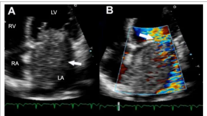
echocardiogram which subsequently showed a large mass within the left atrium. Surgical removal of the mass was recommended, the patient refused surgical intervention. Two years later, the patient returned with symptoms of cardiac insufficiency. Physical exam revealed normal systemic arterial blood pressure, 4+/6+ systolic murmur and 2+/6+ diastolic murmur in the mitral region were noted. Electrocardiogram (ECG) showed left atrium overload. Trans-thoracic echocardiogram showed a large rounded mobile mass with irregular contours, adhering to the inter-atria septum suggestive of left atrial myxoma. The mass was projected toward the mitral valve during ventricular diastole, thus generating flow obstruction (functional stenosis) and poor co-aptation of mitral valve cusps resulting in mitral insufficiency (Figure 1). Contrast echocardiography was performed using an albumin based microbubbles (PESDA - Perfluorocarbon Exposed Sonicated Dextrose and Albumin) and the SONOS 5500 (Philips Medical Systems, Bothell, WA) echocardiograph, with real perfusion imaging in real time (Power Modulation). The use of contrast echocardiograph permitted delineation of the mass borders. For the purpose of vascularization evaluation, a high energy ultra-sound pulse (flash) was used to destroy contrast, followed by the analysis of time-dependent mass re-filling by the microbubbles. As is illustrated in Figure 2, slow contrast filling of the mass was observed suggestive of low vascularization of the tumor mass, typical of benign tumors. The patient was submitted for surgery, confirming a diagnosed left atrial myxoma.
Case 2
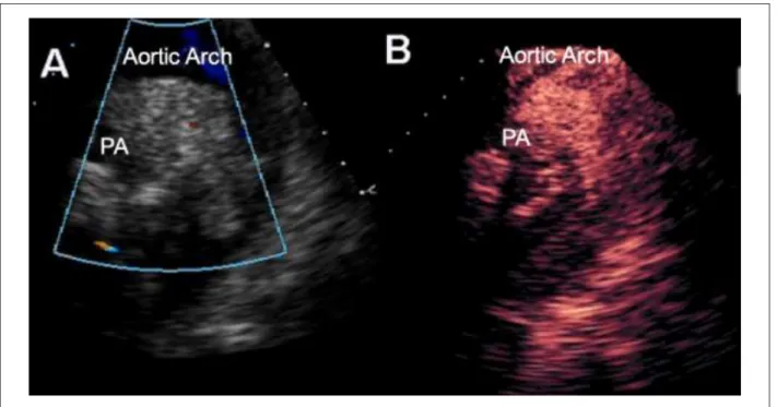
A 42-year old female patient presented for medical services, having a history of pre-cordial pain and dyspnea upon low exertion. Physical exam revealed normal systemic arterial pressure, 2+/6+ systolic murmur in the pulmonary foci, and an un altered pulmonary auscultation. No significant changes were observed on standard 12-lead electrocardiogram. Trans-thoracic echocardiogram showed a localized mass between the aortic arch and pulmonary artery (Figure 3), with irregular borders. An echocardiography with PESDA contrasting agent permitted identification of the rapidly filled with mass with contrast, suggesting an intense vascularization (Figure 4). The patient was submitted for other complementary exams, with resultant diagnosis of pulmonary adenocarcinoma. The mass identified by echocardiography corresponded to a metastatic malignant tumor.
Case 3
Figure 1 -ECG showing large roundish mass in left atrium, adhered to interatrial septum, projecting towards the mitral valve during ventricular diastole and resulting in
Figure 3 -Transthoracic ECG showing irregular contour mass between aortic arch and pulmonary artery (A); Echocardiographic contrast injection demonstrated mass was illed with contrast, which suggests a tumor (B); PA - pulmonary artery.
habit, and myocardial infarction presented for medical services with complaints of dyspnea. Physical examination revealed blood pressure at 90 x 60 mmHg, pulmonary stertoration, 5+/6+ systolic murmur of the mitral valve, hepatomegaly with palpable liver at 4 cm from left costal border, and +/4+ edema in the legs. Twelve lead ECG demonstrated overload of the left chambers and an electrically inactive area in antero-septal wall. ECG showed significant ventricular dysfunction with anterior, septal, and apical akinesia, and imaging suggestive of an apical thrombus. Contrast echocardiography with PESDA permitted better delineation of the adhered apical ventricular mass while demonstrating likewise no filling of the same with contrast, confirming an apical thrombus (Figure 5). An oral anticoagulation was introduced and cardiac insufficiency medications were optimized.
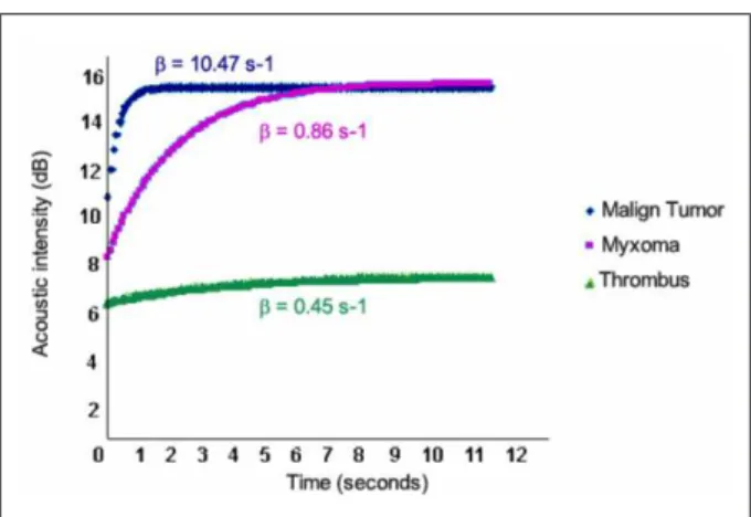
Blood flow quantification inside the mass was performed for all three cases utilizing specific quantification software (Q Lab 4.0, Philips Medical Systems, Bothell, WA). Perfusion analysis is based on the capacity to measure mass refilling velocity by the microbubbles and the maximum intensity of contrast8.
Therefore, the software provides the variable B; representing the mass filling time by contrast related to the degree of vascularization6. The greater the mass vascularization, the
higher the corresponding B value.
Case 1 refers to a myxoma – a benign tumor, with higher prevalence in adult populations9, and accounting for 20-50%
of all cardiac tumors. Contrast perfusion (case 1) analysis revealed a B of 0.86s-1. Case 2 is a secondary malignant
tumor (metastasis of a pulmonary adenocarcinoma), with a B of 10.47s-1. Case 3 is a thrombus with a B value of
0.45s-1 (Figure 6). Therefore, a comparable observation exist
between the type of cardiac mass its vascularization, and the corresponding B value.
Discussion
Current contrast echocardiography indications include left ventricular opacity and delineation of endocardial borders in patients with suboptimal echocardiographic window1. With
the recent development of more persistent micro-bubbles and associated advancements of ultra-sound techniques has contributed to both myocardial and cardiac mass perfusion studies. The concentration of microbubbles in the micro- circulation reflect the blood volume in the different regions of the heart and forms the basis for perfusion evaluation through contrast echocardiography.
Cardiac tumors are very rare with an incidence below 0.1% according to autopsies performed10. Tumors are
classified according to their origin, histology type and growth site. Primary tumors originate start in the heart itself, while secondary tumors originate in other organs. As far as histological patterns, they may be benign or malignant. Finally, as for growth site, they may be classified as intracavitary, intramural, and intrapericardial11. Transthoracic
echocardiographic exceed 90% sensitivity in diagnosing cardiac tumors9,12 and provides information regarding size,
Figure 6 -Curves: Maximum acoustic intensity and mass contrast illing velocity (β) for each of the cases presented; Malign tumor velocity showed to be much higher than benign tumor (myxoma); Thrombus quantiication curve was lattened, thus indicating no time-dependent illing of microbubbles (vascularized mass).
References
1. Mulvagh SL, DeMaria AN, Feinstein SB, Burns PN, Kaul S, Miller JG, et al. Contrast echocardiography: current and future applications. J Am Soc Echocardiogr. 2000;13:331-42.
2. Becker H, Burns P. Handbook of contrast echocardiography – left ventricular function and myocardial perfusion. New York: Springer-Verlag Publishers; 2000. p. 88-108.
3. Mathias W Jr, Arruda AL, Andrade JL, Filho OC, Porter TR. Endocardial border delineation during dobutamine infusion using contrast echocardiography. Echocardiography. 2002;19:109-14.
4. Tsutsui JM, Elhendy A, Xie F, O’Leary E, McGrain AC, Porter TR. Safety of dobutamine stress real-time myocardial contrast echocardiography. J Am Coll Cardiol. 2005;45:1235-42.
5. Tsutsui JM, Elhendy A, Anderson JR, Xie F, McGrain AC, Porter TR. Prognostic value of dobutamine stress myocardial contrast perfusion echocardiography. Circulation. 2005;112:1444-50.
6. Kirkpatrick JN, Wong T, Bednarz JE, Spencer KT, Sugeng L, Ward RP, et al. Differential diagnosis of cardiac masses using contrast echocardiographic perfusion imaging. J Am Coll Cardiol. 2004;43:1412-9.
7. Lepper W, Shivalkar B, Rinkevich D, Belcik T, Wei K. Assessment of
the vascularity of a left ventricular mass using myocardial contrast echocardiography. J Am Soc Echocardiogr. 2002;15:1419-22.
8. Wei K, Jayaweera AR, Firoozan S, Linka A, Skyba DM, Kaul S. Quantification of myocardial blood flow with ultrasound-induced destruction of microbubbles administered as a constant venous infusion. Circulation. 1998;97:473-83.
9. Meng Q, Lai H, Lima J, Tong W, Qian Y, Lai S. Echocardiographic and pathologic characteristics of primary cardiac tumors: a study of 149 cases. Int J Cardiol. 2002;84:69-75.
10. Majano-Lainez RA. Cardiac tumors: a current clinical and pathological perspective. Crit Rev Oncog. 1997;8:293-303.
11. Almeida EC. Tumores do coração. In: Porto CC, ed. Doenças do coração, prevenção e tratamento. Rio de Janeiro: Editora Guanabara Koogan S.A.;1998. p. 986-91.
12. Molina JE, Edwards JE, Ward HB. Primary cardiac tumors: experience at the University of Minnesota. Thorac Cardiovasc Surg. 1990;38:183-91.
13. McAllister HA Jr, Fenoglio JJ Jr. Tumors of the cardiovascular system. In: Atlas of tumor pathology. 2nd series. Fascicle 15. Washington, DC: Armed Forces Institute of Pathology; 1978. p. 81-8.
diagnosis between cardiac benign and malign tumors, and thrombi may be difficult to assess in some cases. Malignant tumors exhibit enriched vascularization allowing rapid cell tumor growth13. Benign tumors (on the other hand) have
less vascularization when compared to malignant tumors with the exception of hemangiomas. Thrombi are generally avascular.