Radiodosimetric considerations on ocular brachytherapy
with iodine-125 and ruthenium/rhodium-106*
Considerações radiodosimétricas da braquiterapia ocular com iodo-125 e rutênio/ródio-106
Arnaldo Prata Mourão1, Tarcísio Passos Ribeiro de Campos2
OBJECTIVE: To analyze dose distribution utilizing plaques with iodine-125 and ruthenium/rhodium-106 in a computational model of the ocular region. MATERIALS AND METHODS: A voxel-based computational model including the different tissues of the ocular region was utilized with the plaque positioned on the sclera. The Monte Carlo code was utilized for simulating irradiation. The dose distribution is demonstrated by isodoses curves. RESULTS: Computational simulations demonstrate the dose distribution inside the ocular bulb as well as in adjacent outside structures. The results have allowed the authors to compare the spatial distribution of doses generated by beta particles and photons. The simulations demonstrated that the utilization of iodine-125 seeds implies a high dose to the crystalline lens, while ruthenium/rhodium-106 results in high dose on the sclera surface. CONCLUSION: The dose to the crystalline lens depends on the tumor position and thickness, the plaque diameter, and the radionuclide utilized. In the present study, the ruthenium/rhodium-106 source is recommended for low tumor thickness. Irradiation with iodine-125 results in higher doses to the crystalline lens than irradiation with ruthenium/rhodium-106. The maximum value for dose to the crystalline lens corresponds to 12.75% of the maximum dose with iodine-125 and only 0.005% for ruthenium/rhodium-106.
Keywords: Brachytherapy; Eye plaque; Ocular brachytherapy; Monte Carlo code.
OBJETIVO: Analisar, por meio de um modelo computacional da região ocular, as características da distribui-ção da dose utilizando placas contendo iodo-125 e rutênio/ródio-106. MATERIAIS E MÉTODOS: Foi utilizado um modelo computacional de voxels da região ocular incluindo os diversos tecidos, com a placa posicionada
sobre a esclera. O código Monte Carlo foi utilizado para simular a irradiação. A distribuição da dose é apre-sentada por curvas de isodoses. RESULTADOS: As simulações computacionais apresentam a distribuição da dose no interior do bulbo e nas estruturas externas. Os resultados permitem comparar a distribuição espacial das doses geradas por partículas beta e por fótons. As simulações mostram que a aplicação de sementes de iodo-125 implica alta dose no cristalino, enquanto o rutênio/ródio-106 produz alta dose na superfície da esclera. CONCLUSÃO: A dose no cristalino depende da espessura do tumor, da posição e do diâmetro da placa, e do radionuclídeo utilizado. No presente estudo, a fonte de rutênio/ródio-106 é recomendada para tumores de dimensões reduzidas. A irradiação com iodo-125 gera doses maiores no cristalino do que a irradiação com rutênio/ródio-106. O valor máximo de dose no cristalino corresponde a 12,75% do valor máximo de dose com iodo-125 e apenas 0,005% para rutênio/ródio-106.
Unitermos: Braquiterapia; Placa ocular; Braquiterapia ocular; Monte Carlo.
Abstract
Resumo
* Study developed at the Nuclear Engineering Department of Universidade Federal de Minas Gerais (UFMG), Belo Horizonte, MG, Brazil.
1. PhD, Professor, Nuclear Engineering Department of Univer-sidade Federal de Minas Gerais (UFMG), Belo Horizonte, MG, Brazil.
2. PhD, Professor, Division of Hospital Engineering of Centro Federal de Educação Tecnológica de Minas Gerais (Cefet-MG), Belo Horizonte, MG, Brazil.
Mailing address: Dr. Arnaldo Prata Mourão Filho. Avenida Amazonas, 5253, Nova Suíça. Belo Horizonte, MG, Brazil, 30480-000. E-mail: [email protected]
Received September 9, 2008. Accepted after revision Decem-ber 12, 2008.
like a round-shaped cap whose concave surface is attached to the sclera. The radio-active material remains encapsuled within the plaque so that the patient contamination is avoided. The outer surface of the plaque has an absorbing shield that absorbs a great amount of radiation which propagates to-wards the opposite direction of the target volume, protecting the tissues lying poste-riorly to the plaque and structures outside the ocular bulb(4,5).
Along years, certain types of radionu-clides have been employed in plaques for ocular brachytherapy. Currently, iodine-125 or ruthenium/rhodium-106 seeds are
Mourão AP, Campos TPR. Radiodosimetric considerations on ocular brachytherapy with iodine-125 and ruthenium/rhodium-106. Radiol Bras. 2009;42(1):43–48.
good solution for the treatment and man-agement of uveal tumors, retinoblastomas and ocular metastases, representing a fea-sible alternative to enucleation. This tech-nique consists of radioactive plaques su-tured onto the external scleral surface, di-rectly above the base of the intraocular tu-mor. The plaque remains positioned until the minimum therapeutic dose is delivered to the tumor volume(1–3). When compared
with ocular teletherapy, brachytherapy al-lows a better spatial dose distribution over the tumor volume with lower radiation deposition on adjacent tissues.
Plaques for ocular brachytherapy look
INTRODUCTION
utilized as radioactive material loaded in a metal matrix. For comparative purposes, the authors have investigated the spatial dose distribution inside the ocular bulb, vitreous body and crystalline lens, as well as external structures, bones, optic nerve and brain. An effective dosimetry is criti-cal in the management of the dose deliv-ered to the tumor as well as in the observa-tion of the dose deposited in surrounding healthy tissues and, consequently, estimat-ing the harmful effects resultestimat-ing from this therapeutic process.
MATERIALS AND METHODS
Aiming at simulating the dose distribu-tion, a computational model of the human eye was developed encompassing three different models as follows: a voxel-based model of structures adjacent to the ocular bulb, an analytical model of the ocular bulb structures, and a voxel-based discretization of the vitreous cavity to allow the observa-tion of the dose within the bulb. A brachytherapy plate was attached to this model of the ocular region for the simula-tion of the dose distribusimula-tion through the Monte-Carlo code (MCNP-v.5).
Computational model of the eye
The voxel-based model of the eye de-fines a volume including all ocular struc-tures. The development of this first model includes the relevant structures situated externally to the ocular globe and the sur-rounding bone structure, and was based on images obtained from the Visible Man
Project(6). A set of 43 axial images of the visible skull was selected and, from these images, only the region of interest corre-sponding to the left ocular bulb was ex-tracted for the model development.
Each of the 43 sections of the ocular region was converted into a gray-scale 82
× 100 pixels matrix measuring 0.5 × 0.5 mm2. The overlapping of the 43 sections generated a non-isotropic volume with 82
× 100 × 43 voxels, defining a volume of 41 × 50 × 38,7 mm3 (x, y, z), correspond-ing to a voxel matrix whose volume ele-ments dimensions were 0.5 × 0.5 × 0.9 mm3. With the aid of anatomic references of the region, this gray-scale voxels-based model was colorized in a manner that each selected color corresponded to a specific tissue, allowing the differentiation among 12 types of tissues present in the region(7,8).
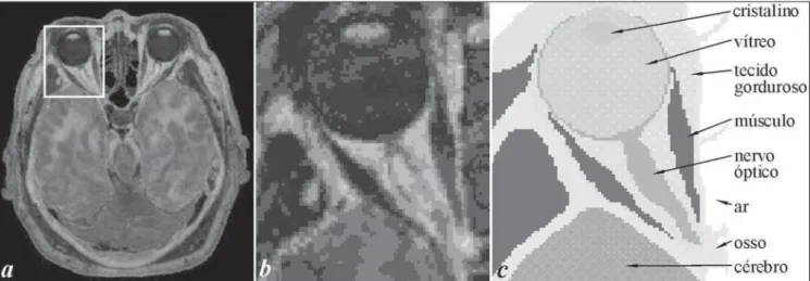
Figure 1a presents the image No.20 of the axial head section with delimitation of the region of interest and the image extracted from this region. This 256-tone gray-scale image of the ocular region is presented on Figure 1b in an 82 × 100 pixels matrix. The same image after colorization according to the tissues present in the region is shown on Figure 1c.
The analytical model of the ocular bulb was developed with basis on anatomical data which have allowed the determination of a set of geometric shapes based on math-ematical equations to reproduce the bulb structures: sclera, choroid, retina, cornea, vitreous body and crystalline lens(9).
The analytical model of the ocular bulb was coupled with the voxel-based model
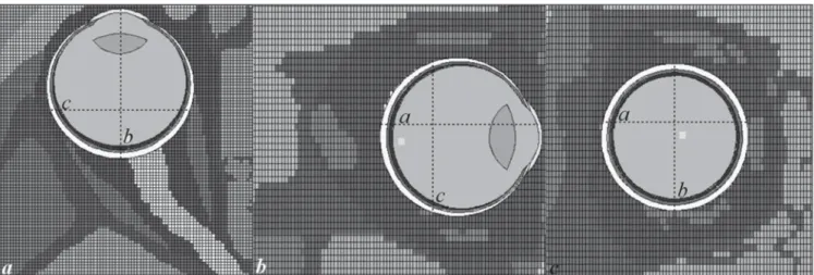
through the Monte-Carlo code (MCNP-v.5) to allow the simulations. Figure 2 pre-sents three sections (axial, sagittal and coronal) of the computation model of the ocular region. On each of these sections, the axis of the positioning of the other two sections is demarcated.
The third model was incorporated into the previously prepared geometrical struc-ture, defining small voxels of 0.5 × 0.5 ×
0.5 mm3 to subdivide the vitreous body region into small cells to allow the mea-surement of the deposited dose and then to obtain the spatial dose distribution within the vitreous body.
Brachytherapy plaques
Two brachytherapy plaques were se-lected, both of them with 15 mm in diam-eter. The model ROPES plaque is utilized for accommodating ten iodine-125 seeds and has an insert with slots where the ra-dioactive seeds are loaded that fits into the stainless steel shell. This shell keeps the seeds positioning and is meant to shield against radiation(10,11). The iodine-125 seed
utilized in the ocular bulb irradiation con-tains a cylindrical-shaped porous ceramic with 3 mm in length saturated with silver iodide containing iodine-125 enclosed by a titanium tube with 4.5 mm in length and 0.8 mm in diameter. The iodine-125 decays by electron capture in tellurium-125 (a stable nucleus), with a supplementary emission of gamma- and x-photons. The iodine-125 decay produces a 185.77 keV energy release. The half-life of an iodine-125 source corresponds to 59.408 days.
Table 1 presents the values of gamma- and x-photons energy released by the iodine-125 decay, as well as each photon emission frequency. Figure 3 presents the spatial distribution of the iodine-125 seeds on the insert of the ROPES plaque.
The other brachytherapy plaque utilized in the computational simulation is made of silver, with 1 mm thickness, containing a ruthenium/rhodium-106 film at 0.1 mm from the concave surface(12).
Ruthenium-106 decays to rhodium-Ruthenium-106 by the emission of beta particle to palladium-106 which is a stable nucleus. The ruthenium-106 decay releases a 39.4 keV energy corresponding to the maximum value of energy emitted by the single beta particle in the decay. The half-life of a ruthenium-106 source corre-sponds to 373.59 days. The rhodium-106 decay releases a 3,542 keV energy. This energy is distributed between the beta-par-ticle and the antineutrino emitted in the decay, so the beta spectrum becomes con-tinuous. The energy of the rhodium-106 beta particle is utilized in the therapeutic process, considering that the ruthenium-106 beta particle presents a poor penetra-tion and does not contribute to the dose deposition on the tumor tissue. The half-life of a rhodium-106 source corresponds to 29.8 s, so the assumed ruthenium/rho-dium-106 source half-life is 373.9 days(13).
Monte Carlo code simulation
The Monte Carlo code is a method uti-lized for simulating the transport of par-ticles such as neutrons, photons and elec-trons and their interactions with the
envi-Figure 2. Section images of the computational ocular region model obtained through the graphic interface of the MCNP-v.5. Axial section (a), sagittal section (b) and coronal section (c).
Table 1 Photons emitted by 125-iodine.
Energy (keV)
3.335
3.606
3.759
3.769
4.030
4.069
4.121
Frequency (%)
0.2300
0.1120
0.6300
5.6000
3.5000
0.4200
0.7000
Energy (keV)
4.173
4.302
4.572
4.829
4.829
26.875
27.202
Frequency (%)
0.0430
1.0100
0.4500
0.1030
0.1600
0.0032
40.6000
Energy (keV)
27.472
30.944
30.995
31.237
31.704
31.774
35.492
Frequency (%)
75.7000
6.8300
13.2000
0.1210
3.8110
0.5800
6.6800
ronment, allowing inclusive the observa-tion of the dose distribuobserva-tion in determined structures. This code has been initially de-veloped to the observation of neutrons transport and later was extended to include other particles such as photons and elec-trons(14,15). The Monte Carlo code runs with
a three-dimensional configuration utilizing a Table with the characteristics of every tissue and material defined through geo-metric cells and has been extensively uti-lized for validating the utilization of brachytherapy with beta particles and pho-ton-emitter radioisotopes(16).
The plates were coupled with the com-putational model of the ocular region on the sclera surface, with its center aligned with the ocular bulb equator on its medial re-gion. This positioning was selected for being considered to be closest to the crys-talline lens. The simulations have taken the dose distribution into consideration be-cause of the photons emitted by the iodine-125 and of beta particles emitted by rho-dium-106.
RESULTS
The plaques simulations were per-formed through the nuclear code MCNP-v.5, obtaining the dose distribution in the ocular region. The simulations for the ru-thenium/rhodium-106 plaque, were aimed only at the rhodium-106 beta emissions, since the contribution of the ruthenium-106 decay to the dose is non-significant because of the low energy release and consequent low beta particles penetration(17).
Therefore, two different simulations were performed for ocular brachytherapy with rhodium-106 in relation to beta par-ticles emission, as follows: one for the ex-ternal and another for the inex-ternal region of the ocular bulb. The simulations for ob-serving the dose distribution due to beta particles were performed with a continuous spectrum of the rhodium-106 beta emis-sions. For the effects of simulation, occur-rence probabilities were considered for each 1 keV variation. As the maximum energy of the rhodium-106 beta particle is 3,541 keV, 3,541 indices of beta decay occurrence probability were generated in this simulation.
Two different simulations were per-formed for ROPES plaque with iodine-125 seeds: one for the internal region of the ocular bulb and another for the external region, for observing the dose generated by the complementary gamma photons and x-photons emitted in the iodine-125 decay by electron capture. Data regarding energy and iodine-125 photons occurrence rates in-cluded on Table 1 were utilized for this simulation.
The doses results in each voxel of the vitreous body, bone structure, brain and optic nerve obtained by simulation on the MCNP-v.5, were utilized as input for the software SISCODES to be transformed into a dose distribution color matrix, allow-ing the observation of the isodoses curves in the ocular region(18). Internal and
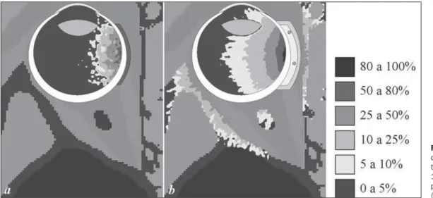
exter-nal simulations were separately performed and later overlapped. Figure 4 presents two images of a same axial section passing on
the center of the ocular bulb and, conse-quently, on the center of the brachytherapy plaque.
The image on Figure 4a presents the perceptual dose distribution due to beta particles emitted by rhodium-106. Based on the legend, both the external and inter-nal dose distribution can be understood, the maximum external dose corresponding to 32.4% of the maximum internal dose. This condition must be taken into consideration in the observation of the dose distribution. The image on Figure 4b presents the perceptual dose distribution due to photons emitted by iodine-125. Based on the leg-end, both the internal and external dose distribution can be understood, the maxi-mum external dose corresponding to 5.77% of the maximum internal dose.
The dose distribution for irradiation with rhodium-106 beta particles is spatially restricted and for this reason the isodose curves are very close in shape and end up mixing with each other. If a dose of 50 Gy is to be obtained on the region correspond-ing to 10% (at a 7 mm depth), the maxi-mum dose delivered near the sclera surface is of 500 Gy. The mean dose deposited on the crystalline lens for this irradiation con-dition is only 2.62 Gy (0.005%). The maxi-mum dose in the external region of the ocular bulb is of 162 Gy, but it is observed in a punctual manner.
For irradiation with iodine-125, the dose distribution generates well-defined isodose curves, which allows an improve-ment in the identification of the dose with depth. If a dose of 50 Gy is to be obtained
on the region corresponding to 50%, the maximum dose delivered near the sclera surface is of 100 Gy. For this condition of ocular bulb irradiation, the maximum dose deposited in the crystalline lens is 12.74 Gy on the nearest border of the plaque. The maximum dose observed in the external region of the ocular bulb is 7.84 Gy.
One of the factors which defines the maximum dose to be delivered is the de-sired dose to the apex of the tumor; for this reason, the thicker the ocular tumor, the higher should be the maximum dose deliv-ered to the sclera, and the increase in maxi-mum dose implies in increase in the dose to external tissues and crystalline lens.
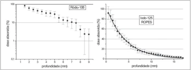
Figure 5 presents two graphs with per-ceptual variation of the dose with depth obtained through simulation on the MCNP-v.5, based on an axis originating in the cen-ter of the plaque concave surface towards the center of the ocular bulb. These graphs depict the variation of dose with depth for the rhodium-106 plaque, and for the ROPES plaque with iodine-125 seeds.
The variation of dose with depth for the rhodium-106 plaque is represented by a typical curve of beta particles penetration which, following a slight decrease, presents a fast decay with depth, achieving null val-ues at about 10 mm. Because of this char-acteristic, beta emitting radioisotopes with high energies are utilized to achieve a pen-etration deep enough to irradiate the tumor tissue. In case of irradiation of the ocular bulb with ruthenium/rhodium-106 plaque, 20% of the value for maximum dose is
achieved at a 5.16 mm depth and 10% at 7.19 mm. From this point on, the dose pre-sents a fast decrease, limiting the applica-tion of irradiaapplica-tion with rhodium-106.
The variation of dose with depth with iodine-125 is represented by a typical curve of photon beam penetration which presents an exponential decay with depth. Consid-ering the high reentrance of photons, the employment of gamma emitting radionu-clides with energy photons in the range of tenths of keV, such as iodine-125, is recom-mended in brachytherapy so as to circum-scribe the dose deposition to the source surroundings. In case of irradiation of the ocular bulb with the ROPES plaque, 50% of the value for the maximum dose is achieved at a 3.16 mm depth, and 30% at 5.25 mm.
DISCUSSION
The results from the simulation of ocu-lar bulb irradiation with the plaque with iodine-125 seeds demonstrate the spatial dose distribution, particularly with the depth in the vitreous body. However, an appropriate tumor dose deposition implies a considerable dose to the crystalline lens. The dose deposited on the crystalline lens primarily depends on the plaque position-ing, its distance from the crystalline lens, the desired dose for tumor apex and, there-fore, the tumor thickness.
According to Nag et al.(19), the
pre-scribed dose for irradiation with plaques with iodine-125 seeds is 85 Gy to the apex
for tumors > 5 mm, and to the base of the tumor in case of lower thickness. Consid-ering a dose of 85 Gy to the apex of a 5 mm tumor, the dose to the crystalline lens ranges from 33.4 to 10.3 Gy, according to the simulation. Considering that recom-mended dose limit for the crystalline lens is 5 Gy, this high dose to the crystalline lens is directly associated with late development of cataract and, consequently, referral for surgery, in this population of patients(19,20).
In simulations with the ruthenium/ rhodium-106 plaque, the adequation of the spatial dose profile, especially in relation to the tumor depth, is not so easily achieved because of the smaller penetration of the beta particles beam. This characteristic al-lows the therapeutic dose concentration on the tumor tissue, generating lower doses to the crystalline lens than those found for the plaque with iodine-125. In spite of this characteristic, the utilization of ruthenium/ rhodium-106 should be recommended for tumors with < 5 mm in thickness and base diameter < 15 mm. Tumors with higher thickness may require higher doses to the sclera surface, generating subsequent com-plications and a low dose to the tumor apex(19,21).
The dose to the scleral surface should be limited to 160 Gy, which means a dose of 50 Gy for 4.06 mm depth for a ruthe-nium/rhodium-106 plaque(19). If a
thera-peutic dose of 55 Gy to the tumor apex is considered for this irradiation, the occur-rence depth is 3.79 mm. So, the selection of an irradiation method, plaque type and
radionuclide depends on the tumor dimen-sions.
For the simulation evaluated in the present study, the irradiation with iodine-125 generates doses to the crystalline lens higher than those of ruthenium/rhodium-106. The dose to the crystalline lens corre-sponds to 12.75% of the maximum dose generated with iodine-125, and only 0.005% for ruthenium/rhodium-106.
REFERENCES
1. Knutsen S, Hafslund R, Monge OR, et al. Dosi-metric verification of a dedicated 3D treatment planning system for episcleral plaque therapy. Int J Radiat Oncol Biol Phys. 2001;51:1159–66. 2. Sauerwein W, Gérard JP. Radiothérapie des
tu-meurs intraoculaires. Câncer/Radiothér. 1999; 3(Suppl 1):102s–6.
3. Dias RS, Giordani AJ, Erwenne CM, et al. Braqui-terapia com rutênio-106 em melanomas uveais – resultados preliminares: experiência uni-institu-cional. Radiol Bras. 2007;40:105–11. 4. Shields CL, Naseripour M, Cater J, et al. Plaque
radiotherapy for large posterior uveal melanomas (> or =8-mm thick) in 354 consecutive patients. Ophthalmology. 2002;109:1838–49.
5. Astrahan MA, Szechter A, Finger PT. Design and dosimetric considerations of a modified COMS
plaque: the reusable “seed-guide” insert. Med Phys. 2005;32:2706–16.
6. National Library of Medicine. The visible man project. [cited 2005 Oct 6]. Available from: http: / / w w w. n l m . n i h . g o v / r e s e a r c h / v i s i b l e / visible_human.html
7. Sobotta J. Atlas de anatomia humana: cabeça, pes-coço, membros superiores. Rio de Janeiro: Gua-nabara Koogan; 1984.
8. Dantas AM. Anatomia funcional do olho e seus anexos. Rio de Janeiro: Revinter; 2002. 9. Mourão AP, Campos TPR. Development of a
hu-man eye model for ophtalmic brachytherapy do-simetry in heterogeneous medium at the uvea. Biomat 2007 – International Symposium on Mathematical and Computational Biology; 2007 Nov 24–29; Armação dos Búzios, RJ, Brasil. 10. Granero D, Pérez-Calatayud J, Ballester F, et al.
Dosimetric study of the 15 mm ROPES eye plaque. Med Phys. 2004;31:3330–6.
11. BEBIG. I-125 ophthalmic seeds. Berlin: Eckert & Ziegler; Rev. 01/2004.
12. BEBIG. Ru-106 ophthalmic plaques. Berlin: Eckert & Ziegler; Rev. 02/2004.
13. Berkeley National Laboratory. Table of isotopes decay data. [cited 2005 Oct 15]. Available from: http://isotopes.lbl.gov/
14. Ballester F, Granero D, Pérez-Calatayud J, et al. Monte Carlo dosimetric study of best industries and Alpha Omega Ir-192 brachytherapy seeds. Med Phys. 2004;31:3298–305.
15. X-5 Monte Carlo Team. MCNP – A general Monte Carlo N-particle transport code, version 5. Los Alamos National Laboratory; 2003.
16. International Commission on Radiation Units & Measurements. Photon, electron, proton and neu-tron interaction data of body tissues. ICRU Re-port No. 46. Bethesda: ICRU; 1991.
17. Mourão AP, Campos TPR. Dosimetric evaluation of the brachytherapy in heterogeneous medium at the uvea. INAC 2007 – International Nuclear Atlantic Conference; 2007 Sept 30–Oct 5; Santos, SP, Brasil.
18. Trindade BM. Desenvolvimento de sistema com-putacional para dosimetria em radioterapia por nêutrons e fótons baseado em método estocástico SISCODES [tese de mestrado]. Belo Horizonte: Universidade Federal de Minas Gerais; 2004. 19. Nag S, Quivey JM, Earle JD, et al. The American
Brachytherapy Society recommendations for brachytherapy of uveal melanomas. Int J Radiat Oncol Biol Phys. 2003;56:544–55.
20. Collaborative Ocular Melanoma Study Group. Incidence of cataract and outcomes after cataract surgery in the first 5 years after iodine 125 brachy-therapy in the Collaborative Ocular Melanoma Study. COMS Report No. 27. Ophthalmology. 2007;114:1363–71.