1
Original Article
Evaluation of Segmentary Contractility in Chagas’
Disease by Using the Integral of the Myocardial
Velocity Gradient (Myocardial Strain) Obtained
Through Tissue Doppler Echocardiography
Carlos Eduardo Suaide Silva, Luiz Darcy Cortez Ferreira, Luciana Braz Peixoto,
Claudia Gianini Monaco, Manuel Adán Gil, Juarez Ortiz, Bárbara Maria Ianni,
José Lázaro Andrade, Wilson Mathias Júnior, Antônio Carlos Pereira Barretto
São Paulo, SP - Brazil
Incor do Hospital das Clínicas da FMUSP and OMNI-CCNI Medicina Diagnóstica
Mailing address: Carlos Eduardo Suaide Silva - Rua Cubatão, 726 Cep 04013-002 - São Paulo, SP, Brazil
E-mail: [email protected] Sent for publication: 07/12/2004 Accepted for publication: 10/29/2004 English version by Stela Maris Costalonga
Objective
To quantify the percentage of contractility of different myo-cardial segments in patients with Chagas’ disease by measuring myocardial strain and to assess the differences in the radial and longitudinal ventricular contractile function in the undetermined and dilated forms of chronic chagasic cardiomyopathy as com-pared with those in a group of healthy individuals.
Methods
The study comprised 39 individuals [20 (51.3%) of the male sex] divided into the following 4 groups: 1) Nl: 17 (43.6%) healthy individuals; 2) Und: 7 (17.9%) patients with the unde-termined form of Chagas’ disease; 3) C1: 7 (17.9%) patients with the chronic form of Chagas’ disease with ejection fraction < 50%; and 4) C2: 8 (20.5%) patients with the chronic form of Chagas’ disease with ejection fraction > 50%. After performing baseline echocardiography, Doppler tissue images were recorded to measure myocardial strain in different segments on longitudinal and transversal parasternal, and apical 2- and 4-chamber views.
Results and Conclusion
The percentage of contractility in the different myocardial segments, both the radial and longitudinal components, is grea-ter in healthy individuals than in patients with the chronic form of Chagas’ disease, and in those with the undetermined form of the disease as compared with that of chronic chagasic patients with EF < 50%. Left ventricular radial contractility is greater than left ventricular longitudinal contractility in all groups (Nl, Und, and Chronic). The data presented allow us to propose a progressive character of myocardial impairment in patients with Chagas’ disease.
Key words
Chagas’ disease, echocardiography, tissue Doppler, myocar-dial strain
Chagas’ disease became known worldwide with the pioneering study of Carlos Chagas, who, in 1909, described, not only the clinical manifestations, but also the etiologic agent, the transmitting agent, and the reservoirs of that disease 1. It is considered a great
public health problem, mainly in developing countries. According to the World Health Organization, 20 million infected individuals live on the American continents, of whom, more than 6 million are in Brazil 2.
The disease may be clinically divided into 2 phases: acute and chronic. After regression of the acute phase, individuals ex-perience a variable period characterized by positive serology and the absence of clinical, electrocardiographic, and radiological ma-nifestations, which is called the undetermined form 3.
The chronic phase occurs in 25 to 30% of infected individuals, affecting mainly male adult individuals in the third or fourth decade of life. Clinically, it may be classified as dilated, arrhythmogenic, thromboembolic, mixed, and silent, and approximately 10% of the patients evolve to severe forms of the disease 3.
Echocardiography represents, currently, one of the most im-portant complementary methods for assessing chronic Chagas’ heart disease, because it allows functional and anatomic evaluation, as well as the evolutionary follow-up of the disease. The echocardio-graphic findings vary from normal examinations to severe systolic and diastolic functional impairment. Approximately 75% of the pa-tients have segmentary contractile alterations, which predominate in the left ventricular postero-inferior wall and apical region.
Recently, the incorporation of the tissue Doppler technique to conventional echocardiography allowed the more detailed eva-luation of the systolic and diastolic myocardial functions in an overall and regional form 4-11.
The major limitations of Doppler tissue imaging are its depen-dency on the angle of incidence of the ultrasound beam in regard to the mobility of the segment to be studied and the fact that it does not differentiate between the presence of an active contraction of a healthy segment and the passive contraction of an akinetic segment, whose mobility depends on the healthy adjacent myocardium.
2
tissue imaging: the measurement of the intramyocardial gradient of velocity or strain rate 12-15.
In general terms, strain represents the deformation of a tissue undergoing the application of a certain force. The concept of strain was described in 1973 by Mirsky and Parmley 16, and it expresses
the local dynamics of myocardial performance. Differently from Dop-pler tissue imaging, strain rate provides information about the local instantaneous measurement of the myocardial compression or ex-pansion rate, independently of the cardiac translation movement 17.
A tendency exists towards considering myocardial thickening, or its radial contractile function, the major component of ventricular systolic function. However, the excursion of the ventricular base towards the apex is known to be approximately 1.5 to 2 cm during systole, while the wall thickening does not exceed 0.5 cm18-20. Nevertheless, the visual analysis is still inadequate to
detect those subtleties, and more accurate data about radial and longitudinal ventricular contractile function are still required.
Muscle may be considered an uncompressible tissue, which means that its longitudinal deformation is inversely proportional to the alterations observed in its thickness, ie, the more the muscle stretches, the thinner it gets; and the more it contracts, the thicker it gets. Thus, by quantifying the stretching/shortening of the myocardial fiber, its degree of relaxation/contractility can be inferred 21.
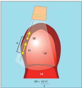
The strain rate is the measurement of the myocardial defor-mation velocity, defined by the formula (V2-V1)/d, where V1 and V2 are the velocities of myocardial shortening (cm/s) in 2 points separated by a d distance (cm) 16(fig. 1). That d distance may be
adjusted by the operator and should usually be approximately 9 mm. Smaller distances increase the degree of noise, while greater distances decrease the resolution of the method.
That measurement provides the intramyocardial gradient of velocities per unit of time and represents the degree of deformity of the fiber. Because the intramyocardial gradient of velocity
re-presents the difference of velocities (cm/s) between 2 points divided by the distance (cm) between those same 2 points, its unit is s-1
[(cm/s)/cm].
Doppler tissue imaging, used in the parasternal views, assesses the radial component of myocardial contractility; when used in the apical views, it assesses the longitudinal contractility of the muscle. Based on those observations, a new technique assessing the gradient of velocity between 2 points close to the myocardium was developed. In other words, it quantifies the stretching/shor-tening of the fiber, and, indirectly, its thickening, which appears to be a much more effective method for assessing myocardial contractility.
Another form of assessing the deformity of the myocardial segments is through the integral of the strain rate. That measu-rement, simply called myocardial strain, provides information about the deformation of the fiber in terms of percentage.
This study aimed at measuring the percentage of contractility of the several myocardial segments in patients with Chagas’ di-sease by measuring the myocardial strain obtained on tissue Doppler echocardiography, and also at defining whether differences exist in the radial and longitudinal myocardial contractile function in the undetermined and chronic forms of the disease when compared with those of a group of healthy individuals.
Methods
This prospective study comprised healthy adult individuals and patients with Chagas’ disease of both sexes, with a satisfactory echocardiographic window, who agreed to sign a written informed consent. Thirty-nine individuals were studied, 20 (51.3%) being of the male sex. They were divided as follows: 1) group Nl, com-prising 17 (43.6%) asymptomatic individuals with normal elec-trocardiograms and echocardiograms, constituting the healthy control group; 2) group Und, comprising 7 (17.9%) asymptomatic individuals with positive serology for Chagas’ disease, with nor-mal electrocardiograms, chest X-rays, contrast studies of the eso-phagus, and contrast enemas, characterizing the undetermined form of Chagas’ disease; 3) group C1, comprising 7 (17.9%) patients with Chagas’ disease in its chronic form, who, in addition to electrocardiographic or radiographic alterations, or both, also had left ventricular ejection fraction < 50% on echocardiograms; and 4) group C2, comprising 8 (20.5%) patients with the chronic form of Chagas’ disease, who had, in addition to electrocardio-graphic or radioelectrocardio-graphic alterations, or both, left ventricular ejection fractions > 50%. For comparing the radial and longitudinal con-tractility, the groups of chronic chagasic patients were considered as a single group (C1+C2). The groups did not differ in regard to age (47.2 ± 9.4 years) or body mass index (25.4 ± 3.5).
The following individuals were excluded from the study: patients with significant valvular or pulmonary diseases, coronary heart disease, individuals with a recent history of drug use or alcoholism, and pregnant women.
After performing the clinical examination and characterizing each group, the patients underwent the echocardiographic study in a device with second harmonic and Doppler color-flow tissue imaging (System V and Vivid Five, GE-Vingmed Ultrasound, Horten, Norway).
The images were obtained with the patient in the left lateral decubitus position. The echocardiogram was recorded in the digi-tal format with the file of static and dynamic images of several
Fig. 1 - Strain rate (SR) measures the intramyocardial gradient of velocity between 2 close points of a muscular segment (V1 and V2) in relation to distance (d) between those points.
SR = V2-V1______
d
d
V2
V1
LV
3
Fig. 3 - Measurement of longitudinal myocardial strain on apical 2-chamber view with the samples positioned in the basal region of the inferior (yellow) and anterior (green) walls. In this case, the percentage of contraction was approximately 18% in the inferior wall and 20% in the anterior.
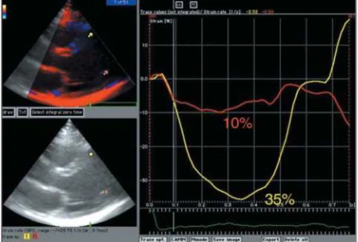
Fig. 2 - Acquisition of radial myocardial strain in the middle segments of the septal (red curve) and posterior (yellow curve) walls acquired on transversal parasternal view of an individual in the healthy group. In this case, the percentage of contraction was 36% in the septal wall and 22% in the posterior wall. Upper left panel = Doppler tissue mode; lower left panel = 2-dimensional mode; right panel = curves of myocardial strain.
echocardiographic sections. The following measurements were ta-ken: dimensions of the aorta (Ao); left atrium (LA); left ventricular diastolic and systolic diameters (LVDD and LVSD); diastolic thick-ness of the ventricular septum and of the left ventricular posterior wall (DTVS and DTLVPW); and left ventricular ejection fraction (EF) according to the Pombo method 22. After performing a complete
baseline echocardiography, Doppler tissue images were recorded for measuring the velocities and myocardial strain. A complete cardiac cycle was recorded and digitally stored in the following 4 different echocardiographic views: longitudinal and transversal paras-ternal, and apical 2- and 4-chamber. In the first 2, the radial component of myocardial contractility (fig. 2) was assessed in the basal and middle segments of the septal and posterior walls (in the longitudinal parasternal view) and in the middle segments of the septal and posterior wall (in the transversal parasternal view). In the apical views, the longitudinal component of myocardial con-tractility was assessed in the basal, middle, and apical segments of the septal, lateral, anterior, and inferior walls (fig. 3).
The moving digital images were transferred to a Macintosh computer (Apple Computer Inc., Cupertino, California, USA) coupled to the echocardiograph. The measurements of myocardial strain
were obtained later by using a specific computer program (EchoPAC GE-Vingmed Ultrasound, Horten, Norway). The distance between the points where the myocardial velocities were collected for calculating strain was fixed as 9.2 mm.
The data were typed in Microsoft Excel 2000 worksheets, and the statistical calculations and analyses were performed with the SigmaStat program for Windows, version 2.0 (Jandel Corpo-ration, 1992-1995). For comparing the values obtained in the different segments assessed in the 4 groups studied, the Kruskal-Wallis test (analog of analysis of variance) was used. When P values < 0.05 were obtained, the Dunn test was used for multiple comparisons between the 4 groups, 2-by-2. For comparing, in each group, the mean values of radial contractility and longitudi-nal contractility, the Mann-Whitney test was used. For all tests used, the alpha error value was fixed as 0.05 or 5%. The values presented represent the median followed by the 25th and 75th percentiles (between parentheses).
The studies were carried out at InCor of the Medical School of the USP and at the laboratory of the OMNI-CCNI Medicina Diagnóstica de São Paulo. The study was approved by the com-mittees on ethics of the 2 institutions.
Results
In regard to the usual echocardiographic measurements, the 4 groups had no statistical differences in the aortic and left atrial diameters, and in the diastolic thickness of the ventricular septum and left ventricular posterior wall. In the groups with the dilated form of chronic chagasic heart disease (C1 and C2), the left ventricular (diastolic and systolic) diameters were greater and the ejection fraction was lower compared with those in the group of healthy individuals and the group of patients with the undetermined form of Chagas’ disease (Nl and Und) (tab. I).
In regard to the myocardial strain measurements, when each segment was individually assessed between the groups, contractility was observed to be significantly smaller in all segments of the septal, posterior, and inferior walls, and in the middle segment of the anterior wall in the C1group as compared with those in the Nl group. The C1 group had a decreased strain in the basal, middle, and apical segments of the septal wall, in the basal and middle segments of the posterior wall, in the middle and apical segments of the inferior wall, and in the middle segment of the anterior wall as compared with those in healthy individuals. Finally, the Und group had a reduced myocardial strain only in the middle segment of the posterior wall. The other segments of the Und, C1, and C2 groups showed no statistical difference in strain mea-surements despite the mild tendency of the median towards being increased in the basal, middle, and apical segments of the septal and lateral walls, and in the basal segments of the inferior and anterior walls in the Und group as compared with those in the healthy individuals group.
4
Table IIb - Myocardial strain measurements in each segment according to the groups involved in the comparison of radial and
longitudinal contractilities
Nl Und C1+C2
Radial 33,7 (30,4;38,2) 32,3 (28,7;33,6) 21,8 (18,5;25,3) Longitudinal 25,9 (24,5;30,7) 28,0 (24,7;29,7) 17,6 (15,7;20,9) p < 0,001 0,053 0,044
The values represent the medians of myocardial strain; the figures between parentheses are the 25th and 75th percentiles, respectively.
The median of the radial strain values (measurements perfor-med in the longitudinal and transversal parasternal views) was 33.7% (30.4%; 38.2%) in healthy individuals; 32.3% (28.7%; 33.6%) in the Und group; 21.7% (19.1%; 23.6%) in the C1 group; and 24.7% (15.2%; 27.8%) in the C2 group (tab. III).
In the assessment of the longitudinal contractile function (measurements performed in the apical 2- and 4-chamber views), the mean percentages of contractility of the segments were as follows: 25.9% (24.5%; 30.7%) in the Nl group; 28% (24.7%; 29.7%) in the Und group; 16.8% (13.2%; 19.2%) in the C1 group; and 19.1% (15.8%; 22.3%) in the C2 group (tab. IV).
Discussion
The left ventricular contractile function in patients with heart disease is routinely assessed by using echocardiography. However, the visual assessment of segmentary contractility is a relative limitation of the method. Although objective indices of ventricular function quantification, such as ejection fraction, end-diastolic volume, systolic volume, and shortening fraction, are routinely used, those parameters are still greatly influenced by the variations in pre- and afterload.
The color kinesis technique may also be used to quantify the contractile function on echocardiography; however, as it depends
on the appropriate identification of the excursion of myocardial segments, its performance in individuals with a little satisfactory echocardiographic window may be difficult 23,24.
Recently, Yamada et al 25 showed that the peak systolic
velo-city measured with Doppler tissue imaging correlated with dP/ dtmax in individuals undergoing cardiac catheterization.
Myocardial strain has also been proposed as an index of con-tractility of the muscle fiber 16.
The Doppler effect was described in 1842 by the Austrian physicist Christian Johann Doppler; its use in medicine, mainly in echocardiography, began approximately 100 years later 26.
When the ultrasound beam reaches a moving structure, such as blood cells, the reflected wave suffers an alteration in its fre-quency, returning to the transducer with a frequency different from that emitted. Thus, when that beam reaches the blood cells that are moving towards the transducer, the frequency of the reflected wave increases, and, therefore, it will be greater than that of the wave originally emitted. On the contrary, if the blood cells are moving away from the transducer, the frequency of the reflected wave will be smaller than that of the wave emitted. Therefore, a variation in frequency occurs, and it may be measured by the equipment, being proportional to blood velocity.
Similarly to the use of Doppler for measuring blood flow velocity inside the heart and vessels, the method allows the identification and measurement of the velocity of the movement of other tissues, such as myocardium. Such a technique is called Doppler tissue imaging (DTI).
Several studies have reported the normal patterns of the systolic and diastolic velocities for each myocardial segment 27-29. The
use of DTI allows the assessment of the overall and regional systolic and diastolic functions 24. However, it has the limitation of
depen-ding on the angle of incidence of the ultrasound beam and of not allowing the assessment of the movement of a segment separated from that of its adjacent segment.
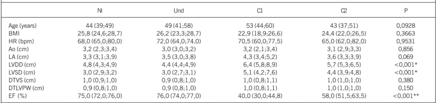
Table I - General characteristics of the patients according to the groups studied
Nl Und C1 C2 P
Age (years) 44 (39;49) 49 (41;58) 53 (44;60) 43 (37;51) 0,0928 BMI 25,8 (24,6;28,7) 26,2 (23,3;28,7) 22,9 (18,9;26,6) 24,4 (22,0;26,5) 0,3663 HR (bpm) 68,0 (65,0;80,0) 72,0 (64,0;74,0) 70,5 (60,0;77,5) 65,0 (62,0;82,0) 0,9531 Ao (cm) 3,2 (2,3;3,4) 3,0 (3,0;3,2) 3,2 (2,1;3,4) 3,1 (2,9;3,3) 0,856 LA (cm) 3,3 (3,1;3,9) 3,5 (3,0;3,8) 4,3 (3,4;5,2) 3,6 (3,3;3,9) 0,069 LVDD (cm) 4,8 (4,3;4,9) 4,4 (4,4;4,9) 6,4 (5,8;8,9) 5,7 (5,3;6,5) <0,001* LVSD (cm) 3,0 (2,9;3,2) 3,0 (2,7;3,1) 5,1 (4,2;7,6) 4,4 (3,9;4,8) <0,001* DTVS (cm) 1,0 (0,9;1,0) 0,9 (0,8;1,0) 1,0 (0,8;1,1) 1,0 (1,0;1,0) 0,380 DTLVPW (cm) 0,9 (0,8;1,0) 0,9 (0,8;1,0) 1,0 (0,8;1,1) 1,0 (1,0;1,0) 0,150 EF (%) 75,0 (72,0;76,0) 76,0 (74,0;77,0) 40,0 (30,0;44,8) 58,0 (51,5;63,5) <0,001**
BMI - body mass index; Ao - aorta; LA - left atrium; LVDD - left ventricular diastolic diameter; LVSD - left ventricular systolic diameter; DTVS - diastolic thickness of the ventricular septum; DTLVPW - diastolic thickness of the left ventricular posterior wall; EF - left ventricular ejection fraction; *C1 > Und, C1 > Nl, C2 > Und, C2 > Nl; **Nl > C1, Nl > C2, Und > C1, Und > C2.
Table IIa - Myocardial strain measurements in each segment according to the groups involved in the comparison of radial and longitudinal contractilities
Nl Und C1 C2 P
MedRadial 33,7 (30,4;38,2) 32,3 (28,7;33,6) 21,7 (19,1;23,6) 24,7 (15,2;27,8) <0,001* MedLong 25,9 (24,5;30,7) 28,0 (24,7;29,7) 16,8 (13,3;19,2) 19,1 (15,8;22,3) <0,001*
5
Table III - Myocardial strain measurements in each segment according to the groups involved for assessing radial contractility
Nl Und C1 C2 P
SepBas PL 32,0 (28,5;33,3) 35,0 (30,0;41,5) 26,0 (20,3;26,0) 31,0 (19,5;39,0) 0,110 SepMed PL 32,0 (28,8;34,7) 32,0 (27,8;38,7) 19,0 (13,0;24,5) 23,0 (17,5;28,5) <0,001* PostBas PL 42,0 (28,8;45,3) 38,0 (29,0;41,5) 19,0 (14,5;30,5) 18,0 (13,0;21,0) <0,001** PostMed PL 32,0 (25,8;38,2) 15,0 (12,8;21,2) 15,0 (9,5;22,0) 14,5 (11,0;21,0) <0,001*** AntMed PT 33,0 (28,8;37,0) 32,0 (29,3;39,0) 32,0 (24,0;37,3) 26,0 (17,5;33,2) 0,430 PostMed PT 35,0 (33,3;43,0) 38,0 (33,5;49,5) 17,0 (11,3;29,8) 23,0 (14,3;31,0) 0,002**** Median 33,7 (30,4;38,2) 32,3 (28,7;33,6) 21,7 (19,1;23,6) 24,7 (15,2;27,8) <0,001**
SepBas - basal segment of the septal wall; SepMed - middle segment of the septal wall; PostBas - basal segment of the posterior wall; PostMed - middle segment of the posterior wall; AntMed - middle segment of the anterior wall; PL - longitudinal parasternal; PT - transversal parasternal; *C1 < Und; C1 < Nl; C2 < Nl; **Nl > C1; Nl > C2; Und > C2; ***Nl > Und; Nl > C1; Nl > C2; **** C1 < Nl; C1 < Und. The values represent the medians of myocardial strain; the figures between parentheses are the 25th and 75th percentiles, respectively.
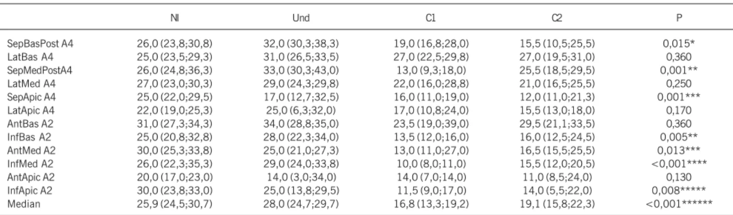
Table IV - Myocardial strain measurements in each segment according to the groups involved for assessing longitudinal contractility
Nl Und C1 C2 P
SepBasPost A4 26,0 (23,8;30,8) 32,0 (30,3;38,3) 19,0 (16,8;28,0) 15,5 (10,5;25,5) 0,015* LatBas A4 25,0 (23,5;29,3) 31,0 (26,5;33,5) 27,0 (22,5;29,8) 27,0 (19,5;31,0) 0,360 SepMedPostA4 26,0 (24,8;36,3) 33,0 (30,3;43,0) 13,0 (9,3;18,0) 25,5 (18,5;29,5) 0,001** LatMed A4 27,0 (23,0;30,3) 29,0 (24,3;29,8) 22,0 (16,0;28,8) 21,0 (16,5;25,5) 0,250 SepApic A4 25,0 (22,0;29,5) 17,0 (12,7;32,5) 16,0 (11,0;19,0) 12,0 (11,0;21,3) 0,001*** LatApic A4 22,0 (19,0;25,3) 25,0 (6,3;32,0) 17,0 (10,8;24,0) 15,5 (13,0;18,0) 0,170 AntBas A2 31,0 (27,3;34,3) 34,0 (28,8;35,0) 23,5 (19,0;39,0) 29,5 (21,1;33,5) 0,360 InfBas A2 25,0 (20,8;32,8) 28,0 (22,3;34,0) 13,5 (12,0;16,0) 16,0 (12,5;24,5) 0,005** AntMed A2 30,0 (25,3;33,8) 25,0 (21,0;27,3) 13,0 (11,0;27,0) 16,5 (15,5;25,5) 0,013*** InfMed A2 26,0 (22,3;35,3) 29,0 (24,0;33,8) 10,0 (8,0;11,0) 15,5 (12,0;20,5) <0,001**** AntApic A2 20,0 (17,0;23,0) 14,0 (3,0;34,0) 14,0 (7,0;14,0) 11,0 (8,5;24,0) 0,130 InfApic A2 30,0 (23,8;33,0) 25,0 (13,8;29,5) 11,5 (9,0;17,0) 14,0 (5,5;22,0) 0,008***** Median 25,9 (24,5;30,7) 28,0 (24,7;29,7) 16,8 (13,3;19,2) 19,1 (15,8;22,3) <0,001******
SepBasPost - basal posterior segment of the septal wall; LatBas - basal segment of the lateral wall; SepMedPost - middle posterior segment of the septal wall; LatMed - middle segment of the lateral wall; SepApic - apical segment of the septal wall; LatApic - apical segment of the lateral wall; AntBas - basal segment of the anterior wall; InfBas - basal segment of the inferior wall; AntMed - middle segment of the anterior wall; InfMed - middle segment of the inferior wall; AntApic - apical segment of the anterior wall; InfApic - apical segment of the inferior wall; A4 - apical 4-chamber; A2 - apical 2-chamber; *C2 < Und; **C1 < Und, C1 < Nl; ***Nl > C1, Nl > C2; ****Nl > C1, Nl > C2, Und > C1, Und > C2; *****Nl > C2; ******Nl > C1, Nl > C2, Und > C1. The values represent the medians of myocardial strain, the figures between parentheses are the 25th and 75th percentiles, respectively.
confirming the segmentary nature of chagasic cardiomyopathy and its predilection for the left ventricular apical and postero-inferior regions. In the C2 group, in which the systolic impairment was milder (ejection fraction > 50%), decreased myocardial strain was observed in a smaller number of segments (middle of the septal wall, basal and middle of the posterior wall, middle and apical of the inferior wall, and middle of the anterior wall).
In the undetermined form, however, an interesting behavior was observed. Although its definition comprised patients with positive serology without alterations on the electrocardiogram or chest X-ray, and, in our study, the ejection fraction in that group was not different from that in the healthy group, the undetermined form behaved in an intermediate manner between normal and the dilated form of chronic chagasic cardiomyopathy. The middle seg-ment of the posterior wall had a decreased contractility as that in the C1 and C2 groups, while some segments showed a tendency towards a greater contractility than those in the group of healthy individuals (mainly the lateral wall and the middle and basal seg-ments of the septal wall). A possible explanation for that finding could be the vicarious nature of healthy segments after myocardial aggression in that disease. Unfortunately, the number of patients in that group was small, and this may have been the cause of the lack of a significant statistical difference.
In the assessment of the radial and longitudinal contractile The strain technique can solve that problem partially. Although
it is even more dependent on the angle of incidence of the beam, the fact of assessing the difference of velocities between 2 close myocardial segments reduces the influence of the cardiac rotation and translation movements and of the movement of adjacent seg-ments (tethering) 30,31.
The measurement of the intramyocardial gradient of velocity, a technique used to calculate the myocardial strain on Doppler echo-cardiography, has already been approached by several authors 32,33.
In our study, we used that technique to quantify the segmentary contractility in healthy individuals and patients with Chagas’ di-sease, in addition to comparing the radial and longitudinal com-ponents of the ventricular contractile function.
The baseline echocardiographic measurements, as expected, showed increased left ventricular diameters in the C1 and C2 groups (dilated form of chronic chagasic cardiomyopathy), and a decreased ejection fraction in the same groups when compared with those in the Nl and Und groups.
6
function, although the left ventricular longitudinal shortening was up to 2 cm and its thickening did not exceed 0.5 cm, both the radial and longitudinal strains were greater in the Nl group as compared with those in the C1 and C2 groups, and in the Und group as compared with those in the C1 group. This, once again, showed the intermediate characteristic of the undetermined form. When each group was individually analyzed, the radial strain was greater than the longitudinal strain in healthy individuals, in pa-tients with the undetermined form, and in the chronic chagasic patients (C1 + C2 groups).
Our study shows the importance of that new technique for quantifying the myocardial contractile function, which can safely identify subtleties not evidenced on visual analysis.
Recently, a study of Doppler tissue imaging 34 revealed
some-thing similar. That study assessed 339 patients, 92 of whom had systolic heart failure, 73 had diastolic heart failure, 68 had isolated diastolic dysfunction on echocardiography, and 106 healthy indi-viduals for control. The authors reported that the systolic and diastolic velocities of the myocardial segments measured with Doppler tissue imaging were significantly decreased in the 3 groups when compared with those in the control group. Moreover, a 4.4-cm/s velocity predicted systolic dysfunction in 92% of the patients with systolic heart failure, in 52% of those with diastolic heart failure, and in 14% of those with diastolic dysfunction. In other words, tissue Doppler enabled the detection of alterations in the systolic function of patients previously labeled as having diastolic heart failure (therefore with normal ejection fraction),
Fig. 4 - Patient from the group with the undetermined form of Chagas’ disease showing normal strain in the septal wall (yellow curve) and abnormal strain in the posterior wall (red curve). No alterations in contractility were observed on visual analysis of the 2-dimensional echocardiogram in the patient.
and even in those with isolated diastolic dysfunction on echocar-diography. The new question would be: does pure diastolic heart failure really exist? Or, are the diagnostic methods used so far not sufficiently sensitive to detect more delicate alterations in myo-cardial function? Returning to our study, quantification of the radi-al and longitudinradi-al contractile function by use of the strain technique enabled the detection of subclinical alterations in myocardial con-tractility, which were not perceptible on visual analysis (fig. 4). This may have a great prognostic significance.
References
1. Chagas C. Estudo sobre a morfologia e o ciclo evolutivo do Schizotrypanum cruzi, n. gen. sp. agente etiológico de uma nova entidade mórbida do homem. Mem Inst Oswaldo Cruz 1909;159-219.
2. Panamerican Health Organization (Tropical Diseases Program). Status of Chagas’ disease in the region of the Americas. Epidemiol Bull PAHO 1984;5:5-9. 3. Maranhão EA, Correia CB, Silva RCB. Cardiopatia Chagásica in Castro I -
Cardio-logia, Princípios e Prática. Porto Alegre: Artes Médicas, 1999:845-65. 4. Waggoner AD, Bierig SM. Tissue Doppler imaging: A useful echocardiographic
me-thod for the cardiac sonographer to assess systolic and diastolic ventricular func-tion. J Am Soc Echocardiogr 2001;14:1143-52.
5. Garcia MJ, Thomas JD, Klein AL. New Doppler echocardiographic applications for the study of diastolic function. J Am Coll Cardiol 1998 Oct;32(4):865-75. 6. Ueno Y, Nakamura Y, Ohbayashi Y, Kinoshita M. Evaluation of left ventricular
systo-lic and diastosysto-lic global function: peak positive and negative myocardial velocity gra-dients in M-mode Doppler tissue imaging. Echocardiography 2002;19(1):15-25. 7. Fedele F, Trambaiolo P, Magni G, De Castro S, Cacciotti L. New modalities of
re-gional and global left ventricular function analysis: state of the art. Am J Cardiol 1998;81(12A):49G-57.
8. Wilkenshoff UM, Hatle L, Sovany A, Wranne B, Sutherland GR. Age-dependent changes in regional diastolic function evaluated by color Doppler myocardial ima-ging: a comparison with pulsed Doppler indexes of global function. J Am Soc Echocardiogr 2001;14(10):959-69.
9. Gulati VK, Katz WE, Follansbee WP, Gorcsan J 3rd.. Mitral annular descent velocity by tissue Doppler echocardiography as an index of global left ventricular function.. Am J Cardiol 1996;77(11):979-84.
10. Edvardsen T, Urheim S, Skulstad H, Steine K, Ihlen H, Smiseth OA. Quantification of left ventricular systolic function by tissue Doppler echocardiography: added va-lue of measuring pre- and postejection velocities in ischemic myocardium. Circula-tion 2002;105(17):2071-7.
11. Rychik J, Tian ZY. Quantitative assessment of myocardial tissue velocities in nor-mal children with Doppler tissue imaging. Am J Cardiol 1996;77(14):1254-7. 12. Abraham TP, Nishimura RA, Holmes DR Jr, Belohlavek M, Seward JB. Strain rate
imaging for assessment of regional myocardial function: results from a clinical mo-del of septal ablation. Circulation 2002;105(12):1403-6.
13. Uematsu M, Miyatake K, Tanaka N, Matsuda H, Sano A, Yamazaki N, Hirama M, Yamagishi M. Myocardial velocity gradient as a new indicator of regional left
ven-tricular contraction: detection by a two-dimensional tissue Doppler imaging tech-nique. J Am Coll Cardiol 1995;26(1):217-23.
14. Stoylen A, Slordahl S, Skjelvan GK, Heimdal A, Skjaerpe T. Strain rate imaging in normal and reduced diastolic function: comparison with pulsed Doppler tissue imaging of the mitral annulus. J Am Soc Echocardiogr 2001;14(4):264-74. 15. Greenberg NL, Firstenberg MS, Castro PL, Main M, Travaglini A, Odabashian JA,
Drinko JK, Rodriguez LL, Thomas JD, Garcia MJ. Doppler-derived myocardial sys-tolic strain rate is a strong index of left ventricular contractility. Circulation 2002;105(1):99-105.
16. Mirsky I, Parmley W. Assessment of passive elastic stiffness for isolated heart mus-cle and the intact heart. Circ Res 1973;33:233-43.
17. Belohlavek M, Pislaru C, Bae R, et al. Real-time strain rate echocardiographic ima-ging: temporal and spatial analysis of postsystolic compression in acutely ische-mic myocardium. J Am Soc Echocardiogr 2001;14:360-9.
18. Lundback S. Cardiac pumping and function of the ventricular septum. Acta Phy-siol Scand Suppl 1986;550:1-101.
19. Alam M, Hoglund C, Thorstrand C. Longitudinal systolic shortening of the left ven-tricle: an echocardiographic study in subjects with and without preserved global function. Clin Physiol 1992;12(4):443-52.
20. Simonson JS, Schiller NB. Descent of the base of the left ventricle: an echocardiogra-phic index of left ventricular function. J Am Soc Echocardiogr 1989;2(1):25-35. 21. Heimdal A, Stoylen A, Torp H, et al. Real-time strain rate imaging of the left
ventri-cle by ultrasound. J Am Soc Echocardiogr 1998;11:1013-9.
22. Pombo JF, Troy BL, Russel RO. Left ventricular volumes and ejection fraction by echocardiography. Circulation, 1972;46:26-35.
23. Vitarelli A, Sciomer S, Penco M, Dagianti A, Pugliese M. Assessment of left ventri-cular dyssynergy by color kinesis. Am J Cardiol 1998;81(12A):86G-9. 24. Perez JE, Waggoner AD, Barzilai B, Melton HE Jr, Miller JG, Sobel BE. On-line
as-sessment of ventricular function by automatic boundary detection and ultrasonic backscatter imaging. J Am Coll Cardiol 1992;19(2):313-20.
25. Yamada H, Oki T, Tabata T, Iuchi A, Ito S. Assessment of left ventricular systolic wall motion velocity with pulsed tissue Doppler imaging: comparison with peak dP/dt of the left ventricular pressure curve. J Am Soc Echocardiogr 1998;11(5):442-9. 26. Satomura S. A study on examining the heart with ultrasonics. I Principles; II
7
27. Palka P, Lange A, Sutherland GR, et al. Doppler tissue imaging: Myocardial wall motion velocities in normal subjects. J Am Soc Ecocardiogr 1995;8:659-668. 28. Galiuto L, Ignone G, DeMaria NA, et al. Contraction and relaxation velocities of the
normal left ventricle using pulsed-wave tissue Doppler echocardiography. Am J Cardiol 1998;81:607-14.
29. Silva CES, Ferreira LDC, Peixoto LB, et al. Estudo das velocidades de contração e relaxamento do miocárdio pela ecocardiografia com Doppler tecidual. Nova alter-nativa na avaliação da função ventricular segmentar. Arq Bras Cardiol 2002;78:200-5.
30. Stoylen A, Heimdal A, Bjornstad K, Wiseth R, Vik-Mo H, Torp H, Angelsen B, Sk-jaerpe T. Strain rate imaging by ultrasonography in the diagnosis of coronary arte-ry disease. J Am Soc Echocardiogr 2000;13(12):1053-64.
31. Heimdal A. Angle dependency of strain rate. In: Doppler based ultrasound imaging methods for noninvasive assessment of viability (dissertation). Trondheim: Nor-wegian University of Science and Technology; 1999, p 55-64.
32. Fleming AD, Xia X, McDicken WN, Sutherland GR, Fenn L. Myocardial velocity gra-dients detected by Doppler imaging. Br J Radiol 1994;67(799):679-88. 33. Miyatake K, Yamagishi M, Tanaka N, Uematsu M, Yamazaki N, Mine Y, Sano A,
Hi-rama M. New method for evaluating left ventricular wall motion by color-coded tis-sue Doppler imaging: in vitro and in vivo studies. J Am Coll Cardiol 1995;25(3):717-24.
34. Yu CM, Lin H, Yang H, Kong SL, Zhang Q, Lee SWL. Progression of systolic abnor-malities in patients with “isolated” diastolic heart failure and diastolic dysfunc-tion. Circulation 2002;105:1195-1201.
35. Dargie, H.J. Effect of carvedilol on outcome after myocardial infarction in patients
with left-ventricular dysfunction: the CAPRICORN randomised trial. Lancet 2001;357:1385-90.
36. Pitt, B.; Remme, W.; Zannad, F.; Neaton, J.; Martinez, F.; Roniker, B. et al. Eple-renone, a selective aldosterone blocker, in patients with left ventricular dysfunction after myocardial infarction. N Engl J Med 2003;348:1309-21.2002;78:200-5. 30. Stoylen A, Heimdal A, Bjornstad K, Wiseth R, Vik-Mo H, Torp H, Angelsen B,
Sk-jaerpe T. Strain rate imaging by ultrasonography in the diagnosis of coronary ar-tery disease. J Am Soc Echocardiogr 2000;13(12):1053-64.
31. Heimdal A. Angle dependency of strain rate. In: Doppler based ultrasound imaging methods for noninvasive assessment of viability (dissertation). Trondheim: Nor-wegian University of Science and Technology; 1999, p 55-64.
32. Fleming AD, Xia X, McDicken WN, Sutherland GR, Fenn L. Myocardial velocity gra-dients detected by Doppler imaging. Br J Radiol 1994;67(799):679-88. 33. Miyatake K, Yamagishi M, Tanaka N, Uematsu M, Yamazaki N, Mine Y, Sano A,
Hi-rama M. New method for evaluating left ventricular wall motion by color-coded tis-sue Doppler imaging: in vitro and in vivo studies. J Am Coll Cardiol 1995;25(3):717-24.
34. Yu CM, Lin H, Yang H, Kong SL, Zhang Q, Lee SWL. Progression of systolic abnor-malities in patients with “isolated” diastolic heart failure and diastolic dysfunc-tion. Circulation 2002;105:1195-1201.
35. Dargie, H.J. Effect of carvedilol on outcome after myocardial infarction in patients with left-ventricular dysfunction: the CAPRICORN randomised trial. Lancet 2001;357:1385-90.