Keywords
Protein isoforms; atrial myosins; heart failure; cardiomyopathy, dilated.
The Role of Titin in the Modulation of Cardiac Function and Its
Pathophysiological Implications
Ricardo Castro-Ferreira, Ricardo Fontes-Carvalho, Inês Falcão-Pires, Adelino F. Leite-Moreira
Serviço de Fisiologia Faculdade de Medicina da Universidade do Porto - PortugalMailing address: Ricardo Luís Castro Silva Ferreira •
Serviço de Fisiologia, Faculdade de Medicina da Universidade do Porto Alameda Prof. Hernâni Monteiro - 4200-219 - Porto
E-mail: [email protected]
Manuscript received September 16, 2009; revised manuscript received November 05, 2009; accepted January 26, 2010.
A
bstract
Titin is a giant sarcomeric protein that extends from the Z-line to the M-line. Due to its location, it represents an important biomechanical sensor, which has a crucial role in the maintenance of the sarcomere structural integrity. Titin works as a “bidireactional spring” that regulates the sarcomeric length and performs adequate adjustments of passive tension whenever the length varies. Therefore, it determines not only ventricular rigidity and diastolic function, but also systolic cardiac function, modulating the Frank-Starling mechanism.
The myocardium expresses two isoforms of this macromolecule: the N2B, more rigid and the isoform N2BA, more compliant. The alterations in the relative expression of the two titin isoforms or alterations in their state of phosphorylation have been implicated in the pathophysiology of several diseases, such as diastolic heart failure, dilated cardiomyopathy, ischemic cardiomyopathy and aortic stenosis. The aim of this study is to describe, in brief, the structure and location of titin, its association with different cardiomyopathies and understand how alterations in this macromolecule influence the pathophysiology of diastolic heart failure, emphasizing the therapeutic potential of the manipulation of this macromolecule.
Introduction
Titin, initially described as connectin, is a giant protein of great elasticity that can be found only in the cardiac and skeletal muscles. Structurally, it is located within the sarcomeres, where it binds to and interacts with other important myofilament proteins, namely actin and myosin.
In the last decade, it has been demonstrated that titin is a structurally complex protein that has an important role in
striated muscle action, particularly in the cardiac muscle. Among its several actions, its role in the maintenance of the structure and architecture of the sarcomere is emphasized, allowing the correct alignment of actin and myosin myofilaments, as well as its “elastic” function, which allows the regulation of the length and distensibility of the sarcomere, thus influencing not only the cardiac diastolic function, but also the systolic function by the Frank-Starling law.
The present review article intends to review the structure and main physiological actions of titin, namely its importance in determining the diastolic and systolic cardiac function and the implications of alterations in this macromolecule in the pathophysiology of heart failure (HF).
Structure and location of titin
Titin is the largest protein found in mammalians, varying from 2970 and 3700 kD, depending on the isoform1. It
is encoded by a single gene, located on the long arm of chromosome 2 in the 2q31 region, consisting of 363 exons. In the heart, titin has two isoforms, N2B and N2BA, which are generated by alternative splicing1.
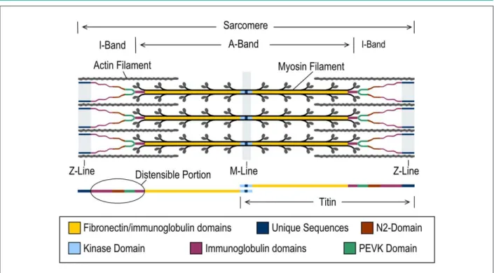
Titin is constituted mostly by two types of domains: fibronectin type III-like (called Fn-3 domains) domains and immunoglobulin-like (Ig) domains (Ig domains). In addition to these domains, titin also contains a kinase domain, located close to the carboxyl-terminal and several unique-sequence regions1-3 (Figures 1 and 2).
Titin is an intrasarcomeric protein, around 1 μm in length, which extends from the Z-line to the M-line2. Thus,
titin encompasses four sarcomere zones, due to which it is traditionally divided in distinct regions, called respectively M-line region, A-band region, I-band region and Z-line region (Figure 1). The Z-line region contains the amino-terminal, whereas the carboxyl-terminal is located in the M-line region. As described next, titin interacts with other proteins in the Z-line region, being also involved in mechanisms of intracellular signaling, functioning as a biomechanical sensor4.
The A-band region contains the majority of the titin molecule, which, in this region, is formed by single repeats of Ig and Fn3 domains and represents a component that constitutes the thick filament (Figure 1). Therefore, this region of titin is not extensible and its composition is similar in the different isoforms of the molecule (N2B and N2BA)5. Contrarily, the
Figure 2 -The N2BA isoform has one more elastic element in series in the
N2 region (segment N2A), conferring it higher levels of elasticity. Therefore, for any length, the passive tension developed by the N2BA isoform is lower than that developed by N2B.
Figure 1 -Titin structure and its position within the sarcomere. Titin crosses the sarcomere from the M-line to the Z-line, where it interacts with several structural proteins capable of responding to muscle stretching. This image shows the representation of the extensible portion of titin overlapping the I-Band.
filament and the Z-line (Figure 3). Structurally, the portion of titin present in the I-band is much more complex than the A-band region, consisting of Ig domains, N2 region and PEVK segment (rich in the amino acids proline (P), glutamate (E), valine (V) and lysine (K))3,6 (Figure 2). Moreover, it is the
I-band region that differentiates the titin isoforms6, as the
sub-region N2 can have two distinct elements: N2A and/or N2B (Figure 2). The N2A element consists of a region with four Ig domains and a unique 106-residue region and the N2B element is constituted by a region with three Ig domains and a unique 572-residue region. The N2A sequences are present in both the cardiac and skeletal muscle, whereas the N2B sequences are exclusively present in the cardiac muscle6.
Elastic properties of titin
The main characteristic of titin is its high degree of elasticity, which allows it to stretch and shorten, thus regulating the sarcomere length and muscle function. Titin constitutes the main determinant of passive tension of cardiomyocytes for physiological sarcomeric lengths, whereas, for supraphysiological lengths, the extracellular matrix collagen becomes more important2.
As mentioned before, the elastic properties of titin are mainly due to the I-band region, as in the A-band region titin is practically inextensible7,8. The elasticity of titin in the
I-band region is given by a molecular structure constituted by several extensible segments, namely by a large number of Ig segments, a PEVK segment and the N2 region (Figures 1, 2 and 3). The behavior of these different segments during muscle stretching and the development of passive tension is complex, as the several segments have different passive tension-length relationships and, additionally, each one stretches at different moments, according to the sarcomere length.
Figure 3 -At the resting sarcomere length (1.9 µm) the elastic elements of titin
are contracted (A). With the increase in length, the immunoglobulin domain is the irst to stretch (by stretching the binding between the Ig) (B). Starting at the length of 2.15 µm, the PVEK and N2 segment start to unfold, allowing
the accommodation of the length increase without an excess development of passive tension (C).
Figure 4 -Passive tension/length relationship of the sarcomere in heart cardiomyocytes that mostly express the N2B or N2BA isoforms. The coexpression of the two
isoforms of titin at varied ratios allows intermedial passive properties to be attained (purple doble arrow), which, therefore, can work as a rigidity adjustment mechanism in long term. Ways to alter the properties of the two isoforms in short term, such as phosphorylation and interaction with calcium, are also demonstrated (blue arrows). (Adapted from Henk and cols., 2004).
With the progressive stretching of the sarcomere, initially the Ig sequence stretches due to the elongation of the binding between the different Ig domains. The PEVK segments only start to elongate at 2.15 μm, and, subsequently, the N2 segments, especially due to the unfolding of its domains8. It
is this complex behavior observed in the extensible region of titin that makes the passive tension-length curve of sarcomere non-linear, that is, as it can be observed in Figure 4, the longer the sarcomere, the higher the inclination of the length-passive
tension curve1-3,7. This behavior does not allow, during the
elongation of the sarcomere that occurs during diastole, the development of an excessive passive tension, which would impair the diastolic function.
Titin is not only the main determinant of passive tension in response to stretching, but also functions as a two-directional elastic coil. That happens because, on the contrary, when the sarcomere length is shortened to lengths that are lower than the resting length, titin also generates an elastic force that has a contrary direction to that of the traditional passive tension. This force, called “restoring force”, allows the rapid re-establishment of the sarcomere length to the resting values, pushing the thick filament away from the Z-line9.
Titin in the cardiac muscle
There are two titin isoforms in the cardiac muscle: the N2B isoform and the N2BA isoform. The N2B is the smaller of the two (2970 kDa) as in the N2 sub-region it is constituted only by the N2B element. In turn, the N2BA isoform is larger (3200 to 3400 kDa) and contains both elements: N2B and N2A. In humans, the N2BA and the N2B isoforms are present in each half of the sarcomere, at a ratio of approximately 30/7010.
different isoforms also determines the elastic recoil after the ventricular contraction. The cardiomyocytes with a high degree of expression of the N2B isoform, which is more rigid, also present a higher restoring force, that is, after the contraction, the sarcomere length returns faster to the resting length. That occurs because, as the N2B isoform is less extensible, it does not only exercise a higher passive tension during stretching, but also generates a higher force to return to the resting length after the contraction. Therefore, in ventricles with high proportions of N2B isoform, there is a higher velocity of elastic recoil, leading to a shortening in the initial diastolic phase duration6.
Titin’s functions on the cardiac muscle
Titin as a determinant of diastolic cardiac function
The main determinants of diastolic cardiac function are the myocardial relaxation and passive ventricular properties. Whereas the myocardial relaxation is modulated by load, by inactivation (associated to calcium kinetics and myofilament dissociation) and by the non-uniformity of the ventricular wall, the myocardial passive properties are influenced by the myocardial rigidity, by the left ventricular wall thickness and by the geometry of the cardiac chambers12.
The diastolic dysfunction is caused by myocardial relaxation retardation and/or alterations in the passive properties of the ventricle, namely by the increase in the ventricular rigidity12.
Influence of titin in ventricular passive properties The increase in ventricular rigidity can be caused by alterations in the intrinsic characteristics of the cardiomyocytes – such as modifications in the constitution of cytoskeleton proteins or the endosarcomeric proteins (such as alterations in titin or alpha-actinin) – or by alterations in the constitution of the extracellular matrix, namely by the increase in the collagen network and extracellular fibrosis12.
Titin is an important determinant of diastolic function, as it influences the cardiomyocyte rigidity, and thus, the passive properties of the ventricle. As seen before, due to its strategic location in the sarcomere and its unique elastic properties, titin is main determinant of passive tension of the cardiomyocyte, for physiological sarcomere lengths7,13,
with the relative contribution of the intermedial filaments and microtubules being < 10%14. For lengths above 2.3 μm,
which are rarely achieved in an intact heart, the passive tension starts to be determined essentially by collagen and the extracellular matrix7,11.
The importance of titin in determining the diastolic function has an impact at the pathophysiological level, considering that, as it will be shown next, it has been demonstrated that patients with diastolic HF present an alteration in the relative proportion of titin isoforms, with an increase in the N2B isoform (more rigid), which seems to explain the increase in the ventricular rigidity observed in these individuals15.
Influence of titin in ventricular relaxation
Titin does not only determine ventricular rigidity. As mentioned before, titin also has elastic properties that function in a two-directional fashion, that is, if on the one hand it generates passive tension when the muscle is stretched, on the other hand, when the sarcomere shortens, titin generates a “restoring force”, which has an opposite direction to that of the passive tension and allows a rapid recovery of the resting length of the sarcomere9.
In hemodynamic terms, this “restoring force” generated by titin allows the increase in the velocity of the ventricular relaxation and the formation of a “ventricular suction” phenomenon. This force allows the increase in the ventricular filling, especially in the initial diastolic phase, which can be particularly important in situations of physical exertion or other tachycardic situations16.
Moreover, even before the start of the diastole, titin also has an effect by determining the end of the myocardial contraction, a process that is also dependent on the sarcomere length. In fact, when the sarcomere shortens to values lower than the resting length, titin will facilitate the process of deactivation of the cross-bridges, influencing and determining the end of the muscle contraction9,16.
Titin as a determinant of systolic cardiac function
Titin is an important determinant of the diastolic cardiac function, but it also influences the systolic function, through the effect it has on the Frank-Starling relationship17.
According to the Law of Frank-Starling, the increase in preload, i.e., the increase in the telediastolic volume improves the systolic function, originating an increase in the ejection volume. Titin, as a determinant of ventricular rigidity and of the passive ventricular properties, influences the telediastolic pressure-volume relationship and thus, the ventricular filling and telediastolic volume18.
At cellular level, the Frank-Starling mechanism is explained by the increase in the sensitivity of the Ca2+ filaments in response
to the increase in sarcomere length19. Titin has a fundamental
role in the regulation of this mechanism, as the passive tension exercised by titin directly determines the increase in the sensitivity of the calcium myofilaments17,20,21. Moreover, titin,
as the structural element of the thick filament in the region of the A-band, is also capable of directly influencing the binding between the actin and myosin filaments, according to the sarcomere length. That is, when the sarcomere length increases, titin seems to potentiate the binding between the actin and the myosin filaments, with the consequent increase in the contraction force22. Therefore, titin is capable of potentiating
the cycle of cross-bridges when the sarcomere is stretched and when it is shortened to lengths below that of the resting length, titin is also capable of inhibiting the formation of cross-bridges16.
Titin in the intracellular signaling: a biomechanical sensor
this biomechanical signal by binding and interacting with several structural and signaling proteins. This signaling occurs mostly in three main regions: the Z-line, the central part of the I-band and the M-line1,6,23
In the region of the Z-line, titin binds to the protein teletonin (T-cap), which functions as a place of interconnection between titin and several other structural and binding molecules23,24.
Among these molecules are the proteins present in the T tubules and the sarcoplasmic reticulum (SR), namely ankyrin-1 (sANK 1), obscurin (RS) and potassium channels (minK/ isk). Therefore, titin influences not only the maintenance of the sarcomere structure, but also the structure and correct organization of T tubules and the sarcoplasmic reticulum23,24.
Protein complexes are formed in the Z-line region, which function as tension signalers and that respond both to the passive force, generated by the titin filaments during stretching and the active force, transmitted by the actin filaments23,24. At this level, there are several proteins
that, by interacting with titin, participate in intracellular signaling cascades, promoting the expression of genes involved in cardiac remodeling. One of these proteins is the MLP (muscle LIM protein – a member of the LIM protein family) and it has been observed that point mutations of this protein can participate in the development of hypertrophic cardiomyopathy, as well as of dilated cardiomyopathy24.
However, the mechanism by which titin, interacting with MLP, is capable of regulating genetic transcription has yet to be established. Apparently, when elevated tensions are generated at the level of the Z-line during the stretching, the separation between the titin and MLP binding occurs and the latter, once free, migrates to the nucleus, where it promotes the transcription of several genes involved in cardiac remodeling24.
At the level of the central region of the I-band, the N2B and N2BA elements function as titin binding sites for several signaling proteins. The N2B-unique sequence, which is found exclusively in the cardiac muscle, binds, through a member of the LIM protein family (proteins that contain the LIM domains – an arrangement of eight cysteine and histidine residues in the configuration (C-X2-C-X16/23-H-X2-C-X2-C-X2-C-X16/21-C-X2/3
-C/D/H)) known as DRAL/FHL 2, to several metabolic enzymes. In states of increased energy need, the function of DRAL/ FHL 2 as binding agent between titin and enzymes such as kinase or adenylate kinase becomes increasingly important, increasing the ATP supply24.
The titin ligands in the I-band region, as well as the proteins associated to them, are also found in the nucleus, where they function as transcription and cell-cycle regulators and they can also be involved in the long-term alterations of the passive properties of cardiomyocytes1. An example is the interaction
with obscurin, a protein with several signaling domains. This titin-obscurin complex responds to the sarcomere stretching and it is involved in the sarcomeric restructuring that occurs, for instance, in muscular adaptation and heart failure2.
The titin region adjacent to the M-line also has sites potentially involved in the sensitivity of myofilaments for Ca2+ and participates extensively in the intracellular
signaling mechanisms2,23.
Other titin functions
In addition to the aforementioned functions, the participation of titin in the process of protein folding, gene expression regulation and several ion channel regulation has started to be investigated23.
Moreover, titin is also important in the process of myofibrillogenesis, forming a type of a structural mold for the correct disposition and organization of actin and myosin filaments25. The loss of titin originates an abnormal
organization of the sarcomere, as recently demonstrated26.
Modulation of titin properties
Titin functions in the sarcomere as a dynamic element, which can be modulated, either at short or long-term, by several factors, allowing the myocardium to modify its compliance in response to the numerous hemodynamic alterations that it is subjected to11,27. As shown in Figure 4, the
rigidity of the cardiac muscle varies according to alterations in the composition and the state of phosphorylation of titin. Such alterations can occur in long term, due to a modification in the relative proportion between the two titin isoforms, or, in short term, due to post-translational alterations, such as the alteration in the state of phosphorylation of titin or its binding to the calcium ion11,27.
Short-term regulatory mechanisms
Alteration in the phosphorylation state of titin
The N2B segment of cardiac titin can be phosphorylated by the protein kinase A (PKA), originating a decrease in its passive tension (Figure 4)28,29. As expected, it was also observed that the
decrease in the passive tension induced by titin phosphorylation by PKA is more pronounced in the cardiac muscle with more elevated levels of N2B segment expression30,31.
The activation of PKA that occurs, for instance, after the beta-adrenergic stimulation, constitutes one of the main mechanisms of short-term physiological regulation of titin distensibility. It is known that the sympathetic stimulation, in addition to its positive chronotropic and inotropic effects, is also capable of improving diastolic function, increasing not only the ventricular relaxation velocity, but also ventricular compliance. This latter effect depends, among other factors, on the phosphorylation of the titin N2B isoform by PKA27,32,33.
The effects of the phosphorylation of the N2B isoform by PKA are also present at the final phase of the systole, as the decrease in the cardiomyocyte rigidity will also lead to a decrease in the restoring force of cardiomyocytes31. This effect,
which is, in theory, of little benefit, can be counterbalanced by the fact that PKA accelerates the myocardial relaxation velocity, through the simultaneous phosphorylation of phospholamban and troponin34. Further studies are necessary to understand
the role of the phosphorylation of titin in cardiac function. More recently, it has also been demonstrated that either the protein kinase G (PKG)35,36, or the alpha isoform of protein
kinase C (PKC)37 equally promotes titin phosphorylation. In the
passive tension35. In the case of phosphorylation by PKC, that
occurs in the PEVK segment of both N2B and N2BA isoforms and the global effect of this phosphorylation is the increase in passive tension35-37.
In summary, titin works as an “adjustable coil”, which, through phosphorylation/dephosphorylation, especially of its N2B segment, allows the adaptation of ventricular function to the body’s necessities.
Regulation of titin by the calcium ion
The calcium ion (Ca2+) has an important role as regulator of
cardiomyocyte distensibility, especially due to the modulation of the interaction between titin and the thin filaments. Studies have shown that the PEVK domain in the extensible region of the N2B isoform binds to F-actin and that this interaction can contribute to the passive rigidity of cardiomyocytes38. Although
Ca2+ alone cannot interfere with this binding, when this ion
binds to protein S100A1, which is found at high concentrations in the myocardium, this interaction is inhibited, decreasing the rigidity of the cardiomyocyte. The observation of the increase in rigidity of the cardiac muscle in parallel to the decrease in the concentration of Ca2+ during diastole supports this
mechanism1,2,23,39.
Mechanisms of long-term regulation of titin
As mentioned before, the cardiomyocytes express the two isoforms of titin, allowing them to reach an intermediate level of passive tension (Figure 3), which can be modified according to the relative proportion of N2B/N2BA expression. A higher expression of the N2B isoform is associated with an increase in the myocardial rigidity, whereas an increase in the N2BA isoform is associated with an increase in its compliance. However, the molecular mechanisms underlying the preferential expression of one of the isoforms remains to be elucidated. It is known that in response to different hemodynamic states, long-term alterations in the expression ratio of the two isoforms of titin can occur, which, as we will see next, take place, for instance, in heart failure15 andaortic stenosis40,41.
It is important to mention that the alteration in the relative proportion of titin isoforms only results in the modification of the elastic properties of titin, without altering its structural properties, as only the portion of titin present in the I-band (that is, the distensible component) undergoes alterations11.
In summary, cardiac function can be altered by alterations in the elastic properties of titin, which can occur in both short and long-term. As shown in Figure 4, the increase in the expression of the N2B isoform is associated with an increase in titin rigidity. This rigidity can be reduced by the phosphorylation of titin by PKA or PKG or increased by the titin-actin interaction, which is inhibited by the Ca2+/
S100A1 complex.
Pathophysiological implications
Titin alterations are involved in the pathophysiology of some cardiac diseases, namely diastolic heart failure (DHF)
and dilated cardiomyopathy (DCM). There is increasing evidence that alterations in the expression ratio of the two titin isoforms, as well as the alteration in its phosphorylation status, are implicated in the development and progression of these diseases.
Alterations in the relative proportion of titin isoforms (N2BA/N2B)
In diastolic dysfunction, myocardial relaxation retardation and/or an increase in ventricular rigidity occur. In a more rigid ventricle, the increase in blood volume during diastole is only achieved at the expense of an increase in the ventricular filling pressure, with an increase in the telediastolic pressure-volume relation, which can originate a clinical picture of diastolic HF. Titin, as the main determinant of passive tension of the cardiomyocyte, determines ventricular rigidity. As recently demonstrated, the cardiomyocytes extracted from endomyocardial biopsies of patients with DHF exhibited a relative increase in the expression of the more rigid titin isoform, the N2B. It was observed that, whereas in a healthy heart the normal N2BA/N2B ratio is around 30/7010, in patients
with DHF, this ratio was 17/83; and in systolic HF, the ratio was 35/6515. However, the same group has recently demonstrated
that the hypophosphorylation of the N2B segment of titin seems to contribute more significantly for the increase in the passive tension verified in cases of DHF41.
An alteration in the ratio between titin isoforms was observed in patients with dilated cardiomyopathy and eccentric remodeling, but here with a relative increase in the expression of the more distensible isoform, N2BA10.
At the initial phase of systolic heart failure, alterations in the isoforms of titin can function as an adaptation mechanism, as the increase in the ventricular compliance allows an increase in the telediastolic volume, and, consequently, an increase in cardiac output, according to the Frank-Starling mechanism32.
Moreover, it is predicted that the lower restoring force developed by the cardiomyocytes during systole (due to the increase in the proportion of the more compliant isoform) would allow a decrease in the final telesystolic volume for any afterload. This decrease would occur due to the reduction in the resistance to cardiomyocyte shortening, thus increasing the ejection volume. In fact, it has been demonstrated that individuals with a higher proportion of N2BA have better tolerance to exercise due to the increase in the oxygen venous pressure10. This effect, which is in theory beneficial,
does not occur in practice, as in the systolic HF, the Frank-Starling mechanism is altered because of the decrease in the sensitivity of myofilaments for Ca2+, namely due to the lower
development of passive tension in the presence of an increase in the N2BA isoform10. Finally, an increase in titin degradation
can occur in the HF heart, which can equally contribute to the attenuation of the Frank-Starling mechanism42. Moreover, this
process subsequently becomes deadaptive, as the increase in the ventricular telediastolic volume results in more stress on the ventricular walls, stimulating and aggravating the process of cardiac remodeling13,32,43.
to an increase in the ventricular rigidity44. This observation,
apparently paradoxical, can be explained by the fact that, in this situation, the increase in ventricular rigidity is due not to alterations at the cardiomyocyte level, but to alterations in the extracellular matrix with an increase in myocardial fibrosis. Here, the increase in titin N2BA isoform (less rigid) can really be a mechanism of compensation to the increase in ventricular rigidity secondary to the fibrosis process. In fact, a decrease in the passive tension of each cardiomyocyte occurs even in ischemic cardiomyopathy, probably due to increase in the N2BA isoform10.
Alterations in the phosphorylation state of titin As mentioned before, titin can also undergo modulation of its rigidity in the presence of post-translational alterations, namely through the phosphorylation of the N2B segment29.
Titin can be phosphorylated by the protein kinase A (PKA) and by the protein kinase G (PKG), leading to a decrease in its rigidity28,29,31,35, thus inducing short-term acute alterations
in ventricular rigidity.
These findings are particularly relevant, as they can also be the target of therapeutic intervention in patients with HF. In fact, the treatment with PKA of cardiomyocytes extracted from patients with diastolic HF allowed a decrease in its passive tension to levels very similar to those found in control individuals30. This decrease in passive tension was directly
proportional to the initial rigidity of the cardiomyocytes, which is in agreement with the idea that the increase in cardiac rigidity is directly related not only to alterations in the relative expression of the isoforms, but also to the state of hypophosphorylation of the rigid isoform of titin.
As expected, due to the lower rigidity of the cardiomyocytes from patients with systolic HF, the same treatment with PKA did not result in such a significant decrease in passive tension15.
Both aortic stenosis and hypertensive cardiomyopathy present diastolic dysfunction, but only the latter presents a substantial increase in myocardial rigidity. In both cases,
the relative expression of the N2BA/N2B isoforms and their degree of total phosphorylation are similar. However, a relative hypophosphorylation of the more rigid isoform, N2B, was observed in hypertensive cardiomyopathy, which has been proposed as the underlying mechanism to the increase in myocardial rigidity and diastolic dysfunction in this disease40,45.
Alterations in titin phosphorylation seem to occur also in dilated cardiomyopathy. In these patients, a decrease in the cardiac PKA activity has been observed, which might be a compensating mechanism in response to the elevated compliance observed in the cardiomyocytes of these patients13.
In summary, titin, through the alteration in the expression of its isoforms and/or its phosphorylation state, seems to be a key element in the pathophysiology of several heart diseases. Moreover, titin can also be a potential target for pharmacological manipulation, thus constituting a new therapeutic target, mainly in heart failure.
Acknowledgements
The authors would like to thank the invaluable help of Pedro Daniel Miranda Couto in preparing the figures.
Potential Conflict of Interest
No potential conflict of interest relevant to this article was reported.
Sources of Funding
This study was partially funded by Fundação para a Ciência e Tecnologia.
Study Association
This study is not associated with any post-graduation program.
References
1. LeWinter MM, Wu Y, Labeit S, Granzier H. Cardiac titin: structure, functions and role in disease. Clin Chim Acta. 2007; 375 (1-2): 1-9.
2. Granzier HL, Labeit S. The giant protein titin: a major player in myocardial mechanics, signaling, and disease. Circ Res. 2004; 94 (3): 284-95.
3. Tskhovrebova L, Trinick J. Properties of titin immunoglobulin and fibronectin-3 domains. J Biol Chem. 2004; 279 (45): 46351-4.
4. Tskhovrebova L, Trinick J. Titin: properties and family relationships. Nat Rev Mol Cell Biol. 2003; 4 (9): 679-89.
5. Maruyama K. Connectin/titin, giant elastic protein of muscle. Faseb J. 1997; 11 (5): 341-5.
6. Granzier H, Labeit S. Cardiac titin: an adjustable multi-functional spring. J Physiol. 2002; 541 (Pt 2): 335-42.
7. Freiburg A, Trombitas K, Hell W, Cazorla O, Fougerousse F, Centner T, et al. Series of exon-skipping events in the elastic spring region of titin as the structural basis for myofibrillar elastic diversity. Circ Res. 2000; 86 (11): 1114-21.
8. Linke WA, Rudy DE, Centner T, Gautel M, Witt C, Labeit S, et al. I-band titin in cardiac muscle is a three-element molecular spring and is critical for maintaining thin filament structure. J Cell Biol. 1999; 146 (3): 631-44.
9. Helmes M, Trombitas K, Granzier H. Titin develops restoring force in rat cardiac myocytes. Circ Res. 1996; 79 (3): 619-26.
10. Nagueh SF, Shah G, Wu Y, Torre-Amione G, King NM, Lahmers S, et al. Altered titin expression, myocardial stiffness, and left ventricular function in patients with dilated cardiomyopathy. Circulation. 2004; 110 (2): 155-62.
11. Cazorla O, Freiburg A, Helmes M, Centner T, McNabb M, Wu Y, et al. Differential expression of cardiac titin isoforms and modulation of cellular stiffness. Circ Res. 2000; 86 (1): 59-67.
12. Leite-Moreira AF. Current perspectives in diastolic dysfunction and diastolic heart failure. Heart. 2006; 92 (5): 712-8.
14. Granzier HL, Irving TC. Passive tension in cardiac muscle: contribution of collagen, titin, microtubules, and intermediate filaments. Biophys J. 1995; 68 (3): 1027-44.
15. van Heerebeek L, Borbely A, Niessen HW, Bronzwaer JG, van der Velden J, Stienen GJ, et al. Myocardial structure and function differ in systolic and diastolic heart failure. Circulation. 2006; 113 (16): 1966-73.
16. Helmes M, Lim CC, Liao R, Bharti A, Cui L, Sawyer DB. Titin determines the Frank-Starling relation in early diastole. J Gen Physiol. 2003; 121 (2): 97-110.
17. Fukuda N, Granzier HL. Titin/connectin-based modulation of the Frank-Starling mechanism of the heart. J Muscle Res Motil. 2005; 26 (6-8): 319-23.
18. Katz AM. Ernest Henry Starling, his predecessors, and the “Law of the Heart”. Circulation. 2002; 106 (23): 2986-92.
19. Kentish JC, Keurs HE, Ricciardi L, Bucx JJ, Noble MI. Comparison between the sarcomere length-force relations of intact and skinned trabeculae from rat right ventricle. Influence of calcium concentrations on these relations. Circ Res. 1986; 58 (6): 755-68.
20. Cazorla O, Wu Y, Irving TC, Granzier H. Titin-based modulation of calcium sensitivity of active tension in mouse skinned cardiac myocytes. Circ Res. 2001; 88 (10): 1028-35.
21. Fukuda N, Wu Y, Farman G, Irving TC, Granzier H. Titin isoform variance and length dependence of activation in skinned bovine cardiac muscle. J Physiol. 2003; 553 (Pt 1): 147-54.
22. Fukuda N, Sasaki D, Ishiwata S, Kurihara S. Length dependence of tension generation in rat skinned cardiac muscle: role of titin in the Frank-Starling mechanism of the heart. Circulation. 2001; 104 (14): 1639-45.
23. Miller MK, Granzier H, Ehler E, Gregorio CC. The sensitive giant: the role of titin-based stretch sensing complexes in the heart. Trends Cell Biol. 2004; 14 (3): 119-26.
24. Linke WA. Sense and stretchability: the role of titin and titin-associated proteins in myocardial stress-sensing and mechanical dysfunction. Cardiovasc Res. 2008; 77 (4): 637-48.
25. Gregorio CC, Granzier H, Sorimachi H, Labeit S. Muscle assembly: a titanic achievement? Curr Opin Cell Biol. 1999; 11 (1): 18-25.
26. Udaka J, Ohmori S, Terui T, Ohtsuki I, Ishiwata S, Kurihara S, et al. Disuse-induced preferential loss of the giant protein titin depresses muscle performance via abnormal sarcomeric organization. J Gen Physiol. 2008; 131 (1): 33-41.
27. Granzier HL, Labeit S. The giant muscle protein titin is an adjustable molecular spring. Exerc Sport Sci Rev. 2006; 34(2): 50-3.
28. Yamasaki R, Wu Y, McNabb M, Greaser M, Labeit S, Granzier H. Protein kinase A phosphorylates titin’s cardiac-specific N2B domain and reduces passive tension in rat cardiac myocytes. Circ Res. 2002; 90 (11): 1181-8.
29. Kruger M, Linke WA. Protein kinase-A phosphorylates titin in human heart muscle and reduces myofibrillar passive tension. J Muscle Res Cell Motil. 2006; 27 (5-7): 435-44.
30. Borbely A, van der Velden J, Papp Z, Bronzwaer JG, Edes I, Stienen GJ, et al. Cardiomyocyte stiffness in diastolic heart failure. Circulation. 2005; 111 (6): 774-81.
31. Fukuda N, Wu Y, Nair P, Granzier HL. Phosphorylation of titin modulates passive stiffness of cardiac muscle in a titin isoform-dependent manner. J Gen Physiol. 2005; 125 (3): 257-71.
32. Lim CC, Sawyer DB. Modulation of cardiac function: titin springs into action. J Gen Physiol. 2005; 125 (3): 249-52.
33. Falcao-Pires I, Fontes-Sousa AP, Bras-Silva C, Leite-Moreira A. Beta-adrenergic stimulation acutely increases myocardial distensibility - A PKA, PKC and Na+/ H+ exchanger mediated effect (abstract). J Am Coll Cardiol. 2007; 49 (9 Suppl. A): 408A.
34. Bers DM. Cardiac excitation-contraction coupling. Nature. 2002; 415 (6868): 198-205.
35. Kruger M, Kotter S, Grutzner A, Lang P, Andresen C, Redfield MM, et al. Protein kinase G modulates human myocardial passive stiffness by phosphorylation of the titin springs. Circ Res. 2009; 104 (1): 87-94.
36. Borbely A, Falcao-Pires I, van Heerebeek L, Hamdani N, Edes I, Gavina C, et al. Hypophosphorylation of the stiff N2B titin isoform raises cardiomyocyte resting tension in failing human myocardium. Circ Res. 2009; 104 (6): 780-6.
37. Hidalgo C, Hudson B, Bogomolovas J, Zhu Y, Anderson B, Greaser M, et al. PKC Phosphorylation of titin’s PEVK element: a novel and conserved pathway for modulating myocardial stiffness. Circ Res. 2009; 105 (7): 631-8.
38. Yamasaki R, Berri M, Wu Y, Trombitas K, McNabb M, Kellermayer MS, et al. Titin-actin interaction in mouse myocardium: passive tension modulation and its regulation by calcium/S100A1. Biophys J. 2001; 81 (4): 2297-313.
39. Stuyvers BD, Miura M, Jin JP, ter Keurs HE. Ca(2+)-dependence of diastolic properties of cardiac sarcomeres: involvement of titin. Prog Biophys Mol Biol. 1998; 69 (2-3): 425-43.
40. Williams L, Howell N, Pagano D, Andreka P, Vertesaljai M, Pecor T, et al. Titin isoform expression in aortic stenosis. Clin Sci (Lond). 2009; 117 (6): 237-42.
41. Borbely A, van Heerebeek L, Paulus WJ. Transcriptional and posttranslational modifications of titin: implications for diastole. Circ Res. 2009; 104 (1): 12-4.
42. Morano I, Hadicke K, Grom S, Koch A, Schwinger RH, Bohm M, et al. Titin, myosin light chains and C-protein in the developing and failing human heart. J Mol Cell Cardiol. 1994; 26 (3): 361-8.
43. Makarenko I, Opitz CA, Leake MC, Neagoe C, Kulke M, Gwathmey JK, et al. Passive stiffness changes caused by upregulation of compliant titin isoforms in human dilated cardiomyopathy hearts. Circ Res. 2004; 95 (7): 708-16.
44. Neagoe C, Kulke M, del Monte F, Gwathmey JK, de Tombe PP, Hajjar RJ, et al. Titin isoform switch in ischemic human heart disease. Circulation. 2002; 106 (11): 1333-41.