UNIVERSIDADE DE LISBOA
FACULDADE DE FARMÁCIA
APOPTOSIS SIGNALING ASSOCIATED WITH
AMYLOID
-INDUCED CELLULAR
DYSFUNCTION AND DEGENERATION
RELEVANCE FOR ALZHEIMER'S DISEASE
Ricardo Jorge Soares Viana
DOUTORAMENTO EM FARMÁCIA
BIOQUÍMICA
UNIVERSIDADE DE LISBOA
FACULDADE DE FARMÁCIA
APOPTOSIS SIGNALING ASSOCIATED WITH
AMYLOID
-INDUCED CELLULAR
DYSFUNCTION AND DEGENERATION
RELEVANCE FOR ALZHEIMER'S DISEASE
Ricardo Jorge Soares Viana
Tese de Doutoramento em Farmácia (Bioquímica), apresentada à
Universidade de Lisboa através da Faculdade de Farmácia
Research advisor:
Professor Cecília M. P. Rodrigues
Lisboa
2010
The studies presented in this thesis were performed at the Research Institute for Medicines and Phamaceutical Sciences (iMed.UL), Faculty of Phamacy, University of Lisbon under the supervision of Professor Cecília M. P. Rodrigues. Part of these studies was also performed at the Department of Pathology, New York University School of Medicine, New York, NY, USA, in collaboration with Professors Agueda Rostagno and Jorge Ghiso.
Ricardo Jorge Soares Viana was the recipient of a Ph.D. fellowship (SFRH/BD/30467/2006) from Fundação para a Ciência e Tecnologia (FCT), Lisbon, Portugal. This work was supported by grants PTDC/BIA-BCM/67922/2006 and PTDC/SAU-FCF/67912/2006 from FCT and FEDER.
De acordo com o disposto no ponto 1 do artigo nº 41 do Regulamento de Estudos Pós-Graduados da Universidade de Lisboa, deliberação nº 93/2006, publicada em Diário da República – II Série nº 153 – 5 de Julho de 2003, o Autor desta dissertação declara que participou na concepção e execução do trabalho experimental, interpretação dos resultados obtidos e redacção dos manuscritos.
A doença de Alzheimer (AD) é uma doença neurodegenerativa devastadora, de causa ainda desconhecida e para a qual não existe cura. Afecta principalmente pessoas idosas e, com o aumento gradual da esperança média de vida, tornou-se um dos mais graves problemas de saúde pública. O período médio de sobrevivência do indivíduo doente, após o diagnóstico, é de apenas 8 anos. Caracteriza-se por originar uma perda progressiva da memória e da capacidade de raciocínio, mudanças de humor, mudanças de personalidade e perda de independência. As mutações responsáveis por AD e pelas formas familiares da doença representam apenas 5% do total de casos. Embora raras, essas mutações estão muito bem estudadas e definidas. As primeiras mutações que foram identificadas situam-se no gene da proteína precursora do péptido β amilóide (Aβ), o qual se localiza no cromossoma 21. As formas mutantes do Aβ, ao contrário da sua forma nativa, têm a particularidade de serem bastante agressivas para as células endoteliais vasculares do cérebro, originando a chamada angiopatia amilóide cerebral. Em termos fisiopatológicos, a AD caracteriza-se, ainda, pela perda massiva de neurónios, o que origina perturbações na função sináptica. Esta perda de neurónios começa no hipocampo, numa área que desempenha uma função primordial na formação de novas memórias, e rapidamente atinge outras zonas do cérebro. As placas amilóides e as tranças neurofibrilhares, resultantes da hiperfosforilação da proteína tau, são os únicos elementos discriminadores da doença, utilizados para realizar o seu diagnóstico definitivo aquando da autópsia.
Estudar e compreender o fenómeno da morte celular programada é de todo imperativo. A apoptose é um processo altamente regulado, que envolve vários organitos e, como tal, pode ser iniciado em diversos locais da célula. Na realidade, cada organito possui sensores que lhes permitem aferir a homeostasia dos processos bioquímicos que regula. Sempre que a homeostasia esteja comprometida, de uma forma que coloque em risco a sobrevivência da célula, os organitos comunicam sinais de morte celular entre si, para que a célula seja removida, sem comprometer as células adjacentes. Assim, a descodificação da complexa rede de sinalização intracelular, responsável pela morte celular programada do tipo apoptótico, pode representar a chave para retardar, ou até prevenir, a neurodegenerescência associada à AD. O conceito de que a morte celular não é meramente aleatória e caótica, mas antes pode seguir um processo programado e regulado, representou um estímulo para desenvolver o trabalho apresentado nesta tese.
Sabendo que a aplicação aguda de formas solúveis do péptido Aβ mimetiza a toxicidade observada na AD, estudaram-se os efeitos do Aβ na morte celular. Inicialmente, procurámos entender o papel da agregação do Aβ na morte celular e, para isso, tirámos partido do facto de os péptidos mutantes de Aβ terem características particulares de agregação, para além de uma afinidade selectiva para as células vasculares. Assim, usando células primárias de endotélio vascular humano, determinámos que o péptido mutante AβE22Q apresenta um estado conformacional intermédio, entre o observado para a formas nativas de Aβ40 e Aβ42. Além disso, mostrámos que, ao contrário das formas nativas, o péptido mutante AβE22Q activa a apoptose nestas células através da translocação da Bax para a mitocôndria, com a consequente libertação do citocromo c para o citosol. Este efeito conseguiu ser revertido com a pré-incubação das células com um agente anti-apoptótico, o ácido tauro-ursodesoxicólico (TUDCA). Contudo, apesar do seu potente efeito anti-apoptótico, o TUDCA não interferiu directamente com as propriedades de agregação características do Aβ in vitro. Estes resultados demonstram que a agregação, por si só, não é suficiente para induzir toxicidade. Sugerem, antes, que os efeitos tóxicos são devidos a conformações específicas que os péptidos adquirem e que estas, sim, conseguem danificar a célula e despoletar a via mitocondrial de apoptose.
Tendo em conta que durante o processo de agregação do péptido Aβ, este forma espécies desnaturadas e que o retículo endoplasmático (ER) é o organito responsável pela conformação espacial das proteínas, estudámos em que medida o Aβ solúvel afecta o ER. A exposição de células PC12 do tipo neuronal ao Aβ provocou uma resposta imediata de sinalização molecular, que envolveu a caspase-12, independentemente da activação da via do stresse do ER e que culminou na morte celular. Assim, observaram-se níveis proteicos diminuídos dos marcadores de stresse do ER, incluindo o GRP94, ATF-6α, CHOP e eIF2α. Este efeito mostrou-se independente de mecanismos de degradação proteica, mediados pelo protessoma ou pela autofagia, tendo sido parcialmente contrariado pelo TUDCA. Por sua vez, com a inibição das calpaínas conseguiu-se bloquear a activação da caspase-12 e a diminuição do ATF-6α. Muito interessante foi, ainda, o facto de ser possível bloquear a diminuição dos níveis da proteína GRP94 através da inibição da via secretória de proteínas, pela utilização da geldanamicina e brefeldina. Este mecanismo de sinalização pelo péptido Aβ coloca o GRP94 no meio extracelular, abrindo uma oportunidade única para o desenvolvimento de novas estratégias terapêuticas.
Para finalizar, tendo em conta que a cinase do terminal amínico da proteína c-Jun (JNK) é um sensor primordial do stresse celular, activado na presença do Aβ, bem como o facto de a JNK activar a caspase-12 e poder ser activada por vias despoletadas no ER, realizámos estudos para compreender o envolvimento desta cinase no contexto de exposição de células PC12 do tipo neuronal ao péptido Aβ solúvel. Os nossos resultados mostraram que a JNK é indispensável à apoptose induzida pelo Aβ e que está localizada no núcleo, no seu pico de actividade. Além disso, através do silenciamento da JNK, confirmámos que a caspase-2 é um alvo específico da JNK, quando activada pelo Aβ. Demonstrámos, ainda, que a caspase-2 activa está localizada no complexo de Golgi, através da clivagem do seu substrato específico, a golgina 160, que é uma proteína estrutural do complexo de Golgi. Através da pré-incubação com TUDCA, conseguiu-se, também aqui, reverter a activação da JNK e a sua translocação para o núcleo, tendo sido ainda possível reverter a activação da caspase-2. Estes resultados sugerem que o complexo de Golgi possa desempenhar um papel fundamental na descodificação da sinalização de morte induzida pelo péptido Aβ.
Os estudos incluídos nesta tese enfatizam a noção de que a AD é uma patologia complexa, que envolve uma rede de sinalização entre vários organitos celulares, para além de contribuirem para uma melhor compreensão da comunicação subcelular, no que respeita aos efeitos tóxicos desencadeados pelo Aβ. A caracterização destas vias de sinalização e de formas de as modular, abrem novas perspectivas para a regulação da disfunção celular e degenerescência induzidas pelo Aβ.
Palavras chave: Ácido tauro-ursodesoxicólico; Apoptose; GRP94; JNK; Organitos
Alzheimer‟s disease (AD) is a devastating neurological disorder characterized by massive loss of neurons and disruption of synaptic function. Cell death appears to involve several organelles, and decoding this complex signaling network of inter-organellar cross-talk might represent the key for delaying or preventing AD.
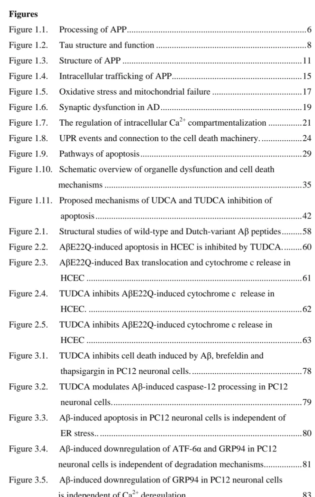
We first compared aggregation/fibrillization properties, secondary structure, and cell death pathways of wild-type amyloid β (Aβ) peptides and AβE22Q Dutch variant, both in the presence and absence of anti-apoptotic tauroursodeoxycholic acid (TUDCA). Our data dissociate the apoptotic properties of Aβ peptides in human cerebral endothelial cells from their distinct mechanisms of aggregation in vitro, while confirming the perspectives for modulation of amyloid-induced mitochondrial apoptosis by TUDCA.
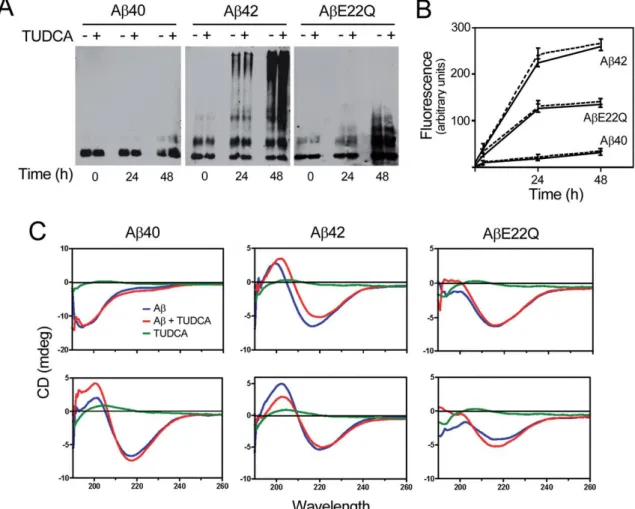
Next, we investigated the ability of the endoplasmic reticulum (ER) to sense stress directly caused by the Aβ toxic core. Aβ exposure triggered an early signaling response by the ER, and caspase-12-mediated apoptosis in PC12 neuronal-like cells, independently of the ER-stress pathway. Indeed, ER stress markers, including GRP94, ATF-6α, CHOP and eIF2α were strongly donwregulated by Aβ, in a protein degradation-independent manner, and partially restored by TUDCA. Calpain inhibition prevented caspase-12 activation and ATF-6α downregulation. However, GRP94 downregulation was associated with protein secretion, which in turn was partially rescued by inhibition of the secretory pathway. Thus, Aβ triggers an ER response beyond the usual ER stress.
Finally, we further investigated the role of the c-Jun N-terminal kinase (JNK) as early stress sensor of Aβ toxicity that is activated by ER pathways. We reported that JNK acted as the proximal stress sensor, which translocated to the nucleus and activated caspase-2 in PC12 neuronal-like cells. Caspase-2 then cleaved golgin-160, suggesting a unique signaling through the Golgi complex. Importantly, TUDCA modulated the JNK/caspase-2 apoptotic pathway triggered by Aβ.
In conclusion, these studies contribute to the understanding of the complex sub-cellular communication involved in Aβ toxicity. Further characterization of these signaling pathways and exact targets are likely to provide new perspectives for modulation of amyloid-induced cellular dysfunction and degeneration.
Keywords: Amyloid-β peptides; Apoptosis; Cell organelles; GRP94; JNK;
Tauroursodeoxycholic acid
Acknowledgements
Não poderia deixar de começar por agradecer à pessoa que possibilitou que este trabalho fosse desenvolvido, a Professora Cecília Rodrigues. Será de todo impossível detalhar uma relação profissional muito frutuosa que durou quatro anos. Contudo, sou impelido a enfatizar uma característica impressionante do seu carácter: o seu sentido de missão! A sua entrega incondicional, aliada à enorme capacidade de trabalho, são traços marcantes da sua pessoa. Posso por isso testemunhar, o quão confortável é para quem está a fazer um doutoramento, saber que se tem sempre a quem recorrer, independentemente dos dias e horas! Assim, cara Professora, como o seu trabalho é feito de partilha, na investigação que faz, e dádiva, nos conhecimentos que transmite como professora, gostava antes de partir de lhe deixar o meu bem-haja e pensar que também lhe deixo a si, alguma coisa de mim.The choice to do a PhD is much more than a simple job for some years. It is a journey that changes the rest of our lives. In many years from now, I am sure I will remember the particular moment of my journey in the New York University, at Professors Agueda Rostagno and Jorge Ghiso laboratory. I was received in the very best warm way and treated as one of the laboratory members. Thank you for letting me be part of the excellence of your laboratory, and also for your humane characteristics that made you so special. I also want to thank Silvia in name of all the laboratory members for making my staying in NY a great time. I could not finish without expressing the feelings I nurture for Milton and André. You were my support, my friends, my "homy" place. You are responsible for the "New Yorker" feeling that I still have today, and I have no words to express how thankful I am.
Para os meus queridos companheiros de laboratório, deixo mais do que um agradecimento. Partilho convosco o sentimento que me percorre quando fecho os olhos e penso em cada um de vós: Felicidade! "Aquele sorriso maroto" invade-me o rosto e
leva-me a viajar pelas nossas leva-memórias. Foram anos intensos que vivemos juntos e nos quais fomos actores principais de momentos memoráveis, dos quais não me poderei esquecer: o apoio incondicional que a Rita e a Filipa me deram em todas as situações no decorrer desta maratona; a minha companheira de percurso, a Márcia; a preocupação carinhosa e subtil da Susana; as brincadeiras com o Rui; as gargalhadas com a Joana; as conversas com a Daniela; a "família" que ganhei com o Pedro; e o ar fresco que a "geração" Benedita, Duarte e Joana Xavier trouxeram, tendo a certeza que irão dar continuidade a este laboratório duma forma ainda melhor do que aquela que já foi conseguida. Quero ainda agradecer à Professora Isabel Moreira da Silva pela forma atenciosa e afável com que sempre me tratou. Quero também agradecer às Professoras Elsa Rodrigues, Maria João Gama e Margarida Castro Caldas, bem como aos membros do laboratório Inês, Maria, Andreia e Miguel pela sempre boa disposição que vos caracteriza e por todas as discussões científicas partilhadas.
Agradeço à minha família Coimbrã. À Universidade de Coimbra na pessoa dos professores da Faculdade de Farmácia, o meu bem-haja! Aos meus amigos Hugo, "Salgados", Ruivo, Rodri, Batista e André, … quero simplesmente abraçar-vos para sentir o calor da vossa amizade que aqui não consigo descrever. Como sabeis, o que criamos em Coimbra não se escreve, vive-se!
Agradeço ao meu irmão Miguel, ao Guilherme e Susana por serem um pilar fundamental da minha vida. Mi, a fé e a confiança que depositas em mim, empurram-me para lugares que não existem e que ainda não foram tocados! Obrigado por me fazeres sentir um ser especial.
Aos meus pais, estou eternamente grato por terem semeado dentro de mim os valores, princípios e moral do meu carácter. Hoje, em tudo o que faço, colho o fruto do vosso trabalho e penso sempre na bênção que tive e tenho de vos ter como meus pais.
Por último, agradeço a quem reconheço o trabalho mais árduo, à minha mulher Charlene. Agradeço todos os dias que passamos juntos; o sofrimento que suportaste para eu prosseguir com o meu sonho; a harmonia que me proporcionas; a felicidade que despertas em mim; a minha personalidade intensa que aguentas com um sorriso; a alegria que me dás por chegar a casa; … todo o equilíbrio que me ofereces. Por tudo isto e muito mais, não consigo expressar em palavras a gratidão que tenho para contigo. Só sentindo a força do brilho dos meus olhos quando te vejo!
Abbreviations
AD Alzheimer‟s disease
AICD amyloid precursor protein intracellular domain
APOE-4 4 allele of the apolipoprotein E APP amyloid precursor protein
ASK1 apoptosis signal-regulating kinase 1
ATF-6 activating transcription factor 6
Aβ amyloid β
AβE22Q mutation in which Gln replaces Glu at position 22 of amyloid β
Bak Bcl-2 antagonist/killer
BAR bifunctional apoptosis regulator
BAX Bcl-2-associated X protein
Bcl-2 B-cell leukemia/lymphoma 2
Bcl-xL B-cell leukemia/lymphoma extra long
BI1 BAX inhibitor 1
C99 β-cleaved C-terminal fragment of amyloid precursor protein
Ca2+ calcium
CHOP CCAAT/enhancer binding protein homologous protein
CTF amyloid precursor protein C-terminal fragment
eIF2α eukaryotic translation initiation factor 2α
ER endoplasmatic reticulum
ERAD endoplasmic reticulum-assisted degradation
FADD Fas associated death domain
GC Golgi complex
GDV granulovacuolar degeneration bodies
GRP78/Bip 78-kDa glucose-regulated protein
GRP94 94-kDa glucose-regulated protein
GSK-3β glycogen synthase kinase 3β
hsp heat shock protein 90
IRE1 inositol-requiring kinase 1
JIP c-Jun N-terminal kinase-interacting protein JNK c-Jun N-terminal kinase
MAPK mitogen-activated protein kinase
MVBs multivesicular bodies
NFTs neurofibrillary tangles
NSR nuclear steroid receptor
PCD programmed cell death
PERK double stranded ribonucleic acid-activated protein kinase- like endoplasmic reticulum kinase
PI3K phosphatidyl inositol 3-kinase
PS1 presenilin-1
PS2 presenilin-2
ROS reactive oxygen species
sAPP soluble amyloid precursor protein
TRAF2 tumor necrosis factor receptor-associated factor 2
TRAIL-R1 tumor necrosis factor-related apoptosis inducing ligand receptor 1
TUDCA tauroursodeoxycholic acid
TUNEL terminal transferasedUTP nick-end labeling
UDCA ursodeoxycholic acid
Publications
The present thesis is based on work that has been published, or submitted for publication, in international peer-reviewed journals:Viana RJ, Nunes AF, Castro RE, Ramalho RM, Meyerson J, Fossati S, Ghiso J,
Rostagno A, Rodrigues CM. Tauroursodeoxycholic acid prevents E22Q Alzheimer's Aβ toxicity in human cerebral endothelial cells. Cellular and Molecular Life Sciences 2009; 66: 1094-1104.
Viana RJ, Ramalho RM, Nunes AF, Steer CJ, Rodrigues CM. Modulation of amyloid-β
peptide-induced toxicity through inhibition of JNK nuclear localization and caspase-2 activation. Journal of Alzheimer's Disease 2010; 22: 557-568.
Viana RJ, Steer CJ, Rodrigues CMP. Amyloid-β peptide-induced secretion of
endoplasmic reticulum chaperone glycoprotein GRP94 in PC12 neuronal-like cells. 2010 (submitted).
The following manuscripts have also been published during the Ph.D. studies:
Ramalho RM, Viana RJ, Low WC, Steer CJ, Rodrigues CM. Bile acids and apoptosis modulation: an emerging role in experimental Alzheimer's disease. Trends in Molecular
Medicine 2008; 14: 54-62.
Ramalho RM, Viana RJ, Castro RE, Steer CJ, Low WC, Rodrigues CM. Apoptosis in transgenic mice expressing the P301L mutated form of human tau. Molecular Medicine 2008; 14: 309-317.
Santos DM, Santos MM, Viana RJ, Castro RE, Moreira R, Rodrigues CM. Naphtho[2,3-d]isoxazole-4,9-dione-3-carboxylates: potent, non-cytotoxic, antiapoptotic agents.
Chemico-Biological Interactions 2009; 180: 175-182.
Amaral JD, Viana RJ, Ramalho RM, Steer CJ, Rodrigues CM. Bile acids: regulation of apoptosis by ursodeoxycholic acid. Journal of Lipid Research 2009; 50: 1721-1734.
Viana RJ, Fonseca MB, Ramalho RM, Nunes AF, Rodrigues CM. Organelle stress
sensors and cell death mechanisms in neurodegenerative diseases. CNS & Neurological
Table of Contents
Resumo ... v Abstract ... xi Acknowledgements ... xv Abbreviations ... xvii Publications ... xix 1 General Introduction ... 1 1.1 Alzheimer's disease ... 3 1.1.1 Epidemiology ... 3 1.1.2 Molecular neuropathology ... 4 1.1.3 Risk factors ... 7 1.1.4 Amyloid precursor protein ... 13 1.2 Cell death mechanisms ... 26 1.2.1 Death receptor and mitochondrial pathways of apoptosis ... 28 1.2.2 Role of apoptosis in Alzheimer‟s disease ... 30 1.2.3 Role of organelle dysfunction in Alzheimer‟s disease ... 35 1.2.4 Inhibition of neuronal apoptosis by bile acids ... 40 Objectives ... 45 2 Tauroursodeoxycholic acid prevents E22Q Alzheimer‟s amyloid β toxicity inhuman cerebral endothelial cells ... 47 2.1 Abstract ... 49 2.2 Introduction ... 50 2.3 Materials and methods ... 52
2.3.1 Peptide synthesis ... 52 2.3.2 Peptide aggregation ... 52 2.3.3 Peptide structural analysis ... 53 2.3.4 Culture of human brain microvascular endothelial cells ... 54 2.3.5 Induction of apoptosis ... 54 2.3.6 Measurement of cell death ... 54 2.3.7 Immunocytochemistry ... 55 2.3.8 Bax translocation and cytochrome c release ... 56 2.3.9 Statistical analysis ... 57 2.4 Results ... 57
2.4.1 TUDCA does not affect Aβ peptide aggregation and secondary
structure ... 57 2.4.2 TUDCA inhibits AβE22Q-induced apoptosis in brain microvascular
endothelial cells ... 59 2.4.3 TUDCA prevents AβE22Q-induced mitochondrial Bax translocation
and cytochrome c release ... 61 2.5 Discussion ... 63 3 Amyloid β peptide-induced secretion of endoplasmic reticulum chaperone
glycoprotein GRP94 in PC12 neuronal-like cells ... 69 3.1 Abstract ... 71 3.2 Introduction ... 72 3.3 Materials and methods ... 74 3.3.1 Cell culture and culture conditions ... 74 3.3.2 Measurement of cell death ... 75 3.3.3 Immunoblotting ... 75 3.3.4 RNA isolation and RT-PCR ... 76 3.3.5 Heat shock protein GRP94 ELISA assay ... 76 3.3.6 Statistical analysis ... 76
3.4 Results ... 77 3.4.1 Aβ-induced apoptosis is mediated by caspase-12 activation and
modulated by TUDCA in PC12 neuronal-like cells ... 77 3.4.2 Aβ-induced, caspase-12-mediated cell death is independent of ER
stress ... 79 3.4.3 ATF-6α and GRP94 downregulation is independent of protein
degradation ... 80 3.4.4 GRP94 downregulation is independent of Ca2+ deregulation ... 82 3.4.5 Protein secretion inhibitors block GRP94 downregulation ... 83 3.5 Discussion ... 85 4 Modulation of amyloid β peptide-induced toxicity through inhibition of JNK
nuclear localization and caspase-2 activation... 89 4.1 Abstract ... 91 4.2 Introduction ... 92 4.3 Materials and methods ... 93 4.3.1 Cell culture and induction of apoptosis ... 93 4.3.2 Measurement of cell death ... 94 4.3.3 Inhibition of JNK pathway ... 94 4.3.4 Immunoblotting ... 95 4.3.5 Immunocytochemistry ... 95 4.3.6 Short interference-mediated silencing of the jnk gene ... 96 4.3.7 Expression of Golgin-160 cleavage products ... 96 4.3.8 Statistical analysis ... 96 4.4 Results ... 97
4.4.1 Aβ-induced apoptosis is mediated by JNK activation and modulated by TUDCA in PC12 neuronal-like cells ... 97 4.4.2 p-JNK localizes to the nucleus and mediates Aβ-induced caspase-2
4.4.3 Aβ-induced caspase-2 activation mediates cell death via the Golgi complex ... 102 4.5 Discussion ... 104 5 Concluding Remarks ... 109 References ... 117
Figures
Figure 1.1. Processing of APP ... 6 Figure 1.2. Tau structure and function ... 8 Figure 1.3. Structure of APP ... 11 Figure 1.4. Intracellular trafficking of APP... 15 Figure 1.5. Oxidative stress and mitochondrial failure ... 17 Figure 1.6. Synaptic dysfunction in AD ... 19 Figure 1.7. The regulation of intracellular Ca2+ compartmentalization ... 21 Figure 1.8. UPR events and connection to the cell death machinery. ... 24 Figure 1.9. Pathways of apoptosis ... 29 Figure 1.10. Schematic overview of organelle dysfunction and cell death
mechanisms ... 35 Figure 1.11. Proposed mechanisms of UDCA and TUDCA inhibition of
apoptosis ... 42 Figure 2.1. Structural studies of wild-type and Dutch-variant Aβ peptides ... 58 Figure 2.2. AβE22Q-induced apoptosis in HCEC is inhibited by TUDCA. ... 60 Figure 2.3. AβE22Q-induced Bax translocation and cytochrome c release in HCEC ... 61 Figure 2.4. TUDCA inhibits AβE22Q-induced cytochrome c release in
HCEC. ... 62 Figure 2.5. TUDCA inhibits AβE22Q-induced cytochrome c release in
HCEC ... 63 Figure 3.1. TUDCA inhibits cell death induced by Aβ, brefeldin and
thapsigargin in PC12 neuronal cells. ... 78 Figure 3.2. TUDCA modulates Aβ-induced caspase-12 processing in PC12 neuronal cells. ... 79 Figure 3.3. Aβ-induced apoptosis in PC12 neuronal cells is independent of
ER stress.. ... 80 Figure 3.4. Aβ-induced downregulation of ATF-6α and GRP94 in PC12 neuronal cells is independent of degradation mechanisms. ... 81 Figure 3.5. Aβ-induced downregulation of GRP94 in PC12 neuronal cells
Figure 3.6. Aβ-induced downregulation of GRP94 in PC12 neuronal cells
is blocked by protein secretion inhibitors. ... 84 Figure 4.1. TUDCA inhibits apoptosis induced by Aβ in PC12 neuronal cells. .. 97 Figure 4.2. TUDCA modulates Aβ-induced p-JNK and p-c-Jun levels in
PC12 neuronal cells. ... 98 Figure 4.3. Aβ induces p-JNK translocation to the nucleus in PC12 neuronal cells. ... 100 Figure 4.4. TUDCA modulates Aβ-induced active caspase-2 levels in PC12 neuronal cells. ... 101 Figure 4.5. Silencing of JNK inhibits Aβ-induced caspase-2 activation in
PC12 neuronal cells. ... 102 Figure 4.6. Caspase-2 localizes to the Golgi complex and cleaves golgin-160 in Aβ-induced cell death in PC12 neuronal cells. ... 103
1.1 Alzheimer's disease
Alzheimer‟s disease (AD) is a fatal progressive neurodegenerative illness,and the most common form of dementia. It is, therefore, a fundamental disorder of cognitive awareness, integrating reasoning, abstraction, language, and memory, one of the defining components of human consciousness (Nussbaum and Ellis, 2003). This condition was first described by Alois Alzheimer in 1906. He presented a clinical/pathological report of a 51-year old woman, Auguste Deter, who had died of what came to be termed “dementia praecox.” At autopsy, it was noted that there were extracellular deposits, termed “plaques”, and intracellular deposits or “tangles”, in the neocortex of the brain. Tangles are a common hallmark of brain injury and are seen after a variety of other insults, such as stroke and head trauma. Hence, their presence does not give any clue as to the etiology of the cognitive decline seen in AD. On the other hand, the plaque deposits, although also seen with other injuries, do appear to bear a relationship to the underlying cause of the dementia (Holtzman, 2010).
1.1.1 Epidemiology
The onset of AD is inevitably followed by increasing mental and physical incapacitation, including loss of ability to look after one‟s own daily needs, followed by institutionalization and death. AD is the most common cause (60-80%) of dementia worldwide. The prevalence of AD is less than 1% in individuals aged 60-64 years, but the probability of developing the disorder increases with age. The estimated rates of occurrence in various age groups are as follows: 65-74 years, 2.5%; 75-79 years, 4%; 80-84 years, 11%; and 85-93 years, 24%. It has been estimated that 4.6 million new cases will appear every year, at a rate of one new case every 7 seconds. With life expectancy increasing, the numbers are predicted to double every 20 years to 81.1 million by 2040 (Ferri et al., 2005). In Portugal, there are ~ 60,000 patients with AD, although this may represent an under estimation, given the difficulty of diagnosis.
AD is fourth in the list of leading causes of death in industrialized societies, preceded only by heart disease, cancer, and stroke. It affects individuals of all races and ethnic groups, occurring slightly more commonly in women than in men. Although AD is emerging as the most prevalent and socially disruptive illness of aging populations, its causes and cure remain enigmas (Aguzzi and O'Connor, 2010).
1.1.2 Molecular neuropathology
AD is characterized by two neuropathological hallmarks, including extracellular deposits, known as amyloid or “senile” plaques, and intracellular neurofibrillary tangles (NFTs) (Spillantini and Goedert, 1998). In addition to these deposits, the AD brain is characterized by a dramatic loss of neurons and synapses in many areas of the central nervous system, particularly in regions involving higher-order cognitive functions, such as learning and memory. Because memory loss may occur by other causes, definitive diagnosis of AD requires postmortem examination of the brain, which must contain sufficient numbers of plaques and tangles to qualify as affected by AD (Braak and Braak, 1998; Dickson, 1997).
Brain regions, such as the basal forebrain and hippocampus that exhibit plaques, typically possess a reduced number of synapses. Neurites found associated with the plaques are also often damaged, suggesting a toxic effect for amyloid β (Aβ) peptide, the major constituent of amyloid plaques (Mattson, 2004). In addition, the levels of many neurotransmitters are greatly reduced, including serotonin, noradrenaline, dopamine, glutamate, and especially acetylcholine. Reduced neurotransmitter levels are thought to be responsible for the broad and profound clinical symptoms of AD (Nussbaum and Ellis, 2003).
1.1.2.1 Amyloid plaques
Amyloid plaques are primarily composed of a 4.3-kDa amyloid peptide, first isolated from the brains of patients with AD in 1984. The Aβ peptide is a 39-43 amino acid sequence that forms extraneuronal aggregates with a fibrillar, β-pleated structure. Moreover, Aβ peptide accumulation can be observed both in brain and blood vessels.
Aβ is cleaved from the amyloid precursor protein (APP) (Fig. 1.1), a 695-770 amino acid transmembrane protein found in virtually all peripheral and brain cells (Selkoe and Schenk, 2003). Although there is no conclusive evidence of the function of APP, an increasing body of data suggests its involvement in regulating neurite outgrowth, synaptic plasticity, and cell adhesion (Mattson, 2004).
APP is normally cleaved within the Aβ domain by α-secretase, liberating a soluble N-terminal fragment (sAPPα) and a membrane-bound C-terminal fragment (C83). Alternatively, APP can be cleaved by β-secretase at the N-terminus of the Aβ domain,
yielding s-APPβ and C99. The latter membrane-bound fragment, then undergoes intramembrane cleavage by γ-secretase at the C-terminus of Aβ, resulting in the liberation of Aβ into the cell (Yamazaki and Ihara, 1998). The secretion of Aβ follows, allowing the peptide to participate in extracellular aggregation and to become incorporated into growing plaques.
The β-site of APP cleaving enzyme (BACE1) is responsible for the β-secretase activity; γ-secretase is composed of four essential subunits, including presenilin-1 (PS1) or presenilin-2 (PS2), together with nicastrin, anterior pharynx-defective 1 (APH-1), and presenilin enhancer 2 (PEN-2) (Thinakaran and Koo, 2008). The γ-secretase complex cleaves at multiple sites within the transmembrane domain of APP, generating Aβ peptides ranging in length from 38 to 42 residues (Selkoe and Wolfe, 2007). Nearly 90% of secreted Aβ ends at residue 40, giving Aβ40, whereas Aβ42 accounts for only less than 10%, and peptides ending at residues 38 are minor components (Thinakaran and Koo, 2008). Notably, the pattern and distribution of these species varies among the different topographical lesions. Parenchymal deposits consist of Aβ42 as the major component, whereas vascular Aβ, particularly in the large leptomeningeal vessels, is primarily composed of Aβ40 species, organized in large concentric sheets and replacing the smooth muscle cell layer (Roher et al., 1993). Amyloid associated with arterioles and small cortical arteries contains a mixture of Aβ40 and Aβ42, while deposits affecting the capillary network are mainly composed of Aβ42 (Kokjohn and Roher, 2009). The reasons for this selectivity as well as its importance for the pathogenesis of the disease remain largely unclear (Rostagno et al., 2010).
The term “soluble Aβ” is generally applied either to newly generated, cell-secreted Aβ or to that fraction of tissue or synthetic Aβ that is taken into the aqueous phase of a non-detergent containing extraction buffer. “Misfolded” and “aggregated” Aβ are terms used to describe very early, nonspecific changes in Aβ folding states or solubility states, respectively (e.g., aggregated Aβ solutions usually scatter light to a greater extent than do solutions of soluble Aβ). “Oligomeric” Aβ refers to peptide assemblies with limited stoichiometry (e.g., dimers, trimers, etc.), while protofibrils are structures of intermediate order between aggregates and fibrils (Fig. 1.1) (Gandy, 2005).
Querfurth and LaFerla, 2010
Figure 1.1. Processing of APP. (A) Non-amyloidogenic processing of APP, with sequential cleavage by membrane-bound α- and γ-secretase. α-secretase cleaves within the Aβ sequence, thus preventing the generation of intact Aβ peptide. sAPPα ectodomain is secreted to extracellular milieu and C83 is released. C83 is then processed by γ-secretase, releasing extracellular p3 (3 kDa) fragment. Alternatively, amyloidogenic processing starts by β-secretase cleavage at the N-terminus of Aβ, releasing a shorter sAPPβ and C99 fragments. The C99 fragment is subsequently cleaved within the transmembrane domain by γ-secretase to originate Aβ and AICD. AICD is targeted to the nucleus, signaling transcription activation. Soluble Aβ is a hydophobic peptide that is prone to aggregation. (B) Protofibrils (upper left) and annular or porelike profiles (lower left) are transient structures observed during in vitro formation of mature amyloid fibrils. In the right panel, self-association of 2 to 14 Aβ monomers into oligomers is dependent on concentration (right, left immunoblot). In the right immunoblot, oligomerization is promoted by oxidizing conditions (lane 2) and divalent metal conditions (lane 3). AICD, APP intracellular domain; GPI, glycophosphatidylinositol.
Numerous mutations have been identified in the gene encoding APP, which alter its expression or processing (Rocchi et al., 2003). These mutations are associated with early onset AD and are implicated in the autosomal dominant form of the disease, which constitutes ~ 5% of all cases. Patients with Down‟s syndrome, who have three copies of the APP gene, show diffuse Aβ deposits as early as in the second decade of life. These eventually develop into mature neuritic plaques that are indistinguishable from those found in the brains of patients with AD.
1.1.2.2 Neurofibrillary tangles
The chief component of intracellular NFTs is tau, a microtubule-associated protein abundant in six different isoforms in the adult brain. In AD, highly phosphorylated and glycosylated tau protein forms paired, helically wound fragments (paired helical filaments), ~ 10 nm in diameter, which associate to give insoluble tangles in nerve cell bodies and dendrites (Fig. 1.2) (Spillantini and Goedert, 1998). Tangles are a hallmark of many dementia-related diseases, including corticobasal ganglionic degeneration, frontotemporal dementia, myotonic dystrophy, Niemann-Pick disease, and progressive supranuclear palsy. The wide variety of neurological disorders in which NFTs occur suggests that paired helical filament formation is a nonspecific marker of certain types of neuronal injury.
1.1.3 Risk factors
Besides ageing, which is the most obvious risk factor for AD, no specific environmental toxin has been found to be consistently associated with the disease, and there have been no randomized clinical trials as yet to support any specific dietary intervention. Some evidence suggests that dietary intake of homocysteine-related vitamins, vitamin B12 and folate; antioxidants, such as vitamin C and E; unsaturated fatty acids; and also moderate alcohol intake, especially wine, could reduce the risk of AD. Epidemiological studies point to depression, traumatic head injury and cardiovascular and cerebrovascular factors, including cigarette smoking, midlife high blood pressure, obesity, hypercholesterolaemia, atherosclerosis, coronary heart disease and diabetes as increasing disease risk, while anti-inflammatory medications seem to reduce risk. Some studies even suggest a beneficial role of psychosocial factors as though, higher education, physical exercise and mental activity (reviewed in Selkoe and Schenk, 2003). Such
reports may point to a role of previously unconsidered pathways in the etiology of the disease, but the mechanistic interpretation of retrospective epidemiological studies is challenging (Citron, 2010).
Querfurth and LaFerla, 2010
Figure 1.2. Tau structure and function. Tau is a highly soluble microtubule-binding protein that contributes to microtubule assembly and cohesion. Four repeat sequences (R1-R4) make up the microtubule-binding domain (MBD) of tau. Tau is abnormally hyperphosphorylated in AD by numerous kinases, leading to the formation of NFTs. Normal phosphorylation of tau occurs on serine (S; inset) and threonine (T; inset) residues, numbered according to their position in the full tau sequence. Between the N‑terminal portion of the protein (projection domain) and the MBD lies a basic proline-rich region (P); these amino acids are phosphorylated by GSK-3β, CDK5 and its activator subunit p25, or MAPK. Non proline-directed kinases, such as Akt, Fyn, PKA,
CaMKII, and MARK are also shown. By activating these kinases, certain inflammatory cytokines can also trigger tau hyperphosphorylation. Hyperphosphorylated sites specific to paired helical filament tau in AD tend to flank the MBD. Excessive kinase, reduced phosphatase activities, or both cause hyperphosphorylated tau. Hyperphosphorylation of tau, particularly that mediated by MARK, CDK5 and GSK-3β destabilizes microtubules, causing impairments in axonal transport and neuronal dysfunction. CDK5, cyclin-dependent kinase 5; MAPK, mitogen-activated protein kinase; PKA, protein kinase A; CaMKII, calcium-calmodulin protein kinase 2; MARK, microtubule affinity-regulating kinase. From Querfurth and LaFerla, 2010.
1.1.3.1 Genetics
Family history is the second-greatest risk factor for AD after age, and the increasing understanding of its genetics has been vital to the knowledge of the pathogenic mechanisms that cause the disease. Genetically, AD is complex and heterogeneous and appears to follow an age-related dichotomy: rare and highly penetrant early-onset familial AD mutations in different genes are transmitted in an autosomal dominant fashion, and late-onset AD that represents the vast majority of the cases and is absent of obvious familial segregation (Bertram and Tanzi, 2005).
1.1.3.1.1 Sporadic disease
The designation of late-onset AD is used when the onset of the disease occurs at 65 years or older, and represents ≥ 95% of all AD cases. Although genetic analysis of segregation and twin studies have suggested a major role of genetic factors in this form of AD (Mayeux et al., 1991), until now, only one factor has been effectively established, the 4 allele of the apolipoprotein E (APOE-4) gene on chromosome 19q13 (Schmechel et al., 1993; Strittmatter et al., 1993). In contrast to all other association-based observations in AD, the risk outcome of APOE-4 has been consistently replicated by different laboratories that developed several studies transversal to numerous ethnic groups (for meta-analysis, see Farrer et al., 1997). The APOE-4 genotype, which occurs in 16% of the population (Roses, 1997) is the major susceptibility gene identified for both rare early-onset and sporadic late-onset AD (Coon et al., 2007). In addition to the increased risk by 4-allele, it has also been reported in several studies a weak, albeit significant, protective effect for the minor allele, 2. Contrasting to mutations in the known early-onset familial AD genes, APOE-4 is neither necessary nor sufficient to cause AD, since
many 4-positive individuals do not develop AD, and many AD patients do not carry these genes. Instead, APOE-4 operates as a genetic risk modifier by decreasing the age of onset in a dose-dependent manner. Despite its long-known and well-established genetic association, the molecular effects of APOE-4 in AD pathogenesis are not yet fully understood, but are expected to include Aβ-aggregation/clearance and/or cholesterol homeostasis (Bertram and Tanzi, 2005).
1.1.3.1.2 Familial disease
Early-onset familial AD (FAD) represents only a small fraction of ≤ 5% of all AD cases. This designation is used when onset ages are younger than 65 years, showing autosomal dominant transmission within affected families. To date, more than 160 mutations in 3 genes have been reported to cause early-onset FAD. These genes include the APP on chromosome 21 (Goate et al., 1991), PS1 on chromosome 14 (Sherrington et al., 1995), and PS2 on chromosome 1 (Levy-Lahad et al., 1995; Rogaev et al., 1995).
PS1 represents the most frequently mutated gene and accounts for the majority of AD
cases with onset prior to age of 50 (Bertram and Tanzi, 2005). Of the genes directly related to the development of familial AD, PS1 and PS2 mutations affect the levels of Aβ42 production, whereas nucleotide changes within the APP molecule have a differential effect depending on the location of the mutated residue (Fig. 1.3).
Alterations in Aβ domain that result from amino acid replacement in regions adjacent to β-secretase cleavage site change the kinetics of APP enzymatic processing. As a consequence, increased production of Aβ42 and Aβ40 is observed, maintaining the ratio Aβ42/Aβ40 unaffected, which manifests in a classic early-onset AD clinical phenotype (Revesz et al., 2009). Two examples are Swedish double mutation (K670N/M671L) (Mullan et al., 1992) that increases the production of Aβ40 and Aβ42 by > 5-fold, and Italian mutation (A673V) (Di Fede et al., 2009) that shows enhanced production of both Aβ species by ~ 2-fold, maintaining the ratio Aβ42/Aβ40 unaltered. However, different processing is observed for mutations in regions adjacent to the γ-secretase cleavage sites. These mutations are typically associated with increased production of Aβ42 and lower levels of Aβ40, analogous to what is observed for mutations in the presenilin genes (Suzuki et al., 1994). As a result, the ratio of Aβ42/Aβ40 is several fold elevated, reaching in some cases ~ 18-fold values. Amino acid
substitutions within residues 21-23 and 34 of the Aβ peptide are, with few exceptions (Wakutani et al., 2004), associated with prominent vascular compromise.
Gandy, 2005
Figure 1.3. Structure of APP. (A) Schematic structure and topography of APP. (B) Area surrounding the Aβ domain, α-, β- and γ-secretase cleavage sites, and amino acid location in selected FAD missense mutations.
1.1.3.2 Cerebral amyloid angiopathy
Cerebral amyloid angiopathy (CAA) due to Aβ deposition is a key pathological feature of FAD. It is an age-associated condition, characterized by deposition of Aβ in the medial layer of cerebral blood vessels (Coria and Rubio, 1996). It is also a significant cause of intracerebral hemorrhage in patients with AD and certain related disorders (Coria and Rubio, 1996; Melchor et al., 2000). Specific missense mutations in the APP gene occurring inside the Aβ region comprising residues 21-23 is considered a „„hot spot‟‟ for mutations due to the high number of genetic variants reported in this area of the molecule. Mutations within this amino acid cluster typically show strong vascular compromise and primarily associate with CAA, hemorrhagic strokes and dementia. Mutations in the Aβ sequence are concentrated at positions 21-23 and are termed Dutch (E22Q) (Miravalle et al., 2000), Italian (E22K) (Miravalle et al., 2000), Iowa (D23N) (Van Nostrand et al.,
2001) , Arctic (E22G) (Nilsberth et al., 2001) , Flemish (A21G) (Demeester et al., 2001) and New Italian (L34V) (Obici et al., 2005) mutations. In general, all mutants are thought to have stronger neurotoxicity than wild-type Aβ, which may correlate with their ability to form protofibrils. However, not all mutants have the same capacity to induce neurotoxic effects. This suggests that the substitution of different amino acids may confer distinct structural properties, influencing the onset and aggressiveness of the disease.
The first mutation inside Aβ domain was described in a condition known as hereditary cerebral hemorrhage with amyloidosis, Dutch type (HCHWA-D), an autosomal dominant disorder clinically defined by recurrent strokes, vascular dementia, and fatal cerebral bleeding in the fifth to sixth decades of life. In this missense mutation, a Gln replaces a Glu residue at position 22 (AβE22Q) resulting from a single nucleotide transversion (G for C) at codon 693 (Levy et al., 1990). The clinical manifestations for the carriers of this mutation include periodic episodes of cerebral hemorrhages that are associated with massive amyloid deposition in the walls of leptomeningeal and cortical arteries and arterioles, as well as in vessels in the brainstem and cerebellum (Bornebroek et al., 1999). Besides the vascular involvement, Dutch familial cases also develop parenchymal amyloid deposits resembling the diffuse preamyloid lesions seen in AD. However, dense-core plaques and NFTs are rare or even not present at all (Levy et al., 1990). The non-fibrillar diffuse plaques show differences in the extent of fibrillary material, ranging from fine diffuse plaques in young patients to more fibrillar, dense content in old patients (Maat-Schieman et al., 2000). Thus, the non-fibrillar diffuse plaques, variably associated with reactive astrocytes and activated microglia (Maat-Schieman et al., 2004) evolve into more fibrillar, dense lesions (Maat-(Maat-Schieman et al., 2000). Nevertheless, the degree of dementia appears to be independent of plaque involvement and neurofibrillary degeneration, which is absent or limited, and contrastingly correlates with the severity of CAA (Natte et al., 2001).
Aβ40 is the predominant Aβ species of HCHWA-D vascular amyloid, with wild-type and Dutch-wild-type mutated sequences present at a 1:1 molar ratio. In contrast, Aβ42 is only a minor component of the vascular deposits (Herzig et al., 2004) and is uniquely associated with species bearing the mutation, and not with those originated from the normal allele (Prelli et al., 1990). So far, it has not been possible to identify Aβ42 wild- type or carrying the E22Q substitution in the cortex by Western blot, and ELISA results show an Aβ42/Aβ40 considerably lower than in AD cases (Herzig et al., 2004).
In vitro cell culture studies show that E22Q has potent toxic effects on
cerebrovascular endothelial and smooth muscle cells (Davis and Van Nostrand, 1996; Miravalle et al., 2000), likely reflecting the in vivo hemorrhage-associated phenotype. The E22Q mutant is also a potent angiogenesis inhibitor, disturbing the fibroblast growth factor 2 (FGF-2) signaling pathway, downregulating phosphorylation of the FGF-2 receptor, and the Akt survival signaling (Solito et al., 2009). Altogheter, these effects lead to endothelial dysfunction and probably contribute, solely or partially, to the disturbances in membrane permeability and hemorrhagic manifestations in the family members (Rostagno et al., 2010).
1.1.4 Amyloid precursor protein
APP in mammals belongs to a family of conserved type I membrane proteins that include APP (Goldgaber et al., 1987), APP like protein 1 (APLP1) (Wasco et al., 1992) and 2 (APLP2) (Slunt et al., 1994). Importantly, the Aβ peptide domain is not conserved and is only present in APP and not in any of the other APP-related genes. Although APP family is abundantly expressed in the brain, and APLP1 expression is restricted to neurons (Lorent et al., 1995), APP and APLP2 can also be found in the majority of other tissues as well. The human APP gene is located on the long arm of chromosome 21 (Lamb et al., 1993). The expression results in alternative splicing that generates APP mRNAs encoding isoforms that go from 365 to 770 amino acid residues. The most important Aβ peptide encoding proteins have 695, 751 and 770 amino acids, and are denominate as APP695, APP751 and APP770 (Zheng and Koo, 2006).
The exact physiological role of APP is not clearly understood. However, several functions have been recognized based on the variety of APP extracellular subdomains, such as cell surface receptor (Lorenzo et al., 2000), cell adhesion (Soba et al., 2005), neurite outgrowth and synaptogenesis (Herard et al., 2006). Furthermore, the APP intracellular domain presents a high degree of sequence conservation between the intracellular domain of APP, APLP1, and APLP2, predicting that it is a critical domain for regulation of APP function (Zheng and Koo, 2006).
In fact, APP can be phosphorylated at multiple sites in both extracellular and intracellular domains (Hung and Selkoe, 1994). Among these, the phosphorylation at the threonine residue (Thr668) in APP intracellular domain has received most attention. Several kinases have been implicated in this phosphorylation event, including the
cyclin-dependent kinase 5 (CDK5), c-Jun N-terminal kinase 3 (JNK3), and glycogen synthase kinase 3β (GSK-3β) (Iijima et al., 2000; Kimberly et al., 2005; Muresan and Muresan, 2005a). APP Thr668 phosphorylation has been described to control APP localization to the growth cones and neurites (Muresan and Muresan, 2005a). Notably, Thr668 phosphorylated APP is preferentially transported to the nerve terminals (Muresan and Muresan, 2005b), and Thr668 phosphorylated APP fragments are increased in AD, but not in control subjects (Lee et al., 2003). This evidence supports the theory that APP Thr668 phosphorylation participate in AD pathogenesis by controling Aβ production. The preceding facts relate to the positive or beneficial functions of APP. Interestingly, there is also extensive research about the cytotoxic apoptotic properties of APP, particularly when APP or the β-cleaved C-terminal fragment of APP ("C99" or "C100") are upregulated (Yankner et al., 1989). Overexpression of the C100 APP C-terminal fragment (CTF) is related with neuronal degeneration (Oster-Granite et al., 1996), possibly through alteration of APP signal transduction. Furthermore, APP CTF may be cytotoxic through amyloid precursor protein intracellular domain (AICD), and appears to require an intact caspase site within the cytosolic tail (Lu et al., 2003).
Finally, APP has also been strongly involved in axonal transport regulation, at least in part through adaptor protein JIP-1 (Sisodia, 2002), a member of the c-Jun N-terminal kinase-interacting protein (JIP) family. This fact has particular significance, since APP is transported in axons via the fast anterograde transport machinery, and protein processing or modifications are known to take place during transit in axons (Zheng and Koo, 2006).
1.1.4.1 Subcellular trafficking, maturation and processing
APP processing takes place in various organelles, during its normal secretory pathway, and also at the cell surface. In both neuronal and non-neuronal cells, APP is recognized to be transported through the secretory pathway, a continuum transport in separate membrane-enclosed organelles that ultimately reach the cell surface (Fig. 1.4). Throughout this secretory transport, post-translational modifications of the newly synthesized APP proteins are prone to occur, which may influence APP cleavage and Aβ production.
During its trafficking through the different subcellular organelles, APP can be processed originating different APP fragments. Although most cell types seem to
metabolize APP using the same basic molecular mechanisms, the relative influence of the different pathways is considerably different, particularly between neurons and non-neuronal cell lines (Hartmann, 1999).
Thinakaran and Koo, 2008
Figure 1.4. Intracellular trafficking of APP. After synthesis, APP enters the endoplasmatic reticulum followed by the Golgi complex, and is transported via the constitutive secretory pathway to the cell membrane (black bars) in the basolateral surface of the cell (route 1). Once in the plasma membrane, APP is rapidly internalized (route 2), and then shuttled through endocytic and recycling compartments back to the cell surface (route 3), or degraded in the lysosome. Amyloidogenic processing involves transit through the endocytic organelles, where APP encounters β- and γ-secretases. Non degradable lysosomal Aβ42 is recycled back to the cell surface. In AD, this process is enhanced by the overproduction of Aβ42 in the endossomes, or the protection of APOE4 over Aβ42 against lysosomal degradation, resulting in accumulation of excessive Aβ42 (toxic forms) at the cell membrane. Non-amyloidogenic processing is thought to be located near the plasma membrane, where α-secretase is present.
Most of the APP processing occurs after complete maturation of the protein, even though some immature APP may also be cleaved by secretases at a low rate in the endoplasmatic reticulum (ER) or the cis-Golgi complex subcellular compartments. Mature APP is processed rapidly, with turnover of ~ 30-45 min, as it is transported to or from the cell surface via the secretory or endocytic pathways, respectively (Koo et al., 1996; Kuentzel et al., 1993; Yamazaki et al., 1996). Moreover, only small amounts of APP were detected at the cell surface when compared to the total cellular pool (Kuentzel et al., 1993). This is consequence of rapid removal of APP at the cell surface. APP half-life was reported to be shorter than 10 min (Lai et al., 1995), either by APP proteolytic
cleavage or endocytosis. Around 30% of cell surface APP is processed to sAPP and secreted (Koo et al., 1996), while the remaining cell surface CTFs may be cleaved by γ-secretase locally, or in the endocytic pathway in endosomes, or further degraded in lysosomes (Kaether et al., 2006). Both cell surface CTF cleavage product and unprocessed full-length APP are re-internalized through coated pits and vesicles by receptor-mediated endocytosis (Yamazaki et al., 1996). If endocytosed, internalized APP half life is ~ 30 min (Koo et al., 1996), with a pool of endosomal APP being delivered to lysosomes.
1.1.4.2 Pathological role of intracellular Aβ
There is substantial evidence from transgenic mouse models that intracellular Aβ initiates cellular dysfunction, before it accumulates in extracellular plaques (Hsia et al., 1999). Moreover, a recent study characterizing intracellular accumulation of Aβ in humans, including patients with AD, concluded that intracellular Aβ was abundantly present, but did not correlate with plaque load or NFT formation (Wegiel et al., 2007). In addition, the disease-associated isoform Aβ42 seems to be more prone to intracellular accumulation than Aβ40. Also, intracellular Aβ occurs most frequently in the hippocampus and entorhinal cortex, which are the brain regions to be affected first in AD (Gouras et al., 2000). Expression of APOE-4 also increases intracellular Aβ (Zerbinatti et al., 2006). Within neurons, Aβ42 seems to localize in multivesicular bodies (MVBs), which are considered late endosomes and are generated from the early endosome system. Immunogold electron microscopy in brains of patients with AD demonstrated that Aβ42 is localized to the external membrane of MVBs (Takahashi et al., 2002). The accumulation of non-fibrillar Aβ within neuronal MVBs has since been shown in APPxPS1 transgenic mice (Langui et al., 2004), and Aβ-containing MVBs were frequently in the perinuclear region. Neurons from APPxPS1 transgenic mice also exhibited Aβ-positive granules within the peri-nuclear region of the cell body, which was double labeled largely with lamp 2, a lysosomal membrane protein, cathepsin D, another lysosomal hydrolase, and MG160, a Golgi complex marker (Langui et al., 2004).
Recent studies have demonstrated that Aβ accumulation within MVBs is pathological, leading to disrupted MVB organization through impairment of the ubiquitin-proteasome system (Almeida et al., 2006). Inhibition of the proteasome by Aβ has been demonstrated in animal models and cell lines (Almeida et al., 2006; Oh et al.,
2005). Proteasome inhibition in the 3xTg-AD mice showed oligomeric Aβ accumulation within neuronal cell bodies (Oddo et al., 2006; Tseng et al., 2008). Taken together, these findings suggest that oligomeric Aβ accumulation within neuronal cell bodies has pathological consequences, such as proteasome impairment that leads to the increase of tau protein. In addition, proteasome inhibition, both in vivo and in vitro, result inelevated Aβ levels, suggestive that the proteasome degrades Aβ, and that Aβ must be within the cytosolic compartment for this degradation to occur (LaFerla et al., 2007).
In Tg2576 mice, accumulation of Aβ has also been reported to take place in mitochondria (Manczak et al., 2006), where all subunits of the γ-secretase have been located (Hansson et al., 2004). Progressive accumulation of intracellular Aβ in mitochondria is associated with reduced enzymatic activity of respiratory chain complexes III and IV, and decreased rate of oxygen consumption (Fig. 1.5) (Caspersen et al., 2005). These observations facilitate the understanding of the large number of mitochondrial alterations described in AD (Keil et al., 2006).
Querfurth and LaFerla, 2010
Figure 1.5. Oxidative stress and mitochondrial failure. Mitochondrial dysfunction has been implicated in the etiology and development of AD. APP may be targeted to mitochondria and
interfere with protein import. Mitochondria have been reported to contain active γ-secretase complexes, which are involved in cleaving APP to form Aβ, and contain also PS1. ROS and reactive nitrogen species (RNS) are generated from respiratory complexes I, II, and III. The resulting free radicals attack the cell and organelle membrane lipids, increasing the levels of toxic aldehydes hydroxynonenal (HNE) and malondialdehyde, and inhibit the mitochondrial aldehyde dehydrogenase. Oxidative damage to membrane-bound, ion-specific ATPases and stimulation of calcium (Ca2+) entry mechanisms, such as glutamate N-methyl-D-aspartate (NMDA) receptors, membrane-attack complex of complement, and ion-selective amyloid pore formation, cause cytosolic and mitochondrial Ca2+ overload. Cellular Aβ binds to alcohol dehydrogenase and directly targets the respiratory chain complex IV (cytochrome c oxidase), and key Krebs-cycle enzymes, leading to increased ROS levels and ATP depletion. Aβ also promotes damage in mitochondrial DNA (mtDNA), which results in augmented dysfunction in respiratory chain complexes and further increase in ROS. Lipid peroxidation products also promote tau phosphorylation and aggregation, which in turn inhibit complex I. As the mitochondrial membrane potential collapses and the mitochondrial permeability-transition pore (MPP) opens, caspases are activated and ROS are released to the cytosol. Cytosolic ROS/RNS in turn were demonstrated to activate KATP channels, which causes alterations in mitochondrial membrane
potential (Ψm) and further augments ROS levels. Aβ also induces the stress activated protein kinases p38 and JNK, as well as p53, which are further linked with apoptosis. Substrate deficiencies, notably NADH and glucose, combine with electron transport uncoupling to further diminish ATP production. GLUT1, 4 , glucose transporter 1, 4.
Intraneuronal Aβ is associated with synaptic dysfunction (Fig. 1.6), which could underlie cognitive deficits. The 3xTg-AD mouse model of AD exhibits intraneuronal accumulation of Aβ at 4 months of age, when cognitive deficits are first detected (Billings et al., 2005). Accordingly, these mice show no plaque formation, little somatodendritic tau and no hyperphosphorylated tau species. Finally, the depletion of intraneuronal Aβ with immunotherapy restores cognition to control non-transgenic levels (Billings et al., 2005).
Querfurth and LaFerla, 2010
Figure 1.6. Synaptic dysfunction in AD. Aβ is thought to have a physiological role in modulating synaptic activity, the disruption of which probably underlies cognitive dysfunction. Excess build-up of Aβ and synaptotoxic Aβ oligomers induce neurotransmitter receptor internalization and inhibition. Aβligomers, impair synaptic plasticity by altering the balance between long-term potentiation (LTP) and long-term depression (LTD) and reducing the numbers of dendritic spines. At high concentrations, oligomers may suppress basal synaptic transmission. Because Aβ oligomers can also enhance LTD through activation of glutamate receptors, stimulation of glutamate receptors results in excitotoxicity that, under conditions of reduced energy availability or increased oxidative stress, leads to Ca2+ influx into postsynaptic regions of dendrites. This, in turn, can trigger apoptosis. Aβ facilitates endocytosis of NMDA receptors and AMPA. Aβ also binds to the receptors of p75 neurotrophin (p75NTr) and brain-derived neurotrophic factor (BDNF) also known as tyrosine kinase B receptor (trkBr), exacerbating a situation in which levels of BDNF and nerve growth factor (NGF) are already suppressed. Aβ impairs nicotinic acetylcholine receptor (nAChr) signaling and acetylcholine (Ach) release from the presynaptic terminal. Thus, ACh levels are markedly reduced in AD. Levels of nAChR are diminished in the AD brain, in part by Aβ-induced internalization. pCaMKII, phosphorylated Ca2+ calmodulin-dependent protein kinase 2; pCREB phosphorylated cyclic AMP response-element-binding protein; VGCC voltage-gated Ca2+ channel.
A large body of evidence indicates that the accumulation of intracellular Aβ induces important expression of ER stress markers. Immunohistochemical studies showed that neurons in postmortem brain samples of AD patients present staining of phosphorylated (activated) unfolded protein response (UPR) kinases, such as double stranded ribonucleic acid-activated protein kinase-like ER kinase (PERK, also known as EIF2AK3) and inositol-requiring kinase 1 (IRE1, also known as ERN1) (Hoozemans et al., 2009). These proteins were clearly upregulated in hippocampal neurons in AD, particularly in neurons containing granulovacuolar degeneration. Interestingly, pPERK-positive neurons also presented abundant staining for GSK-3β. This is a relevant observation since it points out that ER stress may trigger the expression of GSK-3β, a well-known tau kinase, and in that way enhance NFT formation (Resende et al., 2008).
1.1.4.2.1 Endoplasmic reticulum stress
The ER fulfills multiple cellular functions (reviewed in Xu et al., 2005). The lumen of the ER is an exceptional compartment, holding the highest concentration of Ca2+ within the cell, due to active transport of Ca2+ ions by Ca2+ ATPases (Fig. 1.7). The lumen is an oxidative environment, important for generation of disulfide bonds and proper folding of proteins destined for secretion or display on the cell surface. Because of its role in protein folding and transport, the ER is also rich in Ca2+-dependent molecular chaperones, such as 78-kDa glucose-regulated protein (GRP78), also known as Bip (GRP78/Bip), 94-kDa glucose-regulated protein (GRP94), and calreticulin, which stabilize protein folding intermediates (reviewed in Orrenius et al., 2003). Importantly, Ca2+ is a vital second messenger associated with the most fundamental molecular pathways within the cell. Thus, its intracellular free levels are tightly regulated by the ER to avoid cell death induced by intracellular Ca2+ dysregulation.
GRP94 has been extensively linked to cellular Ca2+ homoeostasis (Macer and Koch, 1988). One of the major characteristics that GRP94 shares with other ER stress proteins is that its expression is induced trough a transcriptional feedback loop (Little et al., 1994), when cells are challenged with Ca2+ ionophores (Drummond et al., 1987). GRP94 binds Ca2+ and is one of approximately six luminal proteins that serve as the major Ca2+ buffers of the ER (Cala and Jones, 1994; Macer and Koch, 1988; Nigam et al., 1994). Purified GRP94 is predicted to bind 28 mol of Ca2+/mol of protein by one estimate (Macer and Koch, 1988) and 16-20 mol/mol by another, with a few high-affinity
(Kd 1-5 μM) binding sites and other lower-affinity sites (Kd ∼ 600 μM). Furthermore, GRP94 affects the biosynthesis of several secreted and membrane-bound proteins, such as immunoglobulins (Melnick et al., 1994) or Toll-like receptors (Randow and Seed, 2001), and acts as a peptide-binding protein (Blachere et al., 1997; Srivastava and Udono, 1994). Its peopensity to bind several different peptides has been used to enhance immune responses. When injected into mice, GRP94-bound peptides are transferred on to major histocompatability complex (MHC) class I proteins that then activate peptide-specific T-cells (Berwin et al., 2002; Li et al., 2005; Suto and Srivastava, 1995).
Orrenius, 2003
Figure 1.7. The regulation of intracellular Ca2+ compartmentalization. Cellular Ca2+ influx into the cytosol through the plasma membrane is a tightly control process, both temporally and spatially, that occurs largely by receptor-operated (for example, glutamate receptors), voltage-sensitive and store-operated channels. Once inside the cell, Ca2+ can either interact with Ca2+ -binding proteins or become sequestered into the ER or mitochondria. At rest, cytosolic Ca2+ levels are maintained at relatively low levels, relative to the extracellular space and the intracellular stores by the presence of Ca2+-binding buffering proteins, via the extrusion of cytosolic Ca2+ across the plasma membrane through Ca2+ ATPase (PMCA) pumps and exchangers, and also due to sequestration into intracellular stores. Then, after activation (electrical, synaptic or receptor (R) mediated), they rise rapidly to the low micromolar range. The ER is the largest Ca2+ intracellular store via the pumping of cytosolic calcium of