DEPRIVATION IN
PSYCHOBIOLOGICAL DEVELOPMENT
PAN AMERICAN HEALTH ORGANIZATION Pan American Sanitary Bureau, Regional Office of the
DEPRIVATION IN PSYCHOBIOLOGICAL
DEVELOPMENT
Proceedings of the Special Session
held during the Fourth Meeting
of the
PAHO Advisory Committee on Medical Research
16 June 1965
Scientific Publication No. 134
PAN AMERICAN HEALTH ORGANIZATION Pan American Sanitary Bureau, Regional Office of the
WORLD HEALTH ORGANIZATION 525 Twenty-third Street, N.W. Washington, D.C. 20037, U.S.A.
NOTE
PAHO ADVISORY COMMITTEE ON MEDICAL RESEARCH
Dr. Hernán Alessandri
Ex Decano, Facultad de Medicina Universidad de Chile
Santiago, Chile
Dr. Roberto Caldeyro-Barcia Jefe, Servicio de Fisiología Obstétrica
Facultad de Medicina Montevideo, Uruguay
Dr. Carlos Chagas
Diretor, Instituto de Biofisica Universidade do Brasil Rio de Janeiro, Brasil
Dr. Ignacio Chávez
Rector, Universidad Nacional Autó-noma de México
México, D.F., México
Dr. Rene Dubos Professor and Member The Rockefeller Institute New York, New York
Dr. Bernardo A. Houssay Director, Instituto de Biología y
Medicina Experimental Buenos Aires, Argentina
Dr. Alberto Hurtado Decano, Facultad de Medicina
Cayetano Heredia
Universidad Peruana de Ciencias Médicas y Biológicas
Lima, Perú
Dr. Niels K. Jerne
Chairman, Department of Microbiology School of Medicine
University of Pittsburgh Pittsburgh, Pennsylvania
Dr. Horace W. Magoun Dean, Graduate Division University of California Los Angeles, California
Dr. Walsh McDermott
Chairman, Department of Public Health Cornell University Medical College
New York, New York
Dr. Anthony M.-M. Payne
Chairman, Department of Epidemiology and Public Health
Yale University School of Medicine New Haven, Connecticut
Dr. Marcel Roche
Director, Instituto Venezolano de Investigaciones .Científicas Caracas, Venezuela
Dr. James A. Shannon
Director, National Institutes of Health Bethesda, Maryland
Dr. J. C. Waterlow
Tropical Metabolism Research Unit University of the West Indies Kingston, Jamaica
Professor Abel Wolman
Emeritus Professor of Sanitary Engineer-ing and Water Resources
The Johns Hopkins University Baltimore, Maryland
SECRETARIAT
Office of Research Coordination
Dr. Raymond B. Allen Dr. Mauricio Martins da Silva
Chief Deputy Chief
Mr. Louis Munan
Research Scientist
PAN AMERICAN HEALTH ORGANIZATION Pan American Sanitary Bureau
Dr. Abraham Horwitz, Director
· . ':
tt -}:: ;
-Special Session on DEPRIVATION IN
PSYCHOBIOLOGICAL DEVELOPMENT
Moderator: Dr. H. W. Magoun
PARTICIPANTS
Dr. Herbert G. Birch
Department of Pediatrics, Albert Einstein College of Medicine, New York, New York
Dr. Joaquín Cravioto
Departamento de Nutrición, Hospital Infantil de México, México, D.F., México
Dr. Raúl Hernández Peón
Instituto de Investigaciones Cere-brales, Secretaría de Salubridad y Asistencia, México, D.F., México
Dr. Holger Hydén
Institute of Neurobiology, University of Goteborg, Goteborg, Sweden
Dr. Stephen A. Richardson
Association for the Aid of Crippled Children, New York, New York
Dr. William R. Thompson
Psychobiological Laboratory, Wesle-yan University, Middletown, Connec-ticut
Dr. E. M. Widdowson
Department of Experimental Medi-cine, Cambridge University Medical School, Cambridge, England
Dr. Edward F. Zigler
CONTENTS
Page
Opening Statement Horace W. Magoun 1
Genic Stimulation of Neurons and Glia in Learning With Synthesis of Adenine- 3 Uracil-Rich RNA Holger Hydén
Current Concepts in the Neurophysiology of Learning Raul Hernández Peón 18
Nutritional Deprivation in Psychobiological Development: Studies in Animals 27
E. M. Widdowson
Nutritional Deprivation in Psychobiological Development: Studies in Man 38
Joaquín Cravioto
Psychosocial and Cultural Deprivation in Psychobiological Development: 55 Psychosocial Aspects S. A. Richardson
Discussion Edward F. Zigler 66
Psychosocial and Cultural Deprivation in Psychobiological Development: 71 Dimensions of Early Experience William R. Thonmpson
Research Needs and Opportunities in Latin America for Studying Deprivation
in Psychobiological Development H. G. Birch 77
OPENING STATEMENT
Horace W. Magoun, Moderator
The special program adopted for this meeting on Deprivation in Psychobiological Development is a direct outgrowth and, to a degree, an elaboration of aspects of last year's special program, which was devoted to discus-sion of the environmental determinants of community well-being. It was chaired by Dr. Wolman and keynoted by Dr. Payne. As I recall, Dr. Wolman opened with a quotation from Hippocrates, and when Dr. Payne's turn came he was momentarily at a loss, for he had intended to use the same quotation.
Out of deference to today's speakers, I shall refrain from quoting Hippocrates and so avoid stealing their thunder. Rather, I have selected a text from the English scholar-physician John Locke. In Amsterdam on the last day of the year 1686, Locke finished the fourth and final book of his Essay Concerning Human Under-standing and, with it, inaugurated
contempo-rary study of the subject of today's program. He had been working on this Essay for two
years in Holland, having fled Oxford in 1684 on being accused of behaving fractiously to-ward the English king. Though rumor impli-cated a Dutch mistress, Locke had chosen Hol-land both because the climate suited his health better and because, as he wrote, there was but little beer in France.
In the section "Of Ideas" in his Essay, Locke
said:
Let us then suppose the mind to be, as we say, white paper, void of all characters, without any ideas. How comes it to be furnished? Whence comes it by that vast store which the busy and boundless fancy of man has painted on it with an almost end-less variety? To this I answer in one word, from experience. In that all our knowledge is founded. I think it will be granted easily that if a child were kept in a place where he never saw any other but black and white till he were a man, he would have no more ideas of scarlet or green than he that from his childhood never tasted an oyster or a pineapple has of those particular relishes.
A variety of contemporary studies have begun to move a short way through the great door opened by Locke's Essay to explore the manner
injuries, or other trauma. In 80 per cent of the cases, no such organic pathology could be iden-tified; these latter cases, it was proposed, were functional in origin. Because the major propor-tion of them were found in disadvantaged popu-lations, sociocultural deprivation, a lack of environmental enrichment, and an absence of verbal and cognitive stimulation were all impli-cated in their etiology. Significantly, subsequent provision of these missing environmental factors seemed to go a long way in remedying the deficit.
Second, a presentation on protein-calorie mal-nutrition in last year's meeting of this group pointed out that mental retardation was a con-spicuous feature of such malnourished children; again, succeeding provision of the missing in-put, in the form of protein-rich animal or dairy foods, went a long way toward ameliorating the impairment.
These instances implicate two important fac-tors in normal psychobiological development: first, a rich diet of environmental stimulation; second, a viable protein metabolism, presumably of the brain. They even suggest that modifica-tion of neuro-protein metabolism may consti-tute the mechanism by which environmental stimulation promotes psychobiological develop-ment; for these clinical presentations were not made in a vacuum, but rather against the back-ground of the great current advances in molec-ular and cellmolec-ular biology that have identified, in the mechanisms of genetic biochemistry, the involvement and specification of nucleotide sequences in the DNA of the nucleus of the cell, their transfer to and replication in the RNA of the cytoplasm, and the templates provided by these molecular codes for protein metabolism and enzyme production.
An increasing number of investigators have proposed that nature has not been profligate
enough to have evolved more than a single coding mechanism of this fundamental nature for information storage and retrieval in biology. They suggest that current advances in molecular genetics may be applicable to neural function as well. Their findings imply that afferent stim-ulation may specify the cytoplasmic RNA of neurons of the central nervous system, in a Lamarckian kind of fashion, and thus modify their subsequent protein metabolism and the enzymes they elaborate as transmitter substances that lead to succeeding patterns of neural firing, so as to induce learning and memory in the brain.
Dr. Dubos and I have enjoyed advising Drs. Allen and Martins da Silva on the organization of today's program on this general field. We think they have assembled a remarkably fine group of contributors to this exciting current investigative area who can tell us about the recent advances.
You will note that the initial part of the program is directed toward basic studies and the latter part to the behavioral, social, and cultural aspects of the findings, in both animal and human populations. We are sorry to learn that Dr. Holmberg will be unable, because of illness, to be present. We have prevailed upon Dr. Thompson, who was to be a discussant, to elaborate some of his own valuable material in the program.
GENIC STIMULATION- OF NEURONS AND GLIA IN LEARNING
WITH SYNTHESIS OF ADENINE-URACIL-RICH RNA*
Holger Hydén
Neurons have a large amount of RNA. In learning experiments, a production of RNA and base-ratio changes have been shown to occur in neurons and glia (Hydén and Egyhazi, 1962, 1963, 1964). Physiological and chemical stim-ulation, on the other hand, were found to give an increase of RNA in neurons and a decrease of RNA in glia (Hydén and Pigon, 1960; Egyhazi and Hydén, 1961).
In the present paper the following problems will be discussed, using earlier and new data on RNA obtained from the nervous system.
1. Does the biochemical response of neurons and glia in learning differ from the response after physiological or chemical stimulation? Does the RNA composition reflect nuclear and genic activity when an animal is faced with a situation not encountered before?
2. What may be the mechanism behind in-verse RNA changes between glia and neurons? In this connection I shall take up the question whether a transfer of RNA may occur between glia and neurons, in contradistinction to what happened in learning, where the RNA changes went in the same direction in both neurons and glia.
3. Furthermore, a genetic aspect will be dis-cussed on the basis of RNA data, demonstrat-ing that in brain cells also factors in the en-vironment may lead to a genic stimulation and release of genome activities. A biochemical glia error in a neurological disease may serve to demonstrate a case in which such a genic activity is undesirable and functionally harm-ful.
* The studies reported on here have been supported by the United States Air Force under Grant No. EOAR 63-28 through the European Office, Office of Aerospace Research; The National Multiple Sclerosis Society, ;Néw York; and The Swedish Medical Re-search Council.
Methods of analysis
All data referred to in what follows have been obtained with microchemical technique on isolated neurons or samples of glia (Ed-strcim, 1958 a and b, 1960 a and b, 1964;
each nerve cell could then be easily removed with the aid of the micromanipulator. Twenty-five nuclei were used for each RNA analysis. It was carefully checked that the phenol treat-ment precipitates all RNA in nerve cells. For the quantitative and qualitative analysis of RNA, the micromethods developed at this labo-ratory were used. A detailed technical paper de-scribing the micro-electrophoretic procedure has recently been published (Edstrom, 1964). For each analysis 500 to 700 Mt[/g of RNA were used. The random error in the determination of the RNA in single nerve cells was found to be 4 per cent. The average coefficient of varia-tion of the micro-electrophoresis of the analyt-ical results was 5 per cent for nerve-cell RNA and 7 per cent for yeast RNA. In one case it was possible to compare the result of the micro-electrophoretic separation of hydrolyzed RNA from biological material with that obtained by conventional macrochemical electrophoretic separations. The analysis of nucleolar and ribo-somal RNA of mature starfish oocytes gave the same results with both methods (Edstrom, Grampp, and Schorr, 1961). In model experi-ments on purified samples of RNA, the cor-respondence between macro- and micro-elec-trophoresis is clear (Edstrom, 1964).
The advantage of micro-electrophoresis over macro-electrophoresis is the possibility of ana-lyzing samples at the cellular level. This has proved to be a sine qua non for nerve tissue
since its two cellular components, neurons and glia, differ in amount and composition of RNA. The neuronal and glial RNA were deter-mined on the same dry-weight basis, deterdeter-mined by quantitative X-ray microspectography (Bratt-gard and Hydén, 1952) and using a scanning and computing densitometer (Hydén and Lars-son, 1956 and 1960).
Response of neurons and glia to stimulation and to learning
Base ratios of RNA upon physiological stimulation
To stimulate the neural function, rotatory horizontal and vertical vestibular stimulation
through 1200, changing direction every two seconds, was used. It was found that the so-called Deiters' nerve cells in the vestibular nucleus of the brain stem and nerve cells from the reticular formation increased their RNA content by 5 to 25 per cent. (Deiters' nerve cells, rat, from 680 to 740 ,ucg [horizontal], from 680 to 850 qz¿g [vertical rotation]; nerve cells of nucleus giganto-cellularis, rat, from 540 to 590 íqtg.) (Hydén and Pigon 1960, Hydén and Egyhazi, 1963.) But no significant change in the RNA base ratios could be observed in any of these experiments. The composition re-mained that typical for the cytoplasmic, high-polymer ribosomal RNA, which for Deiters' nerve cells, rat, is A 20.5 G 33.7 C 27.4 U 18.4. The nuclei of nerve cells are usually small in comparison to the bulk of the cytoplasm. In Deiters' nerve cells from rats, for example, they contain 30 mtg of RNA compared to the 650 /z/tg of cytoplasmic RNA. This nuclear RNA has the following base ratios: A21.4 G 26.2 C 31.9 U 20.5 (Hydén and Egyhazi, 1962). The explanation is presumably that the nuclear ribosomes and the nucleolar RNA, which is of ribosomal RNA type, dominate.
This means that upon physiological stimula-tion the newly formed RNA of the nerve cell had the same base composition as the original RNA, being of the ribosomal RNA type.
Base ratios of RNA upon chemical induction of RNA synthesis in neurons
Tricyano-amino propene (triap) adminis-tered to animals at 20 mg per kg has been found to cause an increase within one hour in the amount of RNA per nerve cell, the respira-tory enzyme activities, and total dry weight including proteins. The base ratios of the total nerve-cell RNA were found to change signifi-cantly (Egyhazi and Hydén, 1961).
TABLE 1. Microelectrophoretic Analyses of Composition of RNA in Deiters' Nerve Cells of Rabbits Treated with 20 mg/kg of Tricyano-amino Propene and Killed 1 Hour Later (Purine and Pyrimidine Bases in Molar Proportions in Percentages of Sum). Total Amount of RNA Increased Significantly from 1550 (±78) ,tag/cell to 2120 (±106) tug/cell
CONTROLS TRIAP A RNA FRACTION
Guanine 19.7 - 0.37 20.5 - 0.31 22.5 + 1.61
Adenine 33.5 - 0.39 34.6 -+ 0.28 37.7 + 1.66
Cytosine 28.8 - 0.36 26.7 + 0.24 21.0 + 2.03
Uracil 18.0 - 0.18 18.2 + 0.20 18.8 - 0.91
G + C 1.65 ± 0.023 1.58 + 0.018 1.42 ± 0.090
A U U 1.09 0.021 1.13 0.021 1.20
A 1.09U0.021 1.13 ± 0.021 1.20 0.103
U
No. animals:
No. cells: 4905
11
1200
(From Egyhazi and Hydén, 1961)
A more informative insight into the changes occur-ring in the RNA composition is obtained if the base-ratio altebase-rations are referred to the change in the amount of RNA (called here the ARNA fraction or ARNA) and not to the final amount. This means, for instance, in the case of an increase of the RNA content with a simultaneous change in the base ratios (as in the case above after injection of tricyano-amino propene), that the chemical change of RNA is de-scribed as a synthesis of a newly formed amount of RNA (ARNA) with a different base-ratio composi-tion from that of the original RNA. Similarly, a decrease in the amount of RNA with a simultaneous change in base-ratio composition can be considered a loss of an RNA fraction (ARNA) in such a way that the composition of the ARNA removed differs from that of the original RNA.
Table 1 shows that the increase of the guanine and decrease of the cytosine value is characteristic for the final RNA. This is more accentuated if the changes are computed on the newly formed XRNA fraction, as described above. In both cases (final RNA and ARNA) the ratio A +U does not differ from 1.7 in
A+U
the control nerve cell, being 1.6 and 1.4 respec-tively. This means that the newly synthesized RNA is of the ribosomal type.
As was pointed out above, the amount of RNA in the nucleus of these big nerve cells is
only a fraction of the RNA total, 55 /tug of 1550 [tpg, or 3.5 per cent. Considering, how-ever, the importance of the nucleus for protein synthesis in the cell, the nuclear RNA com-position was analyzed in control nerve cells and in the cells after induction of RNA synthesis by triap. This was done by removing the nu-cleus from each cell by microdissection and pooling the RNA collected from 25 nuclei for each electrophoretic analysis.
Table 2 demonstrates the findings. An aver-age loss of 16 per cent of RNA from the nucleus had occurred. If this loss (ARNA,
column 4) is considered to represent an RNA constituent of the nucleus, it has an unusually high amount of cytosine and uracil. The ratio G + C is 0.73, compared to 1.34 for the con-A+U
trol nuclear RNA and 1.52 for the total nuclear RNA of the triap nerve cells.
TABLE 2. Microelectrophoretic Analyses of Composition of Nuclear RNA in Deiters' Nerve Cells of Rabbits Treated with 20 mg/kg of Tricyano-amino Propene and Killed 1 Hour Later (Purine and Pyrimidine Bases in Molar Proportions in Percentages of Sum). Total Amount of RNA Decreased Significantly from 56 (±2.7) /ggg/nucleus to 47 (-1.9) ggzg/nucleus
CONTROLS TRI-A-P A RNA FRACTION
Adenine 21.3 ± 0.42 21.5 -+ 0.54 20.3 - 3.75
Guanine 26.6 - 0.27 30.0 -t 0.45 9.4 + 7.10
Cytosine 30.8 - 0.38 30.4 - 0.49 32.8 - 3.56
Uracil 21.3 - 0.43 18.1 +- 0.44 37.5 ±-+ 7.36
1.34 + 0.022
G+C
A+U A U
1.00 + 0.028
No. animals: 22
No. nuclei: 275
(From Hydén and Egyhazi, 1962)
1.52 - 0.040
1.19 - 0.041
0.73 ±- 0.172
0.54 - 0.148
26 380
(A20.3 G 9.4 C 32.8 U 37.5) represented a "messenger" RNA. Furthermore, since the nucleolus is the main site of the ribosomal RNA synthesis in the nucleus and has an RNA of ribosomal type, the finding of a more ribosomal character in the nuclei is not surprising.
In using phenylcyclopropylamine, a powerful in-hibitor of monoaminoxidase, 0.3 mg/kg body weight, an increase in neuronal RNA of 30 per cent per neuron was obtained in one hour and a decrease in glial RNA of almost 50 per cent (Hydén and Egyhazi, to be published). Significant base-ratio changes in-volving guanine and cytosine were found. Moreover, the cytochrome oxidase activity increased in the neurons by 250 per cent and decreased in the glia by 30 per cent. We also used actinomycin D, given intravenously to rabbits in doses of 0.3 mg three to seven hours before the injection of phenylcyclopropyl-amine. We found that the actinomycin not only in-hibited the RNA changes but also produced an RNA loss of 100 gzlg/cell. The base composition of this lost RNA fraction was A 25.7 G 43.3 C 23.0 U 8.0. The conclusion from these experiments was that the RNA production induced by the chemical is imme-diately DNA-dependent.
Furthermore, it is interesting that through the influence of actinomycin there was actually a loss of RNA, the base composition of which was highly asymmetric.
In considering the newly synthesized cyto-plasmic RNA after both physiological
stimula-tion and inducstimula-tion by a chemical agent, it can thus be said that this RNA has the base ratios of a ribosomal RNA.
The emergence of adenine-uracil-rich RNA during learning experiments
Two types of learning experiments were per-formed on rats. Cortex and brain-stem neurons involved in the learned behavior were analyzed. In contrast to the results after physiological and chemical stimulation, the RNA formed in the neurons during learning had high adenine-uracil values in one case and was asymmetric and had high adenine values in the other.
The adenine-uracil-rich RNA fraction found
has a low A+ ratio of 0.70 to 0.90. It could
not be extracted by cold perchloric acid, acetic acid, or water. The same was the case with the asymmetric, adenine-rich RNA. It is therefore likely that this fraction consists of polymer
RNA and not of small molecules.
In mammalian cells, nuclear and cytoplasmic RNA fractions have been described that stimulated the incorporation of amino acids. They furthermore
G+C
1963). The question of protein synthesis in mam-malian cells and its dependence on messenger RNA with a slow turnover or on rapidly synthesized RNA in the nucleus or cytoplasm is, however, far from
settled.
In the case of the data presented here, work now in progress will elucidate possible template activity and other characteristics of the different RNA func-tions of neurons and glia.
In the first experiment, right-handed rats were induced to use the left hand in retrieving food from far down a narrow glass tube (Hydén and Egyhazi, 1964). Training periods of 2 X 25 minutes per day were given. The neurons of both sides of the cortex, and from those areas whose destruction prohibited transfer of handed-ness, were analyzed. These neurons have a large nucleus compared to the cytoplasm. Therefore, the analytical result will mainly re-flect nuclear RNA. Eighty-eight rats were used for the analysis of 14,000 cortical neurons. The advantage with this learning experiment is that the controls are present in the same brain. Therefore, a paired t-analysis could be
per-formed on the results from the neurons of both sides. Other control experiments were also performed.
A significant increase in the amount of RNA per cell from the learning side of the cortex occurred. In an extension of the published work, the amount of RNA was found to have increased from 220 st[/g of RNA per 10 nerve cells to 310 ,atg.
When the base ratios of the neuronal RNA of the control side were compared with that of
G±C
the learning side, the ratio A+ was found to have decreased significantly from 1.72 to
1.51.
In the present paper a detailed analysis has been performed to investigate whether the ex-tent and type of RNA change could be corre-lated to the learning process in the individual case. In Table 3, therefore, the data are divided in two groups. The cell material from the cortex of animals 1 and 2 was taken on the rising part of the learning curve on the third
TABLE 3. Characteristics of RNA Formed per Neuron During Transfer of Handedness, Correlated to Training Periods and Performance of Animals
NUMBER OF RELATIVE INCREASE OF
DAYS OF TRAINING SUCCESSFUL TOTAL RNA PER NEURON
ANIMAL (2 X 25 min/day) REACHES (per cent) A RNA COMPOSITION
1 3 107 33 A 25.5
G 36.1 C 9.7 U 28.7
2 5 163 23 A 24.5
G 35.7 C 11.7 U 28.1
3 8 625 63 A 26.2
G 34.9 C 16.1 U 22.8
4 9 1041 105 A 21.0
to the fifth day-that is, during an early part of the learning period. The material from the other two rats was taken on the asymptotic part of the curve on the ninth to the tenth day. In this case the animals had already reached the maximal number of successful performances per training period on the sixth to seventh day.
The increase in RNA content per neuron of the three-to-five-day animals lies at 25-30 per cent. Qualitatively, the RNA formed in the neurons-the ARNA-is characterized by a DNA-like base-ratio composition with adenine and uracil values around 26. (Rat DNA has the following base composition: A 28.6 G 21.4 C 20.4 U 28.4.) The cytosine values were re-markably low. The results were statistically significant.
This result is no longer obtained from the animals that had trained for nine to ten days and performed with a maximal number of reaches and movements-seventy to eighty reaches per period of twenty-five minutes. The RNA result deviated both quantitatively and qualitatively from those of Group One. The relative RNA increase per neuron in the learn-ing cortex was 60 to 100 per cent. The base-ratio composition of the RNA formed (áRNA)
is similar to that of ribosomal RNA, especially in the case of animal number 4.
While the relative RNA increase per neuron was small during the early and acute part of the learning process, the nuclear RNA formed had a DNA-like base-ratio composition. Thus, a stimulation of the genome seems to occur early in a learning situation the animal has not encountered before. By contrast, when the ani-mals performed well according to criterion, this RNA with high adenine-uracil values was replaced by an RNA with a ribosomal base composition. At this stage of the learning, the relative increase of áRNA was greater than during the first stage. Thus, a differentiated formation of RNA occurs during a learning period in the neurons engaged, and the begin-ning seems to be characterized by a genic stimulation, to judge by the character of the RNA formed.
In the second learning experiment young rats had to learn how to balance on a thin, one-meter-long steel wire strung at a 45-degree angle between the floor and a small platform with food on it (Hydén and Egyhazi, 1962 and 1963). They were trained for periods of 45 minutes a day. Seventy-eight rats were used for the analysis of 12,000 nerve cells. Vestibular Deiter's nerve cells clearly involved in this
balance experiment were analyzed. The cyto-plasm of these nerve cells is large in comparison to the nucleus. If a whole cell is analyzed, the characteristics of nuclear RNA will therefore drown in the bulk of the cytoplasmic RNA, since the ratio of nuclear to cytoplasmic RNA is 1:50. No base-ratio changes were detected during learning in the cytoplasmic RNA, al-though an increase from 680 to 750 pp¿g of RNA was determined. Consequently, the nuclei were isolated and the base ratios of the nuclear RNA investigated. Then a clear increase in the ratio A/U of the nuclear RNA was found (1.06 to 1.32), but no significant change in the ratio G+-C
A + U.
Since no control neurons can be obtained from the same brain in such an experiment, four different types of control experiments were performed, with physiological stimulation and stress involving the vestibular pathways. No significant changes in the A/U ratio were found in these controls, although a significant increase in RNA per neuron could be determined. This signifies that the increase in adenine and de-crease of uracil occurring was specific for the
learning experiment. What may be the sig-nificance of the synthesis of such a nuclear RNA fraction, with such high adenine values?
The conclusion is, therefore, that the nuclear RNA with high A/U ratio found in the second type of learning experiment in rats was chromo-somal RNA.
Table 4 presents earlier and new data ob-tained on both nerve-cell nuclei and glia in this learning experiment. The increase in the total amount of nuclear RNA of the neurons was estimated at around 20 per cent, as was also the increase of glial RNA. The ARNA fraction of the nerve-cell nuclei is seen to have a very high adenine and a low uracil content.
G+C
It may be noted that the ratio G + oC f 1.25
does not differ significantly from the corre-sponding control value of 1.38. The enormous increase in the A/U ratio, from 1.06 to 5.9, is, however, striking.
It should be observed, however, that this A/U value of 5.9 contains a great error owing to the two errors in U in control and learning. But since there is a significant change in A/U between control and learning-1.06 and 1.32, respectively-and a sig-nificant increase and decrease of adenine and uracil, respectively, in the ARNA fraction, the A/U value of ARNA must be high. In order to obtain a more reliable value of this latter A/U value a greater number of measurements would have been needed.
These are not available at present, since the data used in this case were not originally prepared for an analysis of the ARNA fraction.
This may be compared to the A/U ratio of the RNA produced in the Balbiani rings in chromosomes of Chironomus, which was found to be around 2 (Edstróm and Beermann, 1962). Since control experiments to the rat learning experiments demonstrated the significance of the increased adenine and decreased uracil values of the newly formed XRNA in the nucleus, it seems justifiable to assume that this RNA is of chromosomal origin.
As Table 4 shows, the neuronal glia reacted in the following way during learning: The adenine content of the ARNA fraction in-creased by 70 per cent, and the cytosine con-tent is markedly decreased in comparison to the controls. The glial XRNA is very similar to the adenine-rich, asymmetric RNA being formed in the neurons during learning and has a GA
+-
ratio of 0.7. The high A/U glial ratioA+-U
of 2.70 indicates that the ARNA being formed in the glia during learning is of chromosomal origin in analogy to the ARNA formed in the
neuron.
TABLE 4. Microelectrophoretic Analyses of Composition of Nuclear RNA in Neurons and Glial RNA of
Deiters' Nucleus in a Learning Experiment in Rats (Purine and Pyrimidine Bases in Molar Proportions in Percentages of Sum)
NUCLEUS
, RNA
GLIA
A RNA
CONTROL LEARNING FRACTION CONTROL LEARNING FRACTION
Adenine 21.4 +0.44 24.1 ± 0.39 38.1 ± 3.25 25.3 ± 0.16 28.3 - 0.45 43.0 ±2.83
Guanine 26.2 ± 0.45 26.7 - 0.87 28.8 ± 5.75 29.0 ± 0.24 28.8 ± 0.31 27.0 2.24 Cytosine 31.9 ± 0.77 31.0 ± 0.96 26.7 ± 6.95 26.5 - 0.43 24.3 ± 0.36 14.0 + 3.12 Uracil 20.5 ± 1.01 18.2 ± 1.11 6.4 ± 8.08 ' 19.2 - 0.27 18.6 ± 0.21 16.0 ± 1.88
G+C 1.38 ± 0.042 1.37 0.049 1.25 ± 0.218 1.25 + 0.030 1.13 ± 0.028 0.69 0.076 A+U
A 1.06 + 0.056 1.32 ± 0.084 5.9 - 7.5 1.32 - 0.020 1.52 ± 0.030 2.70 ± 0.364
U
No. animals: 5 8
No. cells: 500 900
(From Hydén and Egyhazi, 1962 and 1963)
No. glia samples:
5
33
6
A conclusion is therefore that factors in the environment (that is, the new learning situa-tion not encountered before by the animal) produce stimulation of the genome of the glia and the neurons engaged and that this response can be characterized in biochemical terms.
Analysis of the RNA response in two learn-ing situations has thus shown that neurons and glia engaged in the behavior to be established differ from the response in physiological and chemical stimulation with respect to two im-portant parameters. First, the content of RNA increases in both neurons and glia during learning. In physiological and chemical stimu-lation, the RNA changes were inverse. Second, adenine-uracil-rich, asymmetric RNA is formed in both glia and neurons during learning. In the cases of stimulation, the RNA being formed had ribosomal RNA characteristics with respect to base ratios.
By inference, the adenine-uracil-rich asym-metric RNA formed in learning is assumed to be of chromosomal type.
Is there a transfer of RNA between glia and their neurons?
The discussion of the results presented above led to the conclusion that during physiological and chemical stimulation RNA of a ribosomal character is formed both in glia and in neurons. In the two types of learning experiments, on the other hand, the RNA synthesized during the course of the learning was of an asymmetric adenine-rich type, probably a chromosomal RNA, to judge by the base-ratio composition.
This finding leads to the problem of the relationship between the neuron and its sur-rounding glia at a miolecular lvei. It is well established that the morphological relationship between them is a most intimate one. Glial membranes can even invaginate the nerve-cell cytoplasm (Hess, 1958). Furthermore, all parts of the neuronal surface not covered by synapses are covered by glial membranes. From a bio-chemical point of view, kinetic observations of enzyme activities (Hydén and Lange, 1960 and 1962; Hamberger and Hydén, 1963;
Ham-berger, 1963) have led to the conclusion that the neuron and its glia are coupled energetically and that they form a functional unit of the central nervous system.
Following physiological and chemical stimu-lation, the RNA and protein content and the respiratory enzyme activities were found to
increase significantly in the neuron but to de-crease in the glia. A study of the kinetics of
enzyme reactions demonstrated that the neuron is the demanding and dominating partner of the functional unit (Hydén and Lange, 1962). At increased function it rapidly doubled the rate of oxygen consumption. The glia did not change in this respect but resorted partly to anaerobic glycolysis to cover their energy de-mands. It was but a short step from these measurements to supposing the existence of a mechanism transferring macromolecules be-tween the glia and the neuron. Speculations on a transfer of RNA between neurons or between glia and neurons have been published recently (Roberts, 1964; Schmitt, 1964; Lan-dauer, 1964). In what follows, the discussion of such a mechanism will be based on the data obtained in induced RNA synthesis.
I have chosen the results obtained on ves-tibular nerve and glial cells after the adminis-tration of triap to rabbits (Egyhazi and Hydén, 1961; Hydén and Egyhazi, 1962). The animals were killed one hour after the intravenous injection of 20 mg triap per kg body weight. Quantitatively, an increase of 570 jjt/g of RNA to the existing 1500 ,/a/g per nerve cell, and a decrease of 55 /tig per glial sample from the control value of 123 LxLg, were found. These are dramatic changes and from a quantitative point of view sufficiently large to serve as a basis for such a discussion.
It was concluded above that the ARNA frac-tion synthesized was ribosomal in character. Table 5 lists the data from both the glia and the neurons, including the calculated composi-tion of the corresponding XRNA fraccomposi-tions.
Second, the amount of glial RNA is around one tenth of that per nerve cell determined on the basis of the same volume and dry weight. The amount of aRNA fraction determined in the glia is also around one tenth that of the neuronal ARNA fraction. The volume relation-ship of glia to neurons within the lateral ves-tibular nucleus is not known with certainty, but the following may be said: The big Deiters' cells used in our studies are situated some hun-dred microns apart. We are concerned with the so-called neuronal glia enclosing the perikaryon of each nerve cell. Taking the figures calcu-lated by Sholl (1960) and Schadé, van Backer, and Colon (1964) on the size of the territories of individual nerve cells, a volume of 106/[3
for the neuronal glia belonging to each Deiters' nerve cell seems reasonable. On the basis of this figure and a volume of around 105l[3 for
each nerve cell, this would mean that the volume ratio of nerve cell to glia is 1 to 10. It is not unreasonable, therefore, to assume that the loss of 55 X 10 /utg of glial RNA corresponds to the determined increase of 570 ¿qsg of RNA in
each neuron. These experimentally found values could reflect a transfer of RNA from glia to neurons.
Third, a support for this assumption is sup-plied by the calculated base composition of the glial and neuronal ARNA fractions (Table 5). The values are identical. However, experiments using labeled RNA, which at the present time are impossible to carry out, will be decisive in
proving a transfer of RNA from glia to neurons. A concluding remark may be made at this point with respect to the difference in response of neurons and glia in learning and during stimulation. As has been said, the RNA con-tent increased in both glia and neurons during learning; during stimulation, on the other hand, it increased in the neurons but decreased in the glia. One comment is that the possible mechanism of RNA transfer between glia and neurons expressed in inverse RNA changes is linked to and regulated by the level of neural function. In learning, on the other hand, the establishment of the new functional response releases genomic activities. These express
them-TABLE 5. Microelectrophoretic Analyses of Composition of RNA in Nerve Cells and in Glia of Deiters' Nucleus of Rabbits Treated with 20 mg/kg of Tricyano-amino Propene and Killed 1 Hour Later (Purine and Pyrimidine Bases in Molar Proportions in Percentages of Sum). Total Amount of Nerve-Cell RNA Increased Significantly from 1550 (±+78) qu/g/cell to 2120 (+106) gu¿g/cell; Total Amount of Glial RNA Decreased Significantly from 123 (+6.2) lzgg/sample to 68 (-3.4) q,¿g/sample
NEURON GLIA NEURON: GLIA:
A RNA A RNA
CONTROL TRIAP CONTROL TRIAP FRACTION FRACTION
Adenine 19.7 + 0.37 20.5 + 0.31 20.8 + 0.28 20.1 + 0.74 22.5 - 1.61 21.6 - 1.11 Guanine 33.5 - 0.39 34.6 + 0.28 28.8 - 0.64 21.9 - 2.15 37.7 +- 1.66 37.8 + 3.19 Cytosine 28.8 - 0.36 26.7 + 0.24 31.8 + 0.27 38.6 + 2.40 21.0 ±- 2.03 23.0 + 3.33 Uracil 18.0 + 0.18 18.2 - 0.20 18.6 - 0.55 19.4 - 0.65 18.8 - 0.91 17.6 - 1.49
A + U 1.65 + 0.023 1.58 ± 0.018 1.54 + 0.030 1.53 - 0.090 1.42 + 0.090 1.55 - 0.138
A 1.04 0.051 1.20 0.103 1.23 +0.122
-U 1.09 0.021 1.13- 0.021 1.12 0.058 1.04 0.051 1.20 0.103 1.23 0.122
No. animals: 18 20 9
No. cells: 720 800
No. glia samples: 50
(From Egyhazi and Hydén, 1961; Hydé7o and Egyhazi, 1962)
12
selves in their primary RNA products formed in both neurons and glia.
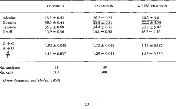
The synthesis of an asymmetric, adenine-rich RNA in Parkinson's disease
In biopsy material from globus pallidus of patients suffering from Parkinson's disease, the big nerve cells and the surrounding glia were analyzed with respect to RNA (Gomirato and Hydén, 1963).
At a very early stage of the disease, the pres-ence of a highly aberrant RNA was found in the glia. The adenine value was highly increased and those of guanine and uracil decreased, and these changes persisted. The neuronal RNA was less changed. Later during the development of the disease, and from the time the overt clinical symptoms emerged and progressed, similar but not identical changes were charac-teristic also for the neuronal RNA. The amount of RNA per nerve cell and glial samples was significantly increased, averaging 145 /i/.g RNA/nerve cell (control 116) and 34 //xg RNA/glial sample (control 17). These striking base-ratio changes and quantitative RNA changes were found in the biopsies whether the patient had been ill for one year or twenty. Due regard was paid to the time relationship between clinical symptoms and biochemical changes in the neurons and glia in the two sides of the same brain. The conclusion of these analyses was that a biochemical error arises in the glia at a very early stage of Parkinson's disease and involves the synthesis of polynucle-otides. The nerve-cell RNA changes were probably secondary to their nature.
Let us discuss the characteristics of the RNA changes occurring in the glia during the course of the disease. In Table 6 the observed base ratios of the controls and the biopsies are seen in the second and third columns. The change in the base ratios is calculated on the increased amount of RNA (ARNA, column 4). Clearly an RNA of a highly asymmetric type has been formed in the glia. Especially striking is the unusually high increase in adenine. The ratio
A + C is 0.79, compared with 1.69 for the A+U
control glia. The A/U ratio is 3.18, compared to 1.05 for the control glia RNA.
In the neurons of globus pallidus, the RNA base-ratio changes are less striking (Table 7). The composition of the total RNA shows a significant increase of adenine and decrease of guanine. If the change in composition is re-ferred to the increased amount of RNA (zRNA, column 4), in analogy to the treat-ment of the glia data above, this neuronal RNA is characterized by a decreased +C
A+U ratio and a high adenine value, as was also the case with the glial RNA. The neuronal changes are less conspicuous, however.
All data indicate the glia as being the type of brain cell in which, in Parkinson's disease, the change in RNA formation begins and is most pronounced. An explanation for this may be the fact that the glia has less RNA content relative to that of the neuron. Therefore, a change in the nuclear RNA formation is more
easily traced in the glia. To judge by the base ratios and the high adenine content, the Parkin-son RNA resembles chromosomal RNA of the type that has been found in Chironomus and in starfish.
It may be that Parkinson's disease represents a type of disorder in which factors in the en-vironment (infections, for example) at a crucial period of the life cycle initiate the release of undesirable genomic activities that will lead to the biochemical error in the glia. A more hazardous explanation, for which there is at present no evidence, would be that the ARNA fraction reflected a virus infection.
Summary
TABLE 6. Microelectrophoretic Analyses of Composition of Glial RNA in Globus Pallidus of 6 Cases of Parkinson's Disease (Purine and Pyrimidine Bases in Molar Proportions in Percentages of Sum). Amount of RNA Increased from 17 (±1.7 ,Lg/glial sample (Controls) to 34 (±2.2) Uítg/glial sample (Parkinson)
CONTROLS PARKINSON A RNA FRACTION
Adenine 19.0 ± 0.78 30.8 ± 0.70 42.4 ± 3.19
Guanine 29.1 - 0.15 20.3 + 0.90 11.5 + 2.75
Cytosine 33.7 ± 0.72 33.2 ± 1.70 32.8 ± 3.45
Uracil 18.2 ± 0.36 15.7 ± 0.60 13.3 ± 1.25
AG ++ U 1.69 ± 0.044 1.15 - 0.047 0.79 ± 0.093
A+U
A -
~1.05
- 0.048 1.96 ± 0.087 ± 3.18 0.384No. analyses:
No. cells: 4,60023
32 4,800
(Prom Gomirato and Hydén, 1963)
TABLE 7. Microelectrophoretic Analyses of Composition of RNA in Nerve Cells in Globus Pallidus of 6 Cases of Parkinson's Disease (Purine and Pyrimidine Bases in Molar Proportions in Percentages of Sum). Amount of RNA Increased from 116 (±5.3) gqig/nerve cell (Controls) to 145 (±6.3) /gjg/nerve cell
(Parkinson)
CONTROLS PARKINSON A RNA FRACTION
Adenine 18.3 _ 0.42 20.7 ± 0.65 30.3 ± 5.0
Guanine 30.5 + 0.44 28.8 + 0.25 22.2 ± 3.53
Cytosine 35.3 o0.60 34.4 ± 0.79 30.8 - 5.02
Uracil 15.9 + 0.36 16.1 + 0.38 16.7 ± 2.16
1.92 + 0.038 1.72 ± 0.042 1.13 - 0.185
A+u
A
~A
1.15 + 0.037 1.29 ± 0.051 1.82 + 0.380U
No. analyses: No. cells:
31
310 30039
In a learning situation, the RNA synthesized in neurons and glia involved in the activity leading to the behavioral change was charac-terized in one case by a DNA-like composition and in another case by asymmetry, high adenine
G+C
values, and low A+U+U values compared to those of ribosomal RNA. To judge by these parameters, the ARNA synthesized in cortical and brain-stem neurons and glia in learning is of a chromosomal type. It is to be noted that in learning the trend of the RNA changes is the same in neurons and glia. By contrast, in the cases investigated we found that upon physiological and chemical stimulation the RNA content of the neurons increases and that of the glial RNA decreases. The new RNA formed was characterized by a base-ratio composition
typical for ribosomal RNA.
The RNA response of neurons and glia in a learning situation not encountered before is thus quite specific. It seems that the environ-mental factors instruenviron-mental in learning can produce a stimulation of the genome of the glia and the neurons engaged.
In a case of chemical stimulation, the in-crease in the RNA content of the neuron could be matched by a corresponding decrease in the RNA in the glia immediately surrounding each neuron. The base-ratio composition of the neuronal and glial /RNA was identical. This brings up the possibility of a transfer of RNA between glia and neurons. It must be pointed out that only a successfully carried out experi-ment with labeled RNA may solve this problem definitely.
Finally, a case of biochemical error involving the RNA of the glia (Parkinson's disease) is discussed. The characteristics of the ARNA produced in the glia at an early stage of the disease are taken to reflect a release of unde-sirable genomic activities at a crucial period of the life cycle.
Roche: In the stimulation experiments, the change is not a difference between the control and the experiment?
Hydén: No, it is the composition of the
additional RNA being formed by the cell under the influence of the chemical substance.
To summarize these results, they seem to mean specific biochemical response in neurons and glia at an early stage of learning, and a response that is different from the RNA re-sponse to other physiological and chemical stimulations.
In the transfer-of-handedness experiments, the results were rather clear-cut with respect to the composition of the RNA being formed. The high adenine-to-uracil ratio that we found in the balancing experiment when we analyzed the nuclear RNA produced in the vestibular nerve cells cannot be ascribed to a contamina-tion of the RNA being formed by ribosomal RNA. That is impossible. Therefore, in both cortical and brain-stem neurons the RNA analysis suggests that transcription of only one of the two strands of DNA has occurred at specific loci on the establishment of the new behavior. In other words, if an animal is faced with a situation that it has not encountered before and that requires learning, a differential transcription seems to occur at the genetic level in the brain cells engaged, and we are now trying to characterize the RNA synthesized in competition experiments.
Since the tasks we chose were clearly within the capacity of the species, the results point to a selective mechanism that operates in the neurons at the genetic level. The immediate task now is to analyze the end products, the proteins. So let us assume for the sake of dis-cussion that, to speak of a mechanism at the molecular level, proteins in neurons and glia produced by a selective activation of specific gene loci in the learning process facilitate cer-tain neuronal pathways necessary for learning and long-term memory. In other words, a selective mechanism operates.
stimulation to give the maximal results in abstract thinking? At what age for linguistic training? This would mean that the very same potentialities were present in the brains of our ancestors who fought hard with clubs for their mere existence. They were certainly capable of learning Boolean logic. But to some it would seem pessimistic that, intellectually speaking, we should be fixed in a cast given by the genes; the next question, therefore, is whether there might be an instruction mechanism at the molecular level that would persist only during the life cycle and would consist of remaining conformational changes of proteins following instructional changes of RNA by external fac-tors. This mechanism would exist, of course, in addition to the selective genetically pro-grammed activity. But here, it seems at present, all that can be done is to formulate the question. Jerne: All cellular biologists have for a long time admired Dr. Hydén's work and his pene-trating analysis of the RNA turnover in single neurons of the brain, and I have followed with great interest his beautiful experimental work and his intelligent approach to the fascinating problems of memory and learning.
This morning I was particularly interested in his informal remarks about selection versus
instruction both in antibody formation and in learning processes by the central nervous system, and also in his demonstration that learning is accompanied by the appearance of a species of RNA in nerve cells that reflects the nucleotide composition of the DNA already present in the genome.
In this connection, I should like to draw at-tention to a paper by Socrates called "Menon," which was edited by Plato and published in Athens 375 years before Christ. Since Hip-pocrates was quoted last year, it might be appro-priate this year to evoke Socrates in order to confirm again our basis in Greek culture.
The point of view expressed by Socrates in this paper contrasts markedly with the ideas of John Locke, who considered the brain as initially a blank sheet of paper, as in the passage Dr. Magoun quoted. In his paper, Socrates
ap-proaches the question whether learning is pos-sible, particularly whether one can learn the truth. And-rather astonishingly, perhaps-he comes to the conclusion that it is impossible to learn the truth. He says that either the truth is already present in the individual, and in that case it cannot be learned, because one cannot learn what one already knows, or the truth is not already present in the individual, and in that case it cannot be learned, because one could not recognize it if it should arrive. He concludes, therefore, that all learning represents a recollection of elements already present in the individual.
This seems to me quite a remarkable analysis for that stage of Western culture, and I think there is some correspondence between the con-clusion Socrates comes to and the finding of Dr. Hydén that the RNA produced during learning reflects the already existing DNA of the genome, which suggests that learning makes use of elements of nucleotide sequences already present in the cell.
Dubos: Professor Hydén, how do you deal with the fact that a living creature that responds constantly to its environment, to the stimuli that impinge upon it, must always be learning? In the sense in which you use the word learning,
isn't it necessary to assume that what you are doing experimentally is accelerating reactions that are taking place all the time?
To complete my question, would a compara-tive study of animals undergoing sensory de-privation sharpen the differences you have observed?
You asked about deprivation. Fifteen years ago Dr. Brattgard made the following experi-ments at our laboratory for his thesis: He reared young rabbits completely in the dark, and then he analyzed the ganglion cells of the retina. He used the methods we had at that time, which were not so refined-mostly X-ray spectra pho-tometry at a wave length of about 10 to 12 angstrom, and also some simple RNA analysis. What he found was that, in comparison with those of seeing animals, which had been ex-posed to stimuli, the cells were about the same size but had a higher water content, a lower protein content, and a very small content of RNA. His conclusion, of course, was that ade-quate stimulation was necessary for biochemical differentiation of the cells. But this is still with respect to a so-called selective mechanism, Dr. Dubos. The question of the instruction mecha-nism remains.
Dubos: Would it not be easier to account for the results you have presented by assuming that the selective training, the selective process of learning, consists in preventing the expres-sion of some of the DNA formula so as to bring about selectivity? Under the influence of the ordinary stimuli to which we are sub-jected, all sorts of things go on and the result is random distribution, whereas with learning there is selective production.
Hydén: Only experimentation could answer such a question. That is a possibility, of course. It would go well with the current lines of cer-tain electroneurophysiologists-that inhibition is the most important functional part in the brain. I should like to be able to present the results of an animal first at the beginning of its life cycle and then later on, to see whether it would respond with the synthesis of more so-phisticated molecules-let us say RNA-in the neurons or whether, as you say, the mechanism would be selective in the sense that fewer and fewer parts of the genome would be expressed, but at a higher rate. We have taken up this question; we have experiments going on in which, as Kretsch did some years ago, we put one group of rats in small, dimly lighted cages,
within hearing of the other animals, and a sec-ond group in a sort of Luna Park with a lot of smorgasbord that they can help themselves to.
Chagas: Allow me, Dr. Hydén, to add my name to the long list of those who have already congratulated you on your remarkable work.
Last year, I was at a congress on the brain and consciousness, and I was then surprised at the fact that the molecular approach to this problem is still a relative rarity in neurobiology, despite the vivid impression it has generally produced. I myself have a certain doubt about it, concerning the time elements in your theory. Of course, they appear to be quite satisfactory, in principle, for long-term memory; but there are several cases-for example, those that Dr. Jerne has so picturesquely called "Socratic"-in which the time may be too short for the "turnover" or synthetic processes that would have to take place. Experiments in this direc-tion should be of great interest.
Hydén: Do you mean the biosynthesis of RNA and proteins, which is known to take too short a time to account for experimental be-havioral knowledge? With short-term memory, it is known that one can interfere with a learn-ing process, but that after a few hours have elapsed it is no longer possible, and also that the hippocampal area is very important in that respect. We have the old results of Lashley, which show that once a new behavior has been established it is soon delocalized. This term
delocalization is not very well understood, but
it becomes clearer if we view it in terms of millions of cells in which a certain change has occurred, and if memory is broadly defined as change by use. I think the time is really suffi-cient: let us say a couple of minutes for a macro-molecular synthesis. Professor Dubos could comment further on that, 1 am sure.
distribution of the various proteins with time, one could not obtain thereby some evidence about the rate at which those chemical events you speak of take place during the process of learning.
Hydén: There are some new data I that have not discussed because we are in the midst of analyzing them. Dr. Blake Moore has found protein-perhaps a group of proteins, but it can-not be more than a very few-of an acidic type that he considers to be typical for brain. He has found them in many species and has seen that they cross-react. He has purified them, and I have some. I have also got from Dr. Levine, at Brandeis University, antibodies he has pro-duced against what we might call the Moore protein, which is very acidic and moves very fast in an electrophoresis, like that of the albumins.
What I have done to begin with is to see whether it is produced by the neurons or by the glia. I can't answer this question, but I can say that I have been able to localize it in neurons. I am using the microcapillary technique with acrylamide gets for electrophoresis-using that as an immune chemical means both with and without antibodies, to see which band is present and which was removed. It certainly is produced in the neurons. But that would mean a rather limited group of proteins-not 200, if it is now true that that is specific for the proteins. Dr. Moore states that the concentra-tion of it in the brain is ten thousand times greater than in the liver and other organs.
Chagas: I was so impressed by the tech-nique described that I may have failed to grasp its full significance. My question now is whether the difference of values found for different iso-lated neurons is not a result of experimental error. Also to be considered, of course, is
whether the composition of the neurons taken from the same area is more or less constant.
Roche: I gather that analyses of RNA, com-position, and so on in glia and neurons parallel each other-that during the learning process the changes in glia are the same as in neurons. Was this expected, and does it lead to any comment as to their relative or respective function in the learning process?
Hydén: To answer Dr. Chagas first, the coefficient of variation in the values is around 5 per cent, about the same as the experimental error. With precisely his question in mind, we have performed a closer statistical study on one part of the nervous system, the inner ear and the bipolar ganglion cells there, and we found that around 12 per cent constituted the intra-group variation. The experimental error could be kept at 5 to 7 per cent.
Now for Dr. Roche's question about the parallel reaction of neurons and glia. That was not expected in the learning process because of our previous experimental background. When we performed physiological stimulation of the vestibular area, and again in stimulation with chemical compounds, we found inverse changes: when protein synthesis or enzyme activities went up in the neurons, they decreased in the glia. So that was what we expected. But in these learning experiments we have found the results in neurons and in glia to be the same.
CURRENT CONCEPTS IN THE NEUROPHYSIOLOGY OF
LEARNING
Raúl Hernandez Peón
The intriguing nature and potentialities of learning have always aroused the interest of philosophers and scientists. This brief review of the main current neurophysiological findings and concepts of learning is intended to convey basic information to unspecialized scientists, and therefore necessarily involves many omissions. Along with a synthetic description of the neural correlates of learning, some personal hypotheses will be advanced that may foster research in this field.
Learning is one of the most pervasive and fundamental biological processes observed throughout the animal scale. Although it is commonly accepted that learning implies the acquisition of information that tends to modify innate or already acquired performances of the organism, the great number of definitions pro-posed for this term reveals the difficulty in assessing the essential nature of the underlying mechanisms. The enduring changes produced by learning are usually associated with adaptive behavior. But in some instances it may con-tribute to maladjustments of the individual.
Because of the ubiquity of learning in or-ganisms of every grade of evolution, the con-clusion seems warranted that it derives from a fundamental property of living matter and therefore does not necessarily require special complex neuronal circuits. To designate the property concerned with enduring neural changes we have adopted the term plasticity,
proposed by Konorsky (1948), thus establish-ing a distinction between it and another funda-mental property of living cells, excitability,
which is related to very transient and reversible changes produced by the stimulus. Granting that through specialization of function both plasticity and excitability developed more in certain elements of multicellular organisms, there is no doubt that the nervous system is endowed with both properties. In animals with
nervous systems in which "all or none" signals are transmitted, plasticity permits the storage of information delivered by those signals, whereas excitability is concerned with their generation and transmission. Furthermore, it seems obvious that, through the evolution of the nervous sys-tem, some neurons developed longer and more excitable axons for transmission than other short-axon non-propagating elements. From the observation that the former type of neurons do not present enduring plastic changes upon stim-ulation, whereas the latter are essential for those changes, the hypothesis of an evolutionary dif-ferentiation of excitable and plastic neurons within the central nervous system may be safely conjectured.
Since nothing is known about the ultimate nature of plasticity, our knowledge about the neural plastic change associated with learning derives mainly from the indirect changes of excitability currently assessed by electrophysi-ological techniques. For discussion of the main neurophysiological correlates of learning it is necessary to establish a general classification of the fundamental types observed from Protozoa to man. First of all, learning may be divided into negative and positive. Negative learning leads to decrement or disappearance of a re-sponse previously evoked by a given stimulus; positive learning involves the acquisition of a response to a stimulus that did not elicit it before. The elimination of irrelevant responses during negative learning is not a passive phe-nomenon but depends on central processes that actually prevent them from appearing as they otherwise would. This active restraining process has been termed plastic inhibition (Hernández
connec-tions by a process designated plastic association Figure 1. DIAGRAMATIC REPRESENTATION
d'~l AfAI1T W1MIlRAT iPRnrFpR.Po TIMVO.TVET IN
in this modern terminology (Hernández Peón, 1957). Plastic inhibition is probably the pri-mary and most important process in learning; without it animal behavior would be disorgan-ized and adaptation of the organism to the ex-ternal environment would be impossible. Plastic inhibition is so important for normal function-ing of the central nervous system that it may be no exaggeration to say that while plastic in-hibition may develop in the absence of plastic association, the latter is always accompanied by the former.
According to their complexity, the following types of learning may be considered: (a) habit-uation; (b) classical conditioning; (c) instru-mental conditioning, or trial-and-success learn-ing; and (d) latent learning. To these, a separate variety of learning produced in new-born animals or certain species by a single exposure to a stimulus and termed imprinting
may be added. A number of neurophysiological studies carried out during the past decade have contributed to an initial understanding of the functional role of various brain regions in cer-tain types of learning and other related processes taking place before, after, and simultaneously in the central nervous system. The main func-tional operations that take place between the stimulus and the behavioral response are sum-marized in Figure 1. All types of stimuli initiate a series of events starting from sensory recep-tion at the receptor level and followed by sensory transmission along all the levels of the central nervous system, both at specific and at non-specific or polysensory pathways. Afferent impulses are thus conducted to neural systems involved in emotions, motivation, learning, and memory. In turn, the impulses coming from those neural circuits are conveyed to a highly specialized neural system concerned with sensory integration and conscious experience. Finally, after the activation of decision-making
mecha-nisms, the adaptive behavioral responses occur. Habituation. Habituation, the simplest type
of learning (Thorpe, 1950), consists in an enduring, progressively oscillating decrement of
MOST BEHAVIORAL RESPONSES
STIMULI
1
LEARNING SENSORY RECEPTION EMOTIONS
AND ·- AND _ AND
MEMORY TRANSMISSION MOTIVATIONS
1
CONSCIOUS "EXPERIENCE~
1
BEHAVIORAL RESPONSE
responses produced by monotonous repetition of a stimulus that loses significance for the organism. This pervasive phenomenon is ob-served not only in effector responses but also in sensory experiences, as everybody can con-firm in everyday life. A logical question to ask is, What changes occur in the central nervous system during habituation that may account for all its manifestations? Recordings of the elec-trical activity of the neuroaxis in unrestrained animals with electrodes permanently implanted have disclosed neural correlates of habituation both at the specific and at polysensory systems in the brain and spinal cord. The terms afferent neuronal habituation and neuropil habituation,
respectively, have been proposed for designating each process (Hernández Peón, 1960).