IRON
METABOLISM
AND ANEMIA
PAN AMERICAN HEALTH ORGANIZATION Pan American Sanitary Bureau, Regional Office of the
IRON
METABOLISM AND ANEMIA
Proceedings of a symposium held during the Eighth Meeting
of the
PAHO Advisory Committee on Medical Research 9 June 1969
Scientific Publication No. 184
PAN AMERICAN HEALTH ORGANIZATION Pan American Sanitary Bureau, Regional Ofice of the
WORLD HEALTH ORGANIZATION 525 Twenty-third Street, N.W. Washington, D.C. 20037, U.S.A.
PAHO ADVISORY COMMITTEE ON MEDICAL RESEARCH
Dr. Hernán Alessandri
Ex-Decano, Facultad de Medicina Universidad de Chile
Santiago, Chile
Dr. Otto G. Bier
Director, PAHO/WHO Immunology Research and Training Center
Instituto Butantan Sao Paulo, Brazil
Dr. Roberto Caldeyro-Barcia Jefe, Departamento de Fisiopatología Facultad de Medicina
Universidad de la Repúiblica Montevideo, Uruguay
Dr. Philip P. Cohen
Chairman, Department of Physiological Chemistry The University of Wisconsin
Madison, Wisconsin, U.S.A.
Dr. René Dubos Member and Professor The Rockefeller University New York, New York, U.S.A.
Dr. Herman E. Hilleboe
Director, Division of Public Health Practice School of Public Health and Administrative
Medicine Columbia University
New York, New York, U.S.A.
Dr. Bernardo A. Houssay
Director, Instituto de Biología y Medicina Experimental
Buenos Aires, Argentina
Dr. Robert Q. Marston
Director, National Institutes of Health Bethesda, Maryland, U.S.A.
Dr. Walsh McDermott
Chairman, Department of Public Health Cornell University Medical College New York, New York, U.S.A.
Dr. James V. Neel
Chairman, Department of Human Genetics University of Michigan Medical School Ann Arbor, Michigan, U.S.A.
Professor Roger Revelle Harvard University
Center for Population Studies Cambridge, Massachusetts, U.S.A.
Dr. Marcel Roche
Director, Instituto Venezolano de Investigaciones Científicas
Caracas, Venezuela
Dr. John C. Waterlow
Director, Tropical Metabolism Research Unit University of the West Indies
Kingston, Jamaica
Professor Abel Wolman
Emeritus Professor of Sanitary Engineering and Water Resources
The Johns Hopkins University Baltimore, Maryland, U.S.A.
Dr. Salvador Zubirán
Director, Instituto Nacional de la Nutrición México, D.F., Mexico
Dr. M. Martins da Silva, Secretary
Chief, Department of Research Development and Coordination
Pan American Health Organization Washington, D.C., U.S.A.
Symposium on
IRON METABOLISM AND ANEMIA
Moderator: Dr. Marcel Roche
PARTICIPANTS
Dr. Joginder Chopra
Department of Health Services Pan American Health Organization Washington, D.C., U.S.A.
Col. Marcel E. Conrad Department of Hematology
Walter Reed Army Institute of Research Washington, D.C., U.S.A.
Dr. James Cook Department of Medicine
University of Washington School of Medicine Seattle, Washington, U.S.A.
Dr. William H. Crosby Department of Medicine
New England Medical Center Hospitals Boston, Massachusetts, U.S.A.
Dr. Clement A. Finch Division of Hematology
University of Washington School of Medicine Seattle, Washington, U.S.A.
Dr. Yaro Ribeiro Gandra Departamento de Nutrico
Faculdade de Higiene e Saúde Pública Sao Paulo, Brazil
Dr. Pauline M. Harrison Department of Biochemistry The University of Sheffield Sheffield, England
Dr. John J. Kevany
Department of Health Services Pan American Health Organization Washington, D.C., U.S.A.
Dr. Miguel Layrisse Sección de Fisiopatología
Instituto Venezolano de Investigaciones Científicas
Caracas, Venezuela
Dr. Luis Sánchez-Medal
Instituto Nacional de la Nutrición México, D.F., Mexico
Dr. Carl V. Moore
Department of Internal Medicine
Washington University School of Medicine St. Louis, Missouri, U.S.A.
Dr. Marcel Roche
Instituto Venezolano de Investigaciones Científicas
Caracas, Venezuela
Dr. M. Martins da Silva, Secretary
Department of Research Development and Coordination
Pan American Health Organization Washington, D.C., U.S.A.
CONTENTS
Page
Opening Statement M arcel Roche ... 1
The Biochemistry of Iron Pauline M. Harrison ... 2
The Control of Iron Balance by the Intestinal Mucosa William H. Crosby ... 21
The Role of Protein in Iron Absorption Marcel E. Conrad ... 27
Intersubject and Intrasubject Variation of Iron Absorption James D. Cook ... 35
Iron Absorption from Food Miguel Layrisse ... 38
Iron Losses Clement A. Finch ... 43
General Discussion ... 46
Human Iron Requirements Carl V. Moore ... 50
Iron-Deficiency Anemia in Latin American and Caribbean Populations Yaro R. Gandra ... 56
Iron Deficiency in Pregnancy and Infancy Luis Sánchez-Medal ... 65
The Relationship Between Hookworm Infection and Anemia Marcel Roche ... 72
Prevention of Iron-Deficiency Anemia loginder Chopra ... 78
OPENING STATEMENT
Marcel Roche, Moderator
It would be safe to say, without any statistics at hand, that 1,000 million people in the world are iron deficient, and many of them actually have anemia. The problem is most acute in tropical areas, and anemias, particularly iron-deficiency anemias, are rampant in all the tropical and subtropical zones of the Americas.
The attempt in this symposium is to present, in a short time, a general view of iron metabolism, extending from the basic concepts of chemistry and biochemistry to the epidemiological and therapeutic aspects of the problem.
Specifically, we shall start by taking a look at basic aspects of hemo-globin transferrin-ferritin metabolism on the whole. Next, there will be four papers dealing with the question of absorption of iron-possibly the single most important influence on the production of iron-deficiency anemia. The subject will be approached from the point of view of mechanisms at the intestinal level; the effect of protein depletion-obviously very important to us, since many of the populations we are interested in are protein-depleted; intersubject and intrasubject variations; and findings to date on iron absorption from specific staples. After this, the old question of iron requirements will be dealt with.
Turning to epidemiology, the particular aspects of iron deficiency and anemia that bear on the Western Hemisphere will be considered. Also, these matters will be looked at as they affect pregnancy and infancy. The relationship between anemia and hookworm disease will then be touched on briefly. The concluding presentation will round out the matter by dealing with the treatment and prophylaxis of iron deficiency.
THE BIOCHEMISTRY OF
IRON.
Pauline M. Harrison
Structure and function in biological iron compounds
The biochemistry of iron may be divided broadly into three interrelated aspects: the struc-ture and function of biological iron compounds, their synthesis and turnover, and the metabolic and functional relationships among the various iron compounds. The present symposium is largely devoted to human iron metabolism: the body's requirements for iron, its absorption from food, the turnover of iron compounds and the maintenance of an iron balance, and the effects of iron deficiency. This paper will concentrate on the structural and functional aspects of iron compounds, principally hemrnoglobin and myo-globin, ferritin and hemosiderin, and transferrin. Their syntheses and metabolic interrelationships will be touched on briefly.
Free ionic iron does not occur to any signifi-cant extent in living organisms because of its tendency to form complexes with many organic compounds. Since quite low concentrations of ionic iron are toxic, this has the biological ad-vantage of enabling iron to be stored, trans-ported, or utilized in nontoxic forms. The bio-chemistry of iron is therefore the biobio-chemistry of the complexes of which it forms a part. The presence of iron atoms may confer on these complexes certain properties, such as the capacity to combine reversibly with oxygen or the ability to accept and donate electrons. These properties acquired through the presence of iron atoms may in turn be profoundly modified by the environ-ment provided for the iron by the
complex-reversible combination with oxygen being a case in point. The effect of the complex may be to allow the iron to function in a specific and controlled manner.
A neutral iron atom contains 26 electrons, 18 of which occur in the closed shells of the argon core. When ionized, two (ferrous) or three (ferric) of the outer electrons are removed, leav-ing the inner electrons unchanged. The spins of the outer electrons may be aligned in a variety of different ways, giving rise to differences in magnetic properties. Thus ferric iron, Fe3+, has five outer electrons. In high-spin compounds, these have all their spins parallel to one another, giving a large magnetic moment. In low-spin compounds, four of the electrons are aligned in two antiparallel pairs, leaving a single unpaired electron and hence a small magnetic moment. Ferric iron with three unpaired electrons is less common. Ferrous iron normally has either four or no electrons unpaired.
The biological properties of iron complexes may depend not only on the presence of a local-ized center of positive charge but also on the tendency of iron to form directional covalent bonds with organic ligands. These ligands are often, but not always, arranged in the form of an octahedron around the iron atom. The nature and arrangement of the ligands affect the distri-bution of the outer electrons in the iron atom and, in particular, the way their spins are paired, enabling the iron to play a variety of roles. The correlation between electronic configuration and function has been discussed by Eichhorn (43).
N (e.g. histidine or pyrrole), O (e.g. 02 or tyrosine) and S (e.g. thiol). The nature of the chemical groups not in the immediate coordina-tion sphere of the iron atom, but surrounding it, may also be of functional importance. This is shown dramatically by the difference in be-havior of the heme iron in hemoglobin as com-pared with that in free heme. The protein in myoglobin and hemoglobin provides the heme group with a nonpolar environment, which allows the ferrous iron at its center to combine with molecular oxygen without itself becoming oxidized, a property not shared by free heme groups (87, 128) or by free Fe2+.
The iron atoms may also affect the properties of the compounds to which they are attached. Thus iron-transferrin (47) and ferritin (104)
are more stable to heat and other denaturing agents and more resistant to attack by proteolytic enzymes than are their apoproteins. This may help to prevent a build-up in the cell of the protein moiety in amounts greatly in excess of the available iron. The increased stability con-ferred by iron-binding may result from confor-mational changes in the protein. This is prob-ably true for transferrin (47, 94) and for the
bacterial nonheme iron protein, rubredoxin, in which an iron atom, coordinated to four thiol groups, forms a bridge between remote parts of the primary structure in a manner analagous to that of a disulphide bond (11). The addition
of heme to globin alters the conformation of the latter, increasing its helix content by 10 to 20 per cent (26, 76). The binding of a gaseous
ligand to hemoglobin iron causes a physiologi-cally significant change in quaternary structure
(116), as discussed below. Conformational
changes in the protein of transferrin induced by iron-binding and by binding of the protein to cell membranes may play a role in the control of iron metabolism (53). On the other hand, the addition of 4,000 or more iron atoms to the molecule has little effect on the structure of
apoferritin (51, 68).
Biological iron compounds form a very diverse group, both structurally and functionally. It is convenient to classify them into those that
con-(proto)heme IX
(a)
z
,N-- -- - N
ZC
y '
2
1-
- -
--11
(c)
FIG. 1. (a) The structure of the heme group (ferro-protoporphyrin IX) as found in hemoglobin, myoglobin, catalase, and peroxidase. In cytochrome c a similar group is attached to the protein by addition of two cysteine thiol groups to the two vinyl groups of the protopor-phyrin. The four protoporphyrin nitrogen atoms form a square planar arrangement to which the iron is coordi-nated. The iron atom may also be attached to other groups on either side of this plane. In deoxymyoglobin and deoxyhemoglobin the ferrous iron atom is coordi-nated only to five ligands in a square pyramidal arrange-ment, as shown in (b), with histidine nitrogen occupy-ing position Z. The iron atom is displaced at least 0.3A from the protoporphyrin plane in the direction of the histidine nitrogen. In oxymyoglobin and oxyhemoglobin the oxygen molecule occupies a position on the side of the plane opposite to Z (like Z' in (c)). In cytochrome c the iron atom is attached to six ligands arranged at the corners of an octahedron and is probably centered in the plane of the nitrogens as shown in (c) (31).
tain heme (27, 30, 31, 45, 90, 101, 119) and
those that do not (29, 47, 89, 100, 118, 147).
Free heme (Figure 1) does not occur in quantity in animal tissues; it becomes incorporated into
proteins or is broken down.
Heme proteins include the very widely dis-tributed proteins of the cytochrome system (90, 101), which occur in virtually all organisms
except anaerobic bacteria, and function as an electron transport chain associated with oxidative phosphorylation in mitochondria. Of the cyto-chromes, c, a protein of molecular weight 12,400 containing a single heme, is the best character-ized. Unlike that of hemoglobin, its
porphyrin ring system is covalently linked to the protein (by thioether links with two cysteine residues). The fifth and sixth sites of the octa-hedrally coordinated heme iron are occupied by histidine, and probably methionine, and the heme group is situated in a deep crevice normal to the surface of the molecule (38). Cytochrome c
accepts electrons from cytochrome b and
trans-fers them to cytochrome oxidase, with which it forms an active complex, the iron atoms being alternately oxidized and reduced. Other heme proteins include myoglobin and hemoglobin, dis-cussed below, and the catalases and peroxidases that catalyze the breakdown of peroxides in the presence of a reducing agent (85, 89, 119, 125).
It has been suggested that an intermediate in the peroxidase reaction may be one in which the fifth and sixth iron coordination sites are empty
(127).
Nonheme iron compounds vary in size from the small ferric hydroxamic acid chelates found in aerobic bacterial cells, which appear to be involved in heme synthesis (130) and also
pos-sibly as iron carriers (118), to the large
iron-storage complexes ferritin and hemosiderin. And they vary in function from electron transfer in the ferredoxins (29) (found in plants and
micro-organisms but not in animals) and in a number of iron-flavoproteins (89, 147) to oxygen carriers, as in the hemerythrins of sipunculids, and iron carriers, as in transferrins. The nature of the iron ligands is often difficult to determine in the absence of an easily recognizable group such as heme, and they may be sulphur (in ferredoxin and rubredoxin), oxygen (in ferrioxamine), or nitrogen (both nitrogen and oxygen in trans-ferrin).
The present symposium is largely concerned with the metabolism of iron compounds in humans. Quantitatively, the most important
iron compound in man is hemoglobin (111),
which accounts for some 70 per cent of the total iron (about 4 g in a man weighing 70 kg), as compared to the related muscle protein, myo-globin, which constitutes about 3 per cent. Next to hemoglobin comes storage iron, located in ferritin and hemosiderin and accounting for
about 26 per cent (111). Transferrin, at about
0.1 per cent, is quantitatively less important, but it plays a vital role in iron transport between sites of iron absorption and storage and hemo-globin synthesis, and probably in the control of iron absorption. The remainder of this paper will be confined to a discussion of these com-pounds. For other iron compounds the reader is referred to recent reviews (27, 29, 30, 31, 45, 47, 89, 90, 100, 101, 111, 118, 119, 147).
Oxygen carriers:
Hemoglobin and myoglobin
Hemoglobin occurs in the red cells of all verte-brates. In higher vertebrates it is typically a tetramer of four polypeptide chains and four heme groups. The molecule is a compact ellip-soid measuring 64 x 55 x 50 A (129) and having a molecular weight of about 65,000. Hemoglo-bin also occurs in many invertebrates, although its polymeric form may vary, e.g. a monomer with a single heme is found in the marine
anne-lid worm Glycera dibranchíata (122) and in
larvae of the insect Chironomus thumni (78),
while polymeric forms with molecular weights of 3,000,000-the erythrocruorins-occur in the
worms Lumbricus terrestis and Arenicola marina
(97). A leghemoglobin has been reported in
the root nodules of leguminous plants (138).
The importance of a supply of oxygen for the survival of man and other animals need hardly be emphasized. Since the body has a limited capacity for storing oxygen, a steady supply and an efficient means of circulating it to the tissues are vital. Hemoglobin fulfills the latter role efficiently both by increasing the ca-pacity of the blood for oxygen by a factor of some seventyfold, and by binding oxygen strongly at the partial pressure of alveolar oxygen and unloading it readily at the reduced PO2 of
the tissues (144).
The relative ease with which tetrameric hemo-globin yields its oxygen at lower oxygen tensions as compared with monomeric myoglobin and monomeric forms of hemoglobin is illustrated by the oxygen dissociation curves in Figure 2. These curves suggest that the combination of oxygen
1ool
60~
40 A NORMAL UMAN UOO
ec 20 MYOGLOSIN
t 20
,I /
.
0 20 40 60 a0 100 120 OXYGEN PRESSURE mm Hg
FIG. 2. A: Oxyhemoglobin dissociation curve of human
blood at 38°C, pH 7.40. B: Oxygen dissociation curve of myoglobin under similar conditions. Reproduced by permission of F. J. W. Roughton (144, p. 775).
with the iron atoms in the two proteins is not simply a function of the presence of iron in a particular local environment. The iron atoms in the two proteins are combined in the same pro-toporphyrin ring system, and the hemes have
very similar surroundings (87, 128). The
sig-moid dissociation curve for hemoglobin as against the hyperbolic curve for myoglobin is attributed to the presence of a "heme-heme in-teraction" in tetrameric hemoglobin and the lack of it in monomeric myoglobin. That is to say, in hemoglobin, combination with the four oxy-gen molecules does not occur independently. Another physiologically important property of hemoglobin not shared by myoglobin is that an increased partial pressure of carbon dioxide fa-cilitates the unloading of its oxygen-the oxy-gen dissociation curve is shifted to the right (Bohr effect).
The configuration of deoxymyoglobin based on X-ray diffraction studies of Kendrew and his
colleagues (88,120) is shown in Figure 3. The
coordination of the iron atom is approximately octahedral, but the sixth coordination site is unoccupied by any ligand. The ferrous iron atom is situated about 0.4 A from the center of the protoporphyrin ring, which supplies four pyrrole nitrogen ligands. A fifth nitrogen ligand is supplied by a histidine side chain (F8) of the protein, while another histidine (E7) is situated close to the empty sixth coordination site. In
FiGo. 3. a-carbon skeleton diagram of myoglobin mole-cule showing helical and nonhelical regions and the location of the heme group. Reproduced by permission of R. E. Dickerson, from H. Neurath (ed.), The Proteins,
2d ed., vol. 2, New York, Academic Press, 1964, p. 634.
oxymyoglobin, an oxygen molecule occupies this sixth site without altering the valence of the iron atom, although it changes from high-spin to
zero-spin state. In vitro, the iron atom is readily
oxidized, giving ferrimyoglobin, in which the sixth site is occupied by a water molecule or other ligands, while the position of the iron atom remains as in ferromyoglobin. The ar-rangement of ligands around the heme iron atom in deoxymyoglobin is identical with that
in a-chlorohemin (92). Figure 3 shows the heme group in a crevice in the protein. An analysis of the protein side chains in proximity to the heme in myoglobin and hemoglobin shows that the protein provides it with a
non-polar surrounding (87,128).
Hemoglobin consists of two pairs of chains with different primary structures (25) and can
therefore be designated as a tetramer a2,/6. Each
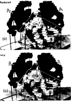
chain, with its attached heme, has a tertiary structure very similar to that of myoglobin. The chains are situated roughly at the corners of a tetrahedron. Models of oxyhemoglobin and de-oxyhemoglobin molecules are shown in Figure 4. These structures show three important features: the chain conformations are unaffected by
Reduced
12
Pi
oxy__ Oa)
FIG. 4. Models of (a) deoxy and (b) oxyhemoglobin at 5.5A resolution. The heme groups are represented by grey discs. The a chains are white and the f, chains black. The a. chain lies at the back of the molecule behind the a, chain. The contact areas between chains are marked as boxes. On combination with oxygen the chains shift relatively by a few A units along the alfiS2
and a2gfl contacts, while contacts aifi, and a2f,82 alter
very little. The P chains move closer together on oxy-genation. Photographs kindly supplied by M. F. Perutz.
bination with oxygen (at least at the resolution, 5.5A, of the models); the relative orientations of the four chains change on combination with oxygen; and the heme groups are not close enough for direct interaction, their iron atoms being 25A or more apart (21, 115, 116).
Much experimental and theoretical work has been carried out in attempts to explain the sig-moid shape of the hemoglobin oxygen dissocia-tion curve (7, 8, 142, 171). Kinetic measurements
have shown that, while the rates of combi-nation of hemoglobin with the first three oxy-gens are approximately the same, combination with the fourth oxygen is much more rapid
(145). This suggests that the structural change
associated with the deoxy-oxy transformation
may have occurred before the fourth ligand com-bines. Most of this change occurs along the contact between the a, and fi2 chains shown in
Figure 4, some of the atoms at the contact being displaced relatively by as much as 6A, although distances between the heme iron atoms in these chains are very little affected. The iron atoms in the two f8-chains, however, move about 6A
closer together on oxygenation (115). How the
structural change is triggered off and relayed across the protein is not yet apparent, although this may become clearer when the conforma-tions of both deoxy and oxyhemoglobin have been determined at atomic resolution. The transformation in quaternary structure seems to be a consequence of the change from 5- to 6-coordination of the iron atom rather than of the change in its spin state.
Structure-function relationships in hemoglo-bin are complicated by the fact that in solution a dynamic tetramer-dimer-monomer equilibrium exists (8, 9, 142). Symmetrical splitting into a/3 dimers probably occurs along the afi2
con-tact and also along the a2fl (141), while the
a1,f1 and a2,82 contacts remain unchanged
(Fig-ure 4). It has been suggested that conforma-tional changes in the af3 dimer are of prime
im-portance in hemoglobin oxygenation and that the oxy-dimers induce a transformation to a more reactive conformation in any deoxy-dimers with which they combine (10, 13, 64, 117).
Re-cent kinetic evidence, however, suggests that the tetramer and not the dimer is the prime unit of function, since combination with at least three ligands is necessary to produce a transformation
to the rapidly reacting form (56). The X-ray
crystallographic studies also support the tetra-meric molecule as the functional unit in coop-eration binding effects (128). In any event, the
functional importance of the protein as well as the iron is evident. This is also shown by the fact that modifications in the protein may reduce or destroy both heme-heme interaction and the Bohr effect. Interestingly, these effects are absent on the reaction of ferrihemoglobin with ligands
(8), and ferrihemoglobin has a conformation
The effect of iron salts as stimulators of both
heme and globin synthesis de novo from amino
acids by reticulocytes in vitro is well known (95), while the rate of globin synthesis is
de-creased in the absence of heme or heme
pre-cursors such as iron (131). The observation
that heme and globin are synthesized at approxi-mately the same rates argues for a mechanism
coordinating their syntheses (95, 131). This
may be achieved by an inhibitory effect of hemin on its own synthesis coupled with a stimulatory effect of heme on the formation of globin (28).
Oxygen concentration also plays a regulatory role in hemoglobin synthesis. Globin synthesis was found to be stimulated by low oxygen tensions and inhibited at the higher levels (67).
Inhibi-tion is relieved by the addiInhibi-tion of heme, and the effect is believed to occur at the level of heme synthesis.
The regulatory effect of heme (and of iron as a heme precursor) on hemoglobin synthesis ap-pears to be twofold. It is found to stabilize reticulocyte polyribosomes (131, 162, 163), and
it also appears to promote the assembly of newly synthesized a and , globin chains (156).
Iron stores:
Ferritin and hemosiderin
Storage iron, amounting to about 700 to 1,000 mg in a normal man (111), represents a mobile
reserve that can be drawn upon in iron de-ficiency or after hemorrhage, thus allowing sup-plies of "functional" iron compounds, such as hemoglobin, to be maintained or rapidly re-placed (22). Iron deficiency in a clinical sense
occurs only when iron stores are depleted. In iron overload, storage iron is increased, while hemoglobin iron usually remains normal. Iron released from hemoglobin in the normal break-down of red cells is also stored temporarily in reticuloendothelial cells from which it can sub-sequently be released and reutilized for hemo-globin synthesis (121). Iron is stored in two
forms, ferritin and hemosiderin, and can be
mobilized from both (22, 150). The formation
of ferritin, which occurs in response to the pres-ence of iron (48, 58), may play a part in the
mechanism regulating iron absorption in mu-cosal cells (33, 37).
In ferritin, the iron is associated with a well-defined protein moiety, apoferritin, forming a
soluble red-brown complex (57, 61). The term
hemosiderin was first applied to microscopically visible, Prussian-blue-staining, insoluble gran-ules isolated from the liver and the spleen (35, 59, 106). It has also been used to denote massive
iron-rich deposits seen in the electron micro-scope (16,132). The need for a soluble and also
nontoxic form of iron store seems to be gener-ally widespread among living organisms. Ferri-tin has been found not only in many vertebrates (109) and invertebrates (140, 160) but also in
the plant kingdom (79, 139, 148), including
fungi (126). It would therefore seem to be of
ancient evolutionary origin. Hemosiderin or he-mosiderin-like deposits have been found in both
vertebrates and invertebrates (35, 159). Under
normal conditions in man and in experimental animals, most storage iron is in the form of ferri-tin, but in iron overload the amount of hemo-siderin may greatly outstrip that of ferritin (108, 149,154).
Ferritin isolated from tissues (principally liver, spleen, and bone marrow) may contain a variable amount of iron, although this is usually around 20 per cent of its dry weight (57), or
about 3,000 iron atoms per molecule. Each preparation contains a spectrum of molecules of different iron content (143), ranging from
iron-free molecules to those containing up to 4,000 or 5,000 iron atoms (50, 70). The distribution of
iron content among the molecules varies from one individual to another and depends on the state of iron-loading. In anemic animals, how-ever, iron-free apoferritin is not present in quan-tity (59, 60). It is apparently degraded when
not required to store iron, whereas iron stimu-lates its synthesis. Ferritin preparations consist mainly of monomeric molecules, but some poly-mers are also present-about 10 to 15 per cent by weight of the preparation (39, 155, 166). On
ion-exchange chromatography (155, 157),
hetero-geneity has been observed in ferritin, but not
in apoferritin (155). No differences could be
The iron of ferritin occurs in a "micelle" with a maximal diameter of about 70. (46, 50, 69, 91), or less in molecules of low iron content (50, 66). Its composition corresponds roughly to
the formula (FeOOH)8 (FeO:OPO lH2) (70,
110), but the phosphate present does not seem to
be an integral part ofu the "icelle and may be largely confined to its surface (51, 72).
Ferri-tin and its protein-free micelles give X-ray (51, 72, 158) and electron diffraction patterns (65)
typical of crystalline material of small particle size. The micelles, which can be seen without staining in the electron microscope, sometimes have the appearance of being subdivided into a few smaller crystallites (46), although this could possibly be an artifact. The appearance of four crystallites or "tetrads" in a micelle of average over-all diameter of about 60Á is taken by many electron microscopists to be diagnostic of ferri-tin. Ferritin can also be recognized-and dis-tinguished from hemosiderin-by its tendency to form close-packed monolayers on electron microscope grids and by the fact that its micelles are surrounded by protein shells, which are not electron dense, and which prevent the micelles from coming into contact (Figures 5 and 6).
FIG. 5. Electron micrographs of ferritin (a) and hemo-siderin (b) prepared from the same horse spleen by the methods of Granick (57) and McKay and Fineberg
(106), respectively. The samples were unstained and unshadowed. The iron-containing micelles can be seen as electron-dense (grey) areas. Magnification X 250,000 (F. A. Fischbach, D. W. Gregory, P. M. Harrison and T. G. Hoy).
detected in the amino acid compositions of the
chromatographic fractions (155). The
hetero-geneity may therefore be due to differences in surface conformation, or in bound ferric or other ions. Variations in the electrophoretic mobili-ties of ferritins isolated from different tissues within the same animal have also been found
(5,54,55).
250,000-Several different structures have been proposed to fit the "tetrad" appearance (46, 114).
How-ever, these fit only a small proportion of the views seen in the electron microscope, while in electronmicrographs taken close to true focus many of the micelles look rather uniform in
ap-pearance (65). In ferritin solutions (50) and
in wet crystals (69), the micelles closely
ap-proximate spheres or polyhedra and are of uni-form density.
The ferritin micelle diffraction patterns differ from those of the well-known ferric oxide or
oxyhydroxide minerals (65, 72). Three
alterna-tive atomic structures have been proposed for the ferritin iron cores (72) or for closely related synthetic "hydrous ferric oxides," which give
similar diffraction patterns (24, 158). These
vary in the coordination of the oxygen atoms
(O=, OH- or H20) around the ferric iron-they
may be all octahedral (158), all tetrahedral (24),
or mixed octahedral and tetrahedral (72). Ow-ing to the poor quality of the diffraction patterns, it is not easy to decide between these alterna-tives. From measurements of the magnetic sus-ceptibility of ferritin iron, values of about 3.8 Bohr magnetons were obtained for the magnetic moment. This value is close to that expected for ferric iron with three unpaired electrons (instead of the more usual five or one unpaired electron) and a square planar arrangement for
the iron oxygen ligands (59, 110). Recently,
however, a value of 5.08 ^gt has been reported
(20), with evidence for antiferromagnetic order-ing in the crystallites at low temperatures. This value is closer to that normally observed for high-spin ferric compounds with five unpaired electrons (5.9 ,t).
The protein shell that surrounds the iron-con-taining core of ferritin is roughly spherical, with an average outer diameter of about 122AÁ (maxi-mum 130A) and an inner diameter of about 73A under conditions in which the protein is
hydrated (19, 50, 69). In the electron
micro-scope these dimensions may be reduced by about 15 per cent (166) (Figure 6). Sedimentation studies and X-ray measurements of the molecu-lar weight of apoferritin give values in the range
460,000 to 480,000 (69, 143), using the measured
partial specific volume, 0.747 (143), or 440,000 to
460,000, using V calculated from the amino acid composition (166). The molecular weight based
on the tryptophan content (75, 153) is 420,000 to
460,000, and on light scattering 430,000 (137).
Ferritin containing its full complement of iron has a molecular weight of about 900,000 (50, 70). The protein shell is divided into about
twenty subunits (74, 77), probably arranged at
the apexes of a pentagonal dodecahedron (69)
(Figure 7). This arrangement allows for the presence of channels connecting the inside and outside of the molecule. These channels are about 10A to 15A wide (134, and T. G. Hoy
and P. M. Harrison, unpublished observations), thus allowing for the passage of hydrated iron atoms in and out of the molecule. Properties that depend on the size and/or external surface of the molecule-viscosity (104), electrophoretic
mobility (105), crystal packing (68), gel
filtra-tion (6), antigenicity (105)-show that the
pro-tein is essentially unchanged on binding iron in
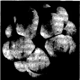
FIG. 7. Model of an apoferritin molecule, reproduced by
permission of G. H. Haggis (66). It shows the
arrange-ment of twenty protein subunits at the apexes of a pentagonal dodecahedron (69) with a pentagon face at the front of the model. At the center of each face there is a channel that connects the outside of the molecule with the central cavity in the protein and allows the passage of iron in and out of the molecule. The central cavity, which occupies about 22% of the total volume, is not visible in the model, but it can be seen in Fig. 6.
its interior, and this is borne out by a compari-son of X-ray diffraction patterns of ferritin and apoferritin (51, 68, 71). Nevertheless, the
pres-ence of the iron-containing core renders the pro-tein less susceptible to denaturation (104) and
to attack by proteolytic enzymes (104). The
atomic structure of the iron core is not spe-cifically orientated with respect to the protein
(51). It appears that the cores can grow in
different directions inside the protein shell, their external shape complementing that of the pro-tein when the latter is essentially full (51, 65).
The biosynthesis of ferritin has received con-siderable attention since it was discovered that
de novo synthesis of ferritin protein from amino
acids was induced by administration of iron salts (48, 49). Several workers have confirmed
this finding in whole animals (41, 99, 103), in
tissue slices (146, 172, 173), and in tissue
cul-tures (136). Tracer experiments with `4C-labeled
amino acids show that apoferritin, or possibly a ferritin of very low iron content, is formed first, and that with time the radioactivity passes to iron-rich species, suggesting that the empty shells are gradually being filled (41). Reconstituted
"fcrritin" can also be produced in vitro from
intact apoferritin molecules, ferrous salts, and oxygen or other oxidizing agents (17, 72, 98).
The product resembles ferritin in electron micro-scopic appearance and in its diffraction pattern, despite the absence of phosphate (72). This
ob-servation, together with the results of the tracer experiments, suggests that ferritin may be formed from apoferritin by a similar mechanism
in vivo (40), rather than by aggregation of
apo-ferritin subunits around a preformed iron-core
template (123). The protein shell may itself
assist in the removal of electrons from the Fe2+ ions entering the molecule. Some "isoferritins" may be more active in incorporating iron than others in vivo (54).
The mechanism of induction of ferritin by iron is not yet known, although it seems fairly certain that it does not occur through control of transcription, but .rather at some subsequent
stage (41). Drysdale and Munro (39, 41, 42)
conclude that messenger RNA for ferritin is
stable and that the iron either causes it to be used more efficiently or assists in the release of apoferritin subunits from the ribosomes or their aggregation to completed shells. Iron, however, is not essential for aggregation of apoferritin subunits in vitro (39, 73). Since both ferritin
and hemoglohin are induced by iron and both are found in erythroid cells, it seems likely that their biosynthetic control mechanisms are inter-connected in these cells (44, 103, 161, 174).
Little is known about the mechanism of re-lease of iron from ferritin in vivo. It can be
re-moved as Fe2+ from intact molecules by reduc-ing agents in vitro (18, 61), or, slowly, as Fe3+
by iron chelating agents (124, 169). An in vivo
release mechanism involving xanth;ne oxidase has been proposed (62). Iron that has been more
recently deposited as ferritin following red-cell breakdown is more easily mobilized than iron from older ferritin deposits (121). Possibly this
is because the more recently formed ferritin has a lower iron content and the iron can be more readily removed from only partially filled mole-cules, or it might be due to conformational dif-ferences in the protein. Iron can apparently be released both from intact molecules and from those in which the protein has been degraded
(154).
Hemosiderin is both chemically and metaboli-cally related to ferritin. This term has been used to describe both amorphous and crystalline intracellular deposits seen in the electron micro-scope (16, 133), but it seems certain that the
latter are ferritin. It has been suggested that the term hemosiderin should be restricted to those granules that are water insoluble (151). While
this is a useful means of distinguishing hemo-siderin from ferritin at a preparative level, it obviously cannot apply to iron-rich deposits seen in electron micrographs of tissue sections. Here the term should perhaps be restricted to deposits that are amorphous and in which the iron mi-celles are not clearly surrounded by well-defined
protein shells. Such deposits are, however, of variable appearance, sometimes, but not always, membrane-bound (14, 15, 132). On the basis of
morphological appearance alone, it may not
ways be possible to decide whether they contain ferritin or not.
Isolated hemosiderin granules have variable composition. Their iron, phosphorus, and sul-phur contents are higher than those found in ferritin (35, 106, 107, 135, 150, 168), and they
contain a number of different organic constitu-ents, including protein and small amounts of
apoferritin (107, 135). The magnetic moment
of the iron in hemosiderin is similar to that in ferritin, but with a greater range of values (3.5 to 4.7 ,o (150, 154). X-ray diffraction patterns
obtained from ferritin and hemosiderin extracted from the same normal horse spleen show that the atomic structures of the two mineral com-ponents are similar, although the average par-ticle size observed in hemosiderin is smaller (F. A. Fischbach, D. W. Gregory, P. M. Harri-son, and T. G. Hoy, unpublished observations).
The biological origin of hemosiderin is of some interest. There is evidence that in liver parenchymal cells it is a breakdown product of
ferritin (154), presumably resulting from
di-gestion of the protein with intracellular prote-ases. The X-ray diffraction results mentioned above would be consistent with this conclusion. Hemosiderin-like material can also be produced from ferritin by denaturation followed by trypsin
digestion (102). Studies on human siderotic
livers show that above a certain level of iron-loading the ratio of hemosiderin to ferritin is nearly constant, thus suggesting a dynamic
equi-librium between the two storage forms (108).
In rabbits, the ferritin level reached a maximum limiting value in response to massive doses of iron, whereas the hemosiderin content appeared to be able to rise continuously (154). The
im-plication in this case would be that the rate of ferritin turnover was increased at high iron levels, or that some of the hemosiderin was not formed from ferritin and possibly differed from "normal" hemosiderin. Thus, there may be two mechanisms for "hemosiderin" formation, one operating when the iron-level exceeds the cell's ability for ferritin synthesis and turnover. Since ferritin-like colloidal "iron oxide hydrates" can be produced in vitro in the absence of ferritin
protein (24, 158), two such mechanisms appear
to be plausible.
Iron transport and delivery:
Transferrin
A group of closely related iron-binding glyco-proteins occurs in vertebrate blood serum (trans-ferrin), in mammalian milk (lactotrans(trans-ferrin), and in avian egg white (ovotransferrin or conal-bumin) (47). These proteins are similar in size
and iron-binding properties, although they differ in amino acid composition and carbohydrate content (47). The discussion that follows will
be principally concerned with serum transferrin. Transferrin accounts for 2 to 3 per cent of the dry weight of vertebrate sera. This represents about 100 times the amount of free iron in serum, but only 0.1 per cent of the total body iron (3 to 4 mg transferrin-bound iron in humans). Human serum transferrin has a mo-lecular weight of about 86,000 [86,000-93,000 based on physical measurements (12, 47), 86,000
from iron-binding (82)]. It contains several
di-sulphide bridges but no free thiol groups (47).
Its attached carbohydrate, the function of which is uncertain, consists of four moles of sialic acid, eight moles of N-acetylglucosamine, four moles of galactose, and eight moles of mannose for every 90,000 molecular weight, joined to the protein as two branched glycopeptide chains
(80). This suggests the presence in the protein
of two peptide chains. Only one N-terminal resi-due (valine) has been found until recently (12),
but Jeppsson and Sjoquist have observed N-terminal serine in addition to valine (84), and
they have also reported the splitting of trans-ferrin into two subunits in 8M urea (83). Greene
and Feeney were unable to obtain evidence for dissociation under similar conditions and think that probably all transferrins are monomers
(63.)
Metal-free transferrin is colorless, but when ferric iron is bound in the presence of bicarbon-ate a salmon-pink complex is formed. It also complexes less firmly with other metals, such as copper. Two metal ions are bound per molecule of transferrin. At physiological pH, transferrin
has a very high affinity for iron, the equilibrium binding constants being of the order of 10°for
both iron atoms (2). Electron spin resonance studies have shown that the iron is bound as Fe'+, and that the two binding sites are approxi-mately equivalent and at least 9A apart (2, 4,
167)TT. Ane ph-ysiologialoions, three
pro-tons are displaced and one mole of bicarbonate is bound per iron atom (4). The bicarbonate was previously thought to be coordinated to the iron atoms (167), but recent evidence suggests
it is probably attached to groups on the protein (3) and not directly to the iron. The metal ligands are probably three tyrosine oxygens and two nitrogens fromn the imidazole or guanidyl groups of histidine or arginine, respectively (2,
93, 167). Transferrin will bind iron or copper
slowly in the absence of bicarbonate. Electron spin resonance studies suggest that more pro-tein ligands are available for binding (three or four N and three O ligands) when bicarbonate is absent (1). Evidence of heterogeneity in the two binding sites in the pH range of 4 to 6 has been obtained in the absence of bicarbonate (1), although at pH 7 to 11 the sites were indistin-guishable. Kinetic evidence indicates that the binding of the two atoms is cooperative, chela-tion of the first ion facilitating that of the sec-ond, presumably as a result of a conformational
change in the protein (170). The two binding
sites also appear to be functionally different. Re-lease of bound iron to immature red cells oc-curred more readily from one site than from
the other (52). Exchange of iron atoms between
the two sites occurs only very slowly or not at all (4, 52, 113). As already noted, iron-binding
stabilizes the protein (47) without substantially
altering its shape. However, in the course of binding, alterations occur in the antigenic
struc-ture (94), indicating that some conformational
change has taken place. Possibly the metal stabilizes the protein by forming crosslinks be-tween separated regions of the primary
struc-ture.
The physiological function of transferrin is to transport iron. Most of the body's iron is con-served and reused after the erythrocytes and
other iron-containing cells have been destroyed. Little iron is excreted and only a small propor-tion of dietary iron is absorbed. Transferrin acquires iron, derived from hemoglobin break-down or entering the body through the in-testinal mucosa, and delivers it to the
erythro-n i ti, INnn MrIMrnx fnr ;nnr~n
poietic bone marro for incorporation into new hemoglobin molecules, to other tissues requiring physiologically "active" iron, and to the storage depots. Transferrin also transfers iron from the placenta to the fetus. This protein has been regarded as having an essentially passive role, i.e. it simply provides a convenient means of carrying iron in a nontoxic form. Recent work, however, suggests that it may play an active part in the mechanisms regulating both absorp-tion and delivery of iron (53, 86). These
con-trol mechanisms appear to depend on structural changes in the protein, which result both from its chelation with iron and from its attachment to specific receptor sites on cell surfaces.
Every day plasma transferrin delivers from 30 to 40 mg of iron to the erythropoietic bone marrow-the tissue with by far the highest iron requirement. The protein is not consumed when its iron is released. It becomes available for fur-ther iron transport and is able to deliver about six to ten times its weight of iron in a day. Un-like free iron, which is taken up indiscriminately, transferrin iron is passed selectively to immature red cells, which are still actively synthesizing hemoglobin, rather than to mature cells in which
hemoglobin synthesis has ceased (81, 82, 86).
This suggests there are receptor sites on the sur-faces of immature cells that become lost as the cell ages. These sites must be specific for trans-ferrin protein and not for its iron. The rela-tive ease with which transferrin iron can be transferred to reticulocytes, as compared with its removal in vitro, suggests that a
confor-mational change in the protein occurs on bind-ing. The uptake of iron by reticulocytes seems
to occur as a three-stage process (82, 112,
113): (1) physical absorption of transferrin to
the tertiary structure of the protein; and (3) transfer of iron to the cell.
Stage 1 is reversible, the molecules being com-paratively easily displaced competitively by other molecules. At stage 2, iron-bound molecules have a greater affinity for the receptors than does apotransferrin (82), suggesting that iron-binding
facilitates the conformational change required for attachment to the receptor. Transferrin molecules containing two bound iron atoms re-lease their iron more readily than do those with only one (52). Probably only one of these atoms
is taken up at a time, iron from one site being preferred over that from the other (52). Stages 2
and 3 are dependent on the active metabolism of the cell (81, 112). The iron does not become
free during transfer and cannot be eluted by apotransferrin or other chelators (81).
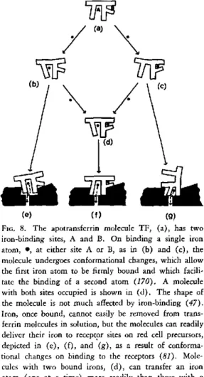
The conformational changes associated with the binding of iron to transferrin and its de-livery to reticulocyte receptors is summarized diagrammatically in Figure 8. As already indi-cated, these changes, and the ways in which the transferrin-binding sites are occupied by iron, may be factors controlling the rate at which iron is released into the serum from intestinal muco-sal cells, and hence iron absorption from the gut, as well as the rate of delivery (53). Under normal physiological conditions, transferrin is only about one third saturated with iron (22).
At high levels of saturation (above 60 per cent), much of the iron is deposited in the liver. The saturation may be normally kept at 30 to 60 per cent, so that the marrow, which has a high affinity for iron, can obtain an adequate supply while other tissues are not overloaded (22).
Neither the percentage saturation of transferrin nor the level of iron in the stores appear to have a controlling influence on iron absorption (34,
164).
Absorption of iron is related to the rate of erythropoiesis, even under conditions in which iron-loading is abnormally high (23, 164) or
low (164), and to the rate of plasma iron turn-over (165). Not all the iron in the mucosa finds
its way into the serum. At high iron levels some of this iron is sequestered in the mucosa in the
(a)
(e) (f (g)
FiG. 8. The apotransferrin molecule TF, (a), has two iron-binding sites, A and B. On binding a single iron atom, *, at either site A or B, as in (b) and (c), the molecule undergoes conformational changes, which allow the first iron atom to be firmly bound and which facili-tate the binding of a second atom (170). A molecule with both sites occupied is shown in (d). The shape of the molecule is not much affected by iron-binding (47). Iron, once bound, cannot easily be removed from trans-ferrin molecules in solution, but the molecules can readily deliver their iron to receptor sites on red cell precursors, depicted in (e), (f), and (g), as a result of conforma-tional changes on binding to the receptors (81). Mole-cules with two bound irons, (d), can transfer an iron atom (one at a time) more readily than those with a single iron atom (52), as can be seen by comparing (f) with (e) or (g). Iron atoms from site A can be rather more readily transferred, (e), than can those from site
B, (g), (53).
form of ferritin (32, 33, 37), which is
synthe-sized in response to the presence of iron (152). Much of this iron is lost to the body when the cells are exfoliated. Mucosal cells can both re-lease iron to the serum and acquire iron from it, depending on the body's requirements. Con-rad, Weintraub, and Crosby (34) proposed that absorption is controlled by the concentration of iron in the intestinal mucosa and that this in turn depends on the amount of "messenger" iron from the plasma entering these cells. This messenger iron would be free to enter the in-testinal cells when not required elsewhere. When
13
erythropoiesis is stimulated, much of the mucosal iron would pass to the plasma and absorption would then be increased.
These ideas have been interpreted by Fletcher and Huehns (53) in terms of the distribution of iron on plasma transferrin. They point out that transferrin consists of four molecular species, two with a single iron atom at different sites (A and B), one with no iron atoms, and one with two iron atoms occupying both sites. The suggested order of ability to deliver iron to the
erythro-blasts (52, 53) would be as follows:
/ AFe
TF
BFe
/AFe
>TF
B0
/Ao
>TF
BFe They propose that "messenger" iron is repre-sented by those molecules that have two bound iron atoms. The amount of this species present would tend to be decreased during high marrow activity and increased when the body's iron stores are large. The quantity of iron entering the mucosa from the plasma would be deter-mined by the number of transferrin molecules carrying two iron atoms, of which that at site B might be the more easily incorporated. Iron re-leased from the mucosal cells to the plasma would, however, have a preference for site A on the transferrin molecule-that is, the same site from which iron is preferentially delivered to the red cell precursors.
These ideas offer a simple explanation of the link between absorption and the body's need for iron. The degree to which transferrin mole-cules are saturated with iron also depends on the rate at which the protein is synthesized and catabolized, both of which have been found to be related to the erythropoietic rate (36, 96).
Sup-pression of transferrin synthesis seems to be
FIG. 9. Diagram of the central role of transferrin in iron metabolism. Plasma transferrin is represented by three species, TF(o)2, TF(Fe,o), and TF(Fe)2, which have their two binding sites occupied by no, one, or two iron atoms, respectively. Differences between the nature of the binding sites (see Fig. 8) have been ignored. The arrows represent the directions of flow of iron atoms in and out of the various compartments. The sizes of the compartments are in no way related to the amounts of
iron they contain.
associated with iron-loading, high hepatic ferri-tin formation, and low erythropoiesis. In iron deficiency, the concentration of transferrin in the plasma is abnormally high (22, 96). Thus,
iron influences the synthesis of transferrin in a way that is different from its effect on ferritin formation and in a way that ensures that the body's main requirement for iron will be met.
The central role of transferrin in the metabo-lism of iron is illustrated schematically in
Fig-ure 9.
REFERENCES
1. AASA, R., and P. AISEN. Electron paramagnetic
resonance study of iron and copper complexes of transferrin. 1 Biol Chem 243: 2399-2404, 1968.
2. AASA, R., B. G. MALMSTROM, P. SALTMAN, and T. VANNGARD. The specific binding of iron (III)
and copper (II) to transferrin and conalbumin.
Biochim Biophys Acta 75: 203-222, 1963.
3. AISEN, P., R. AASA, B. G. MALMSTROM, and
T. VANNGARD. Bicarbonate and the binding of iron
4. AisEN, P., A. LIEBMAN, and H. A. REICH. Studies on the binding of iron to transferrin and conalbumin. 1 Biol Chem 241: 1666-1671, 1966.
5. ALFREY, C. P., E. C. LYNCH, and C. E. WHIT-LEY. Characterization of ferritin isolated from hu-man marrow, spleen, liver, and reticulocytes. 1 Lab Clin Med 70: 419-428, 1967.
6. ANDREWS, P. The gel-filtration behaviour of proteins related to their molecular weights over a wide range. Biochem 1 96: 595-606, 1965.
7. ANTONINI, E. Interrelationship between struc-ture and function in hemoglobin and myoglobin.
Physiol Rev 45: 123-170, 1965.
8. ANTONINI, E. Hemoglobin and its reaction with ligands. Science 158: 1417-1425, 1967.
9. ANTONINI, E., M. BRUNORI, and S. ANDERSON. Studies on the relations between molecular and functional properties of hemoglobin. VII. Kinetic effects of the reversible dissociation of hemoglobin into single chain molecules. 1 Biol Chem 243:
1816-1822, 1968.
10. ANTONINI, E., E. CHIANCONE, and M. BRUNORI. Studies on the relations between molecular and func-tional properties of hemoglobin. VI. Observations on the kinetics of hemoglobin reactions in concen-trated salt solutions. 1 Biol Chem 242: 4360-4366, 1967.
11. BACHMAYER, H., A. M. BENSON, K. T. YASU-NOBU, W. T. GARRARD, and H. R. WHITELEY. Non-heme iron proteins. IV. Structural studies of micro-coccus aerogenes rubredoxin. Biochemistry 7: 986-996, 1968.
12. BEARN, A. G., and W. C. PARKER. Transferrin. In Glycoproteins; Their Composition, Structure, and Function, B.B.A. Library, vol. 5, Amsterdam, Else-vier, pp. 413-433, 1966.
13. BENESCH, R. E., R. BENESCH, and G. MACDUFF. Subunit exchange and ligand binding; a new hy-pothesis for the mechanism of oxygenation of hemo-globin. Proc Nat Acad Sci USA 54: 535-542, 1965.
14. BEssIS, M., and J. BRETON-GORIUS. Accumula-tion de granules ferrugineux dans les mitochondries des érythroblastes. C R Acad Sci 244: 2846-2847, 1957.
15. BEssIs, M., and J. BREToN-GoRIus. Trois as-pects du fer dans les coupes d'organes examinées au microscope electronique (ferritine et derivé, dans les cellules intestinales, les érythroblastes et les cellu-les réticulaires). C R Acad Sci 245: 1271-1272,
1957.
16. BEssIS, M., and J. BRETON-GORIus. Differents aspects du fer dans l'organisme. 1. Ferritine et mi-celles ferrugineuses. II. Differentes formes de l'hémo-sidérine. 1 Biophys Biochem Cytol 6: 231-240, 1959.
17. BIELIC, H-J., and E. BAYER. Synthetisches ferritin, ein Eisen (III)-komplex des apoferritin.
Naturwiss. 42: 125-126, 1955.
18. BIELIG, H-J., and E. BAYER. Eisenaustausch zwischen proteinen; Modellversuche zur eisenre-sorption und speicherung im Tierkorper. Naturwiss.
42: 466-467, 1955.
19. BIELIG, H-J., O. KRATKY, G. ROHNS, and H. WAWRA. Small-angle scattering of apoferritin in solution. Biochim Biophys Acta 112: 110-118, 1966. 20. BLAISE, A., J. CHAPPERT, and J-L. GIRARDET. Observation par mesures magnétiques et effet Moss-bauer d'un antiferromagnétism de grains fins dans la ferritine. C R Acad Sci 261: 2310-2313, 1965.
21. BOLTON, W., J. M. Cox, and M. F. PERUTZ. Structure and function of haemoglobin. IV. A three-dimensional fourier synthesis of horse
deoxyhaemo-globin at 5.5A resolution. 1 Molec Biol 33: 283-297,
1968.
22. BOTHWELL, T. H., and C. A. FINCH. Iron Metabolism. Boston, Little, Brown and Co., 1962.
23. BOTHWELL, T. H., G. PIRzIo-BIRoLI, and C. A. FINCH. Iron absorption. I. Factors influencing ab-sorption. 1 Lab Clin Med 51: 24-36, 1958.
24. BRADY, G. W., C. R. KURKJIAN, E. F. X. LYDEN, M. B. ROBIN, P. SALTMAN, T. SpIRO, and A. TERZIS. The structure of an iron core analog of ferritin. Biochemistry 7: 2185-2191, 1968.
25. BRAUNITZER, G., K. HILSE, V. RUDLOFF, and
N. HILSCHMANN. The hemoglobins. Advances Pro-tein Chem 19: 1-71, 1964.
26. BRESLOW, E., S. BEYCHOK, K. D. HARDMAN,
and F. R. N. GURD. Relative conformation of sperm whale metmyoglobin and apomyoglobin in solution.
1
Biol Chem 240: 304-309, 1965.27. BRILL, A. S., and R. J. P. WILLIAMS. The absorption spectra, magnetic moments, and the binding of iron in haemoproteins. Biochem / 78: 246-253, 1961.
28. BRUNS, G. P., and 1. M. LONDON. The effect of hemin on the synthesis of globin. Biochem Bio-phys Res Commun 18: 236-242, 1965.
29. BUCHANAN, B. B. The chemistry and function of ferredoxin. Structure and Bonding 1: 109-148, 1966.
30. CAUGHEY, W. S. Porphyrin proteins and en-zymes. Ann Rev Biochem 36: 611-644, 1967.
31. CHANCE, B., R. W. ESTABROOK, and T. YONE-TANI (eds.), Hemes and Hemoproteins. New York, Academic Press, 1966.
32. CHARLTON, R. W., P. JACOBS, J. D. TORRANCE, and T. H. BOTHWELL. The role of intestinal mucosa in iron absorption. 1 Clin Invest 44: 543-554, 1965.
33. CONRAD, M. E., and W. H. CROSBY. Intestinal mechanisms controlling iron absorption. Blood 22: 406-415, 1963.
34. CONRAD, M. E., L. R. WEINTRAUB, and W. H.
CROSBY. The role of the intestine in iron kinetics. 1 Clin Invest 43: 963-974, 1964.