Licenciado sob uma Licença Creative Commons DO): http://dx.doi.org. . / - . . .AO
[T]
Functional exercise capacity, lung function and chest wall
deformity in patients with adolescent idiopathic scoliosis
[)]
Capacidade funcional de exercício, função pulmonar e geometria da
caixa torácica em pacientes com escoliose idiopática do adolescente
[A]
Evandro Fornias Sperandio[a], Milena Carlos Vidotto[a], Anderson Sales Alexandre[a], Liu Chiao Yi[a],
Alberto Ofenhejm Gotfryd[b], Victor Zuniga Dourado[a]*
[a] Universidade Federal de São Paulo Unifesp , Santos, SP, Brazil
[b] (ospital Santa Casa de Misericórdia de Santos (SCMS , Santos, SP, Brazil
[R]
Abstract
Introduction: The adolescent idiopathic scoliosis A)S causes changes on the compliance of the chest. These
changes may be associated with impaired lung function and reduced functional exercise capacity of these adolescents. We aimed to evaluate the correlation between functional exercise capacity, lung function and geometry of the chest at different stages of A)S. Materials and methods: The study was carried out in a
cross-sectional design which were evaluated A)S patients at different stages of the disease. For chest wall evalua-tion, were created geometry angles/distances A/D , which were quantified by Software Postural Assessment. The functional exercise capacity was assessed by a portable gas analyzer during the incremental shuttle walk test )SWT . Besides that, manovacuometry and spirometry were also performed. Results: Linear regressions
* EFS: PhD candidate, e-mail: [email protected] MCV: PhD, e-mail: [email protected]
ASA: MSc, e-mail: [email protected] LCY: PhD, e-mail: [email protected]
564
showed that oxygen uptake peak VO was correlated with distance travelled in the )SWT R = . , maxi-mal respiratory pressures, cough peak flow R = . and some thoracic deformity markers D , D and A .
Discussion: We observed that the chest wall alterations, lung function and respiratory muscle strength are
related to the functional exercise capacity and may impair the physical activity performance in A)S patients.
Final considerations: There is correlation between functional exercise capacity, lung function and geometry
of the chest in A)S patients. Our results point to the possible impact of the A)S in the physical activities of these adolescents. Therefore, efforts to prevent the disease progression are extremely important.
[P]
Keywords: Chest. Pulmonary function testing respiratory mechanics. Scoliosis.
]
[B]Resumo
Introdução: A escoliose idiopática do adolescente (EIA) provoca alterações na conformidade da caixa torácica. Essas alterações podem estar associadas ao prejuízo da função pulmonar e à redução da capacidade funcional de exercício desses adolescentes. Materiais e métodos: O estudo foi realizado em delineamento transversal no qual foram avaliadas a correlação entre a capacidade funcional de exercício, função pulmonar e a geometria da caixa torácica de 27 pacientes em diferentes estágios da EIA. Para avaliação da geometria torácica foram criados ângulos/distâncias (A/D) que foram quantificados pelo Software de Avaliação Postural. Foram reali-zadas manovacuometria, espirometria e a capacidade funcional de exercício foi avaliada por meio de um ana-lisador de gases portátil durante o incremental shuttle walk test (ISWT). Resultados: As regressões lineares mostraram que o consumo de oxigênio se correlacionou com a distância caminhada no ISWT (R2 = 0,52), as pressões respiratórias máximas, o pico de fluxo de tosse (R2 = 0,59) e alguns marcadores de deformidade torá-cica (D1, D2 e A6). Discussão: Observamos que a alteração da geometria da caixa torátorá-cica, a função pulmonar e a força dos músculos respiratórios estão associadas à capacidade funcional de exercício e podem prejudicar o desempenho das atividades físicas dos pacientes com EIA. Considerações finais: Existe correlação entre a capacidade funcional de exercício, função pulmonar e a geometria da caixa torácica em pacientes com EIA. Nossos resultados apontam o possível impacto da EIA nas atividades físicas desses adolescentes. [K]
Palavras-chave: Caixa torácica. Teste de função pulmonar. Mecânica respiratória. Escoliose.
Introduction
The adolescent idiopathic scoliosis A)S is the most common type of scoliosis. )ts prevalence is es-timated at between % and % of all adolescents aged to years old , affecting mainly females at a ratio of . : .
Because of the complex interconnections between sternum, ribs and spine, the displacement and rota-tion of the vertebrae have a profound effect on the shape of the chest, creating a convex and a concave side . The rib cage provides the structure that contains the lungs and supports the respiratory muscles. The normal ventilatory mechanics depends on a compliant rib cage and the distortion of the rib cage associated with spinal deformity contributes to
altered ventilatory mechanics , and decreased ability to perform physical activities in E)A subjects . Additionally, chronic muscular weakness may play a role in the lack of muscular and cardiorespira-tory fitness , .
A direct relationship between the decreased maxi-mal aerobic capacity and forced vital capacity has also been reported in A)S patients . )t is also seen that aerobic exercise improves the forced vital capacity and inspiratory capacity of these patients and posi-tively influences the cardiorespiratory fitness .
Methods
The study was conducted in a cross-section de-sign. We enrolled patients with A)S of both gender ag-ing between and years. Patients with previous or current history of heart, lung, or neuromuscular disease and patients who, for any reason, failed to perform the assessments proposed were excluded. Patients were asked about their level of physical ac-tivity in their daily life, and those who reported to be physically active were also excluded. Patients were referred to the Orthopedic Clinic of a local hos-pital, where they underwent radiographic evaluation of Cobb angles. All the study participants signed in-formed consents. The present study was approved by the local ethics committee No. .
)nitially were evaluated patients, one was ex-cluded due to asthma and one for failing to perform the evaluations. Eight of these patients were preop-eratively and nineteen underwent surgical treatment of spinal arthrodesis. All patients were classified as Lenke ), with deviation of the main thoracic curve to the right. The respiratory muscle strength, lung function, exercise capacity and chest wall shape were evaluated.
Anthropometrics
Weight and height were measured by standard techniques. Weight was assessed to the nearest . kg, and height was measured to the nearest . cm. The body mass index was calculated by dividing weight in kilograms by height in square meters kg/m .
Respiratory assessment
The respiratory muscle strength was quantified by measuring the maximum inspiratory pressure M)P and maximum expiratory pressure MEP accord-ing to the Brazilian Thoracic Association statement
. These measurements were performed with the participant properly seated and using a manometer MVD model; Globalmed, São Paulo, SP, Brazil . The M)P and MEP were performed from functional residual capacity.
Spirometry was performed using a handheld spirometer Spiropalm; COSMED, Pavona di Albano, )taly according to the Brazilian Thoracic Association
recommendations . Forced vital capacity FVC , forced expiratory volume in the first second of expi-ration FEV , and the FEV /FVC ratio were quanti-fied and were expressed as absolute values and as percentage of predicted values . Peak cough flow PCF was carried out using the method described by Fiore et al. .
Incremental shuttle walk test
The )SWT was performed according to the meth-ods described by Singh et al. . The walking veloc-ity was imposed by audio signals recorded on a CD. (eart rate (R , blood pressure, and dyspnea and leg fatigue Borg scale were determined before and after each )SWT. The test was performed twice to minimize the learning effect. The interval between tests was set at minutes and/or the return of the aforementioned variables at baseline. The distance walked during the second test was considered for further analysis.
During the second )SWT, the expired gases were collected and analyzed with a portable telemetric gas analyzer K b ; Cosmed, Pavona di Albano, )taly . The calibrations with room air, reference gas, L syringe, and delay were performed following the manufacturer’s recommendations.
Oxygen uptake, VCO , VE, tidal volume VT , re-spiratory rate f , ventilatory equivalents of O VE/ VO and CO VE/VCO , the rate of gas exchange R , (R, and pulse of O PuO as well as other variables obtained by calculations were assessed breath by breath. After collecting these variables, the data were filtered every seconds for further analysis.
Chest wall evaluation
566
scapulae angles ; D xiphoid–last false rib on the right and left side ; D manubrium–last false rib on the right and left side ; and D xiphoid–anterior superior iliac spine on the left and right side . All these angles and distances were created by our team, except the A angle, which was reproduced from the study of Davidson et al. .
Statistical analysis
The data were analyzed descriptively and present-ed as mean and standard deviation when presentpresent-ed symmetrical distribution and as median variance when presented asymmetric distribution. The normal-ity of the variables was investigated by Kolmogorov-Smirnov test. A series of linear regressions were per-formed to assess the correlations between variables. The probability of an alpha error was set at %. right side and posterior view. An Ethylene Vinyl
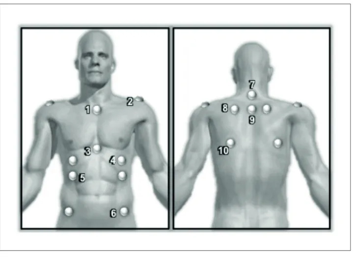
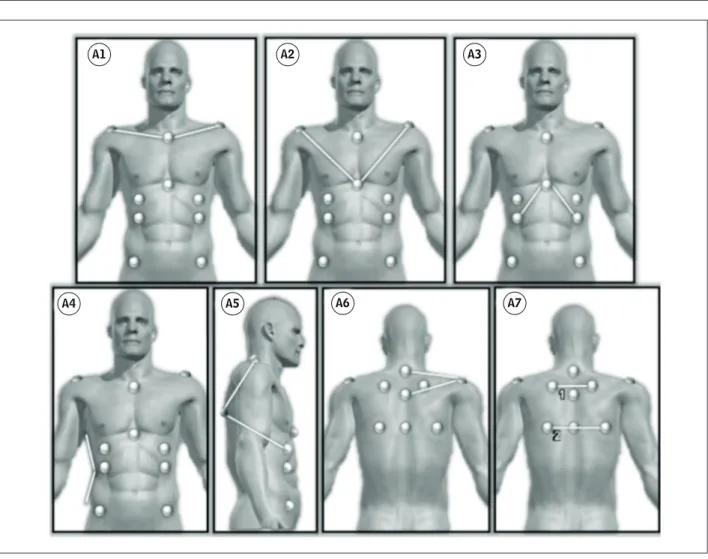
Acetate EVA carpet was used in order to mark the feet position for each photo taken. The anatomical points were marked on the skin by fixing half sphere of styrofoam balls of mm diameter, using double-sided tape. These markings are shown in Figure . The anatomical points used were based on the SAPO protocol, except point and points , and that were created by our research team. We evaluated the thoracic markers by angles A and distances D as follows Figures and , respectively : A right acromion/manubrium/left acromion ; A right ac-romion/xiphoid/left acromion ; A last false right rib/xiphoid/last false left rib ; A angle between the deepest point of the waist and upper and lower edges of the waist ; A inframamilar/inferior angle of the scapula/right and left acromion ; A C /acromion right and left/T ; A angle formed by the intersec-tion of the tangent segments of the upper and lower
Figure 1 – Anatomical points
A1 A2 A3
A7 A6
A5 A4
Figure 2 – Angles
Note: A1 = right acromion/manubrium/left acromion; A2 = right acromion/xiphoid/left acromion; A3 = last false right rib/xiphoid/last false left rib; A4 = angle between the deepest point of the waist and upper and lower edges of the waist; A5 = inframamilar/inferior angle of the scapula/right and left acromion; A6 = C7/acromion right and left/T3; A7 = angle formed by the intersection of the tangent segments of the upper and lower scapulae angles.
D1 D2 D3
Figure 3 – Distances
568
Results
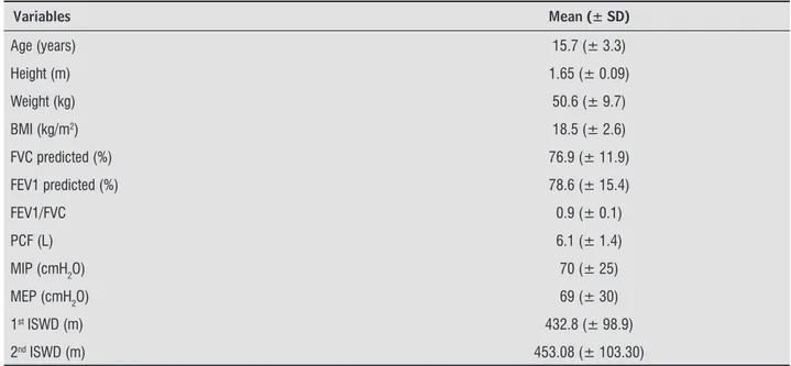
The characteristics of the patients, females, are shown in Table . The average BM) shows that in-dividuals had low weight . ± . . By the average of predicted FVC and FEV /FVC ratio, we could iden-tify a restrictive component in these patients . ± . and . ± . , respectively . Linear regression showed that the highest correlation was obtained between peak VO and )SWD R = . ; p < . . )n addition to this, we found correlation between peak
Table 1 - Demographic, anthropometric, lung function and respiratory muscle strength of the 27 patients
Variables Mean (± SD)
Age (years) 15.7 (± 3.3)
Height (m) 1.65 (± 0.09)
Weight (kg) 50.6 (± 9.7)
BMI (kg/m2) 18.5 (± 2.6)
FVC predicted (%) 76.9 (± 11.9)
FEV1 predicted (%) 78.6 (± 15.4)
FEV1/FVC 0.9 (± 0.1)
PCF (L) 6.1 (± 1.4)
MIP (cmH2O) 70 (± 25)
MEP (cmH2O) 69 (± 30)
1st ISWD (m) 432.8 (± 98.9)
2nd ISWD (m) 453.08 (± 103.30)
Note: Note: BMI = body mass index; FVC = forced vital capacity; FEV1 = forced expiratory volume in 1st second; PCF = peak cough fl ow;
MIP = maximal inspiratory pressure; MEP = maximal expiratory pressure; ISWD = incremental shuttle walk distance.
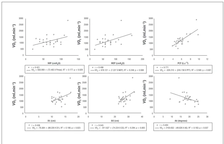
VO and M)P R = . ; p = . , MEP R = . ; p = . , PCF R = . ; p < . , D R = . ; p = . , D R = . ; p = . , and A R = . ; p = . as shown in Figure . We have also obtained correlation between VEmax and M)P R =
. ; p = . , MEP R = . ; p = . , PCF R = . ; p < . , D R = . ; p = . and D R = . ; p = . . The last dependent variable, )SWD, showed correlation with M)P R = . ; p = . and MEP R = . ; p = . , FEV R = . ; p < . and A R = . ; p = . .
Discussion
This study evaluated cardiorespiratory fitness and the chest wall shape of adolescents with E)A. Positive correlations between metabolic variables and thoracic deformity markers, lung function and respiratory muscle strength were established. We observed that the deformation of the rib cage can change the physical performance of these patients.
)n our study there was a correlation between peak VO and chest wall shape, which shows how the deformity can possibly alter the ventilatory efficiency and compromise the physical ability in
between respiratory muscle strength and FVC. The reduction in respiratory muscle strength and pulmo-nary restriction are well-described characteristics in A)S patients , - .
Martínez-Llorens et al. also found positive cor-relation between exercise capacity, represented by the maximum work rate, and respiratory pressures. )n addition, a positive correlation was established
VO 2 (ml.min -1) VO 2 (ml.min -1) VO 2 (ml.min -1)V O2 (ml.min -1) VO 2 (ml.min -1) VO 2 (ml.min -1) D1 (cm)
MIP (cmH2O) MEP (cmH2O) PCF (L.s-1)
D2 (cm) A6 (degrees)
3000 2500 2000 1500 1000 500 0
0 50 100 150
0 5 10 15 20 0 10 20 30 40 0
r = 0.404
VO2 = 2103.652 - (49.626 X A6); R2 = 0.163; p = 0.037
r = 0.771
VO2 = -326.210 + (245.738 X PFT); R2 = 0.595; p = 0.001
r = 0.498
r = 0.543
VO2 = 676.121 + (7.227 X MEP); R2 = 0.248; p = 0.008
VO2 = - 811.627 + (74.224 X D2); R2 = 0.294; p = 0.003
r = 0.436
VO2 = -76.304 + (98.220 X D1); R2 = 0.190; p = 0.023
r = 0.421
VO2 = 650.069 + (72.465 X Pimax); R2 = 0.177; p = 0.029
5 10 15 20 25 30
0 50 100 150 200 0 2 4 6 8 10 12
3000 2500 2000 1500 1000 500 0 3000 2500 2000 1500 1000 500 0 3000 2500 2000 1500 1000 500 0 3000 2500 2000 1500 1000 500 0 3000 2500 2000 1500 1000 500 0
Figure 4 – Significant correlations among oxygen uptake (VO2), maximal inspiratory pressure (MIP), maximal expiratory
pres-sure (MEP), peak cough flow (PCF), distance 1 (D1), distance 2 (D2) and angle 6 (A6)
)n addition, VEmax was also imparied. )n this study
there was a positive correlation between VEmax and
thoracic deformity markers D and D , which con-firms the influence of thoracic deformity ventilation in A)S patients. This finding is consistent with the results of Barrios et al. which found lower VEmax
values relative to peers controls and this variable was correlated negatively with the magnitude of scoliosis curvature, measured by Cobb angle. Another result that suggests the ventilatory inefficiency of these pa-tients is the increased respiratory oxygen equivalent
VE/VO . During the exercise, the respiratory rate is significantly higher in A)S patients than in healthy people , . This increased respiratory rate can be explained as a compensatory mechanism adopted in response to the low tidal volume and the fact that they do not present good efficiency on the variation
of the respiratory pattern. Thus, in case of increased oxygen demand to the body, there is an increase in respiratory rate to compensate the limitation of dia-phragmatic incursion , .
These studies showed that, even with the in-creased respiratory frequency, VEmax is reduced in
patients with A)S, which shows the inefficiency of mechanical ventilation of patients. We may suggest through our results that the reduction in respiratory muscle strength may lead to reduction of VEmax, once
we found correlation between VEmax and respiratory
pressures. Likewise, the VEmax correlated with PCF,
showing that the smaller the VEmax, the lower the
570
of the cause of muscle tone dysfunction is genetic predisposition, which would develop a flexible spine and, therefore, not resist the growth spurt without modification .
The implementation of walking-based aerobic ex-ercises would be rational strategy for the treatment of patients with A)S. dos Santos Alves et al. found a significant improvement in FVC, inspiratory capacity, FEV , and increased MWD after aerobic exercises in patients with A)S. Another similar study observed % improvement in aerobic capacity in the trained group and . % decrease in the control group . )n addiction, the manual therapy aided with Dynamic Brace System has improved the respiratory param-eters and trunk morphology value . These stud-ies show that, in fact, regular aerobic exercises and strength training , play an important role in the treatment of patients with A)S.
This study has limitations that should be de-scribed. We did not inform the Cobb angles of the pa-tients; this is justified by the fact that most patients in the postoperative period did not return to the column clinic for a routine visit and the radiographic imaging for evaluation of the angle. To increase the sample size was necessary to cluster patients in different stages of the disease, both preoperatively and post-operatively. Besides, we did not perform the CPET for comparison of variables obtained during )SWT. (owever, our main objective was to quantify the re-duction in functional exercise capacity ie, ability to walk , and furthermore, the )SWT has been widely correlated with CPET and is suitable for evaluating aerobic capacity in healthy middle-aged and older adults in patients with chronic diseases. )n addi-tion, studies that evaluated the functional exercise capacity of A)S patients used the MWT. Although the MWT is considered intense , the )SWT pro-voked a significantly higher VO , VCO , VEmax and FCmax
compared to MWT .
We can conclude that there is a correlation be-tween peak VO variables, VEmax and )SWD with
maxi-mal respiratory pressures, lung function and chest wall shape A)S patients. These correlations show the influence of the alterations of the chest wall shape in functional exercise capacity and how the disease is likely to limit the activities of daily living of these adolescents. (owever, studies comparing pre and postoperative are needed in order to better clarify the influence of the chest wall deformity on the func-tional exercise capacity.
restriction. Our patients had significantly shallower slope of ΔVT/ΔlnVE, that is, worse breathing pattern during walking. Associated with lower VE and VT at the end of the )SWT, these results clearly show the restrictive ventilatory pattern in response to exercise . This inefficiency, coupled with the low ventila-tory capacity and low VOmax may be responsible for
. Fiore Jr. JF, Chiavegato LD, Denehy L, Paisani DM, Faresin SM. Do directed cough maneuvers improve cough effectiveness in the early period after open heart surgery? Effect of thoracic support and maxi-mal inspiration on cough peak expiratory flow, cough expiratory volume, and thoracic pain. Respir Care.
; : - .
. Singh SJ, Morgan MD, Scott S, Walters D, (ardman AE. Development of a shuttle walking test of disability in patients with chronic airways obstruction. Thorax.
; : - .
. Davidson J, dos Santos AM, Garcia KM, Yi LC, João PC, Miyoshi M(, et al. Photogrammetry: an accurate and reliable tool to detect thoracic musculoskeletal abnormalities in preterm infants. Physiotherapy.
; : - .
. Martínez-L lorens J, Ramírez M, Colomina MJ, Bagó J, Molina A, Cáceres E,et al. Muscle dysfunction and exercise limitation in adolescent idiopathic scoliosis. Eur Respir J. ; : - .
. Kearon C, Vivi ani GR, Killian KJ. Factors influencing work capacity in adolescent idiopathic thoracic sco-liosis. Am Rev Respir Dis. ; : - . . Lisboa C, More no R, Fava M, Ferretti R, Cruz E.
)nspi-ratory muscle function in patients with severe ky-phoscoliosis. Am Rev Respir Dis. ; : - . . Barrios C, Pér ez-Encinas C, Maruenda J), Laquía M.
Significant ventilatory functional restriction in ado-lescents with mild or moderate scoliosis during maxi-mal exercise tolerance test. Spine Phila Pa .
; : - .
. Boyer J, Amin N, Taddonio R, Dozor AJ. Evidence of airway obstruction in children with idiopathic sco-liosis. Chest. ; : - .
. Newton PO, Faro FD, Gollogly S, Betz RR, Lenke LG, Lowe TG. Results of preoperative pulmonary function testing of adolescents with idiopathic scoliosis. A study of six hundred and thirty-one patients. J Bone Joint Surg Am. ; : - .
. Alves VL, Avanzi O. Objective assessment of the car-diorespiratory function of adolescents with idiopathic scoliosis through the six-minute walk test. Spine Ph-ila Pa . ; :E - .
Referências
. Arle t V, Reddi V. Adolescent idiopathic scoliosis. Neu-rosurg Clin N Am. ; : - .
. Rosa les-Olivares LM, García J, Miramontes-Martínez VP, Alpízar-Aguirre A, Arenas-Sordo ML, Reyes-Sán-chez AA. [Surgical treatment for scoliosis. Minimal evolution control at years]. Cir Cir. ; : - . . Koumbour lis AC. Scoliosis and the respiratory system.
Paediatr Respir Rev. ; : - .
. Kotwicki T, Szulc A, Dobosiewicz K, Rapala K. The pathomechanism of idiopathic scoliosis: the impor-tance of physiological thoracic kyphosis. Ortop Trau-matol Rehabil. ; : - .
. Takahash i S, Suzuki N, Asazuma T, Kono K, Ono T, Toya-ma Y. Factors of thoracic cage deformity that affect pulmonary function in adolescent idiopathic thoracic scoliosis. Spine Phila Pa . ; : - . . Czaprows ki D, Kotwicki T, Biernat R, Urniaz J,
Ronikier A. Physical capacity of girls with mild and moderate idiopathic scoliosis: influence of the size, length and number of curvatures. Eur Spine J.
; : - .
. dos Sant os Alves VL, Stirbulov R, Avanzi O. )mpact of a physical rehabilitation program on the respira-tory function of adolescents with idiopathic scoliosis. Chest. ; : - .
. Sperandi o EF, Alexandre AS, Yi LC, Poletto PR, Gotfryd AO, Vidotto MC, et al. Functional aerobic exercise capacity limitation in adolescent idiopathic scoliosis. Spine J.
; : - .
. Garber C E, Blissmer B, Deschenes MR, Franklin BA, Lamonte MJ, Lee )M, et al. American College of Sports Medicine position stand. Quantity and quality of ex-ercise for developing and maintaining cardiorespi-ratory, musculoskeletal, and neuromotor fitness in apparently healthy adults: guidance for prescribing exercise. Med Sci Sports Exerc. ; : - . . Pereira CAC, Neder JA. Diretrizes para testes de função
pulmonar. J Bras Pneumol. ; Supl. :s -s . . Pereira C A, Sato T, Rodrigues SC. New reference values
572
. Negrini S, Donzel li S, Lusini M, Minnella S, Zaina F. The effectiveness of combined bracing and exercise in adolescent idiopathic scoliosis based on SRS and SOSORT criteria: a prospective study. BMC Musculo-skelet Disord. ; : .
. Sperandio EF, Ara ntes RL, Matheus AC, Silva RP, Lauria VT, Romiti M, et al. )ntensity and physiological responses to the -minute walk test in middle-aged and older adults: a comparison with cardiopulmonary exercise testing. Braz J Med Biol Res. ; : - . . Onorati P, Antonu cci R, Valli G, Berton E, De Marco F,
Serra P, et al. Non-invasive evaluation of gas exchange during a shuttle walking test vs. a -min walking test to assess exercise tolerance in COPD patients. Eur J Appl Physiol. ; - : - .
Received: / /
Recebido: / /
Approved: / /
Aprovado: / /
. DiRocco PJ, Vacc aro P. Cardiopulmonary functioning in adolescent patients with mild idiopathic scoliosis. Arch Phys Med Rehabil. ; Pt : - . . Grivas TB, Vasil iadis ES, Mihas C, Savvidou O. The
ef-fect of growth on the correlation between the spinal and rib cage deformity: implications on idiopathic scoliosis pathogenesis. Scoliosis. ; : .
. Leong JC, Lu WW, Luk KD, Karlberg EM. Kinematics of the chest cage and spine during breathing in healthy individuals and in patients with adolescent idiopathic scoliosis. Spine Phila Pa . ; : - . . Lowe TG, Edgar M , Margulies JY, Miller N(, Raso VJ,
Reinker KA, et al. Etiology of idiopathic scoliosis: current trends in research. J Bone Joint Surg Am.
; -A : - .
. Athanasopoulos S , Paxinos T, Tsafantakis E, Zachariou K, Chatziconstantinou S. The effect of aerobic training in girls with idiopathic scoliosis. Scand J Med Sci Sports.
; : - .
. Wnuk B, Frackiew icz J, Durmala J, Czernicki K, Wad-olowski K. Short-term effects of combination of sev-eral physiotherapy methods on the respiratory func-tion – a case report of adolescent idiopathic scoliosis. Stud (ealth Technol )nform. ; : - . . Mc)ntire K, Asher M, Burton D, Liu W. Trunk rotational
strength training for the management of adolescent idiopathic scoliosis A)S . Stud (ealth Technol )nform.