Catheterization of the mesenteric artery to treat portal
vein thrombosis
Cateterismo de artéria mesentérica para tratamento de trombose de veia porta
Guilherme Benjamin Brandão Pitta1, Deise Azevedo Pereira2, Milena de Fátima Queiroz Oliveira2
*
, Eduardo Abadie Guedes2, Joaquim Araújo Sampaio3
Abstract
Portal vein thrombosis is a rare vascular cause of acute abdomen and it is directly related to hereditary or acquired thrombophilias. his article presents the case of a 60-year-old male patient, with clinical signs of mesenteric ischemia that was conirmed by imaging examination. He underwent enterectomy and enteroanastomosis and, after detection of portal vein thrombosis by splenoportography, he was prescribed drug-based treatment with continuous infusion of recombinant tissue plasminogen activator (Alteplase) via selective catheterization of the superior mesenteric artery. his is a treatment innovation. he portal system was successfully recanalized. However, the patient developed abdominal sepsis and required intensive care for 25 days. His clinical status improved and he was discharged with a prescription for oral anticoagulant. his article presents a brief review of the literature and a discussion of portal vein thrombosis.
Keywords: portal vein; thrombosis; thrombophilia; mesenteric ischemia; acute abdomen.
Resumo
A trombose de veia porta é uma causa rara de abdome agudo vascular e está diretamente relacionada a tromboilias hereditárias ou adquiridas. O caso de um paciente de 60 anos, sexo masculino, com quadro clínico de isquemia mesentérica conirmada por exame de imagem é apresentado. Foi submetido a enterectomia e enteroanastomose e, após esplenoportograia que detectou trombose de veia porta, indicou-se tratamento medicamentoso com infusão contínua de ativador tecidual do plasminogênio recombinante (Alteplase) através de cateterismo seletivo da artéria mesentérica superior. Trata-se de um tratamento inovador. Obteve-se sucesso na recanalização do sistema porta. O paciente evoluiu com quadro de sepse abdominal, necessitando de assistência em terapia intensiva por 25 dias. Evoluiu bem e recebeu alta hospitalar com o uso de anticoagulante. O artigo apresenta uma breve revisão de literatura e discussão do caso clínico.
Palavras-chave: veia porta; trombose; tromboilia; isquemia mesentérica; abdome agudo.
1Universidade Estadual de Ciências da Saúde de Alagoas – UNCISAL, Faculdade de Medicina, Maceió, AL, Brazil. 2Universidade Federal de Alagoas – UFAL, Faculdade de Medicina, Maceió, AL, Brazil.
3Hospital Memorial Arthur Ramos – HMAR, Cirurgia Vascular, Maceió, AL, Brazil.
Financial support: None.
Conlicts of interest: No conlicts of interest declared concerning the publication of this article. Submitted: October 27, 2016. Accepted: February 01, 2017.
INTRODUCTION
Portal vein thrombosis (PVT) is an uncommon event among patients who do not have cirrhosis or cancer. Approximately 60% of cases are associated with prothrombotic conditions, especially myeloproliferative diseases and hereditary thrombophilias.1-3 The proportion
of cases deined as idiopathic PVT has reduced after recent identiication of additional hereditary thrombotic
risk factors.4 Although rare, PVT is potentially fatal
when complicated by intestinal ischemia.5
The clinical presentation of PVT ranges from asymptomatic cases, in which obstructions are partial, to liver failure and death, in acute cases.6 Splenoportography
(SPG) and abdominal computed tomography (CT) with contrast are safe methods for diagnosing this disease, while treatment is individualized and may involve anticoagulation, systemic or catheter-guided thrombolysis, and/or surgery, in cases that progress to intestinal necrosis.1,7,8
This article describes the case of a 60-year-old patient with PVT caused by thrombophilia that progressed to intestinal necrosis and presents a brief review of the literature on this disease.
PART I: CLINICAL CASE
A 60-year-old male patient, born and living in Maceió, AL, Brazil, was admitted to the emergency department complaining of diffuse abdominal pains with onset 48 hours previously and of increasing intensity, accompanied by nausea and vomiting. He reported having been treated previously for deep venous thrombosis (DVT) in the left lower limb. He was hypertensive, an irregular user of losartan potassium, and had dyslipidemia and hepatic steatosis, in addition to being an ex-smoker, a social drinker and sedentary. His family history included three sisters and a daughter who had had DVTs.
During the physical examination, he was conscience and agitated, with sudoresis and tachypnea, but without fever, cyanosis, or jaundice, and had hypertension (150×80 mmHg), with pallid (++/4+) and hydrated mucosas. His abdomen was swollen, distended, and tense, while deep palpation provoked pain, of greater intensity at the right iliac fossa, and bowel sounds were reduced in all areas. Extremities were perfused, peripheral pulses were palpable and robust, and he was free from lower limb edema and his calves were normal.
Blood tests and biochemical assays revealed discrete leukocytosis (13,100/mm3) and neutrophilia
(10,083/mm3) and hyperglycemia (146 mg/dL).
All other biochemical assay results were within
normal limits. A CT of the thorax and abdomen with contrast detected PVT (Figure 1) and the patient was immediately transported to the vascular surgery service. An SPG reveled that there was no venous return via the superior mesenteric and splenic veins and showed that the site of occlusion was the origin of the portal vein (Figure 2).
PART II: WHAT WAS DONE
Initial conduct was an exploratory laparotomy to investigate the cause of acute abdomen, which found an extensive area of jejunum-ileum necrosis, with indication for enterectomy and enteroanastomosis. During the procedure, approximately 60% of the
Figure 1. Computed tomography of chest and abdomen, showing
thrombosis in the portal vein (arrowed).
Figure 2. Splenoportography (venous phase) showing absence
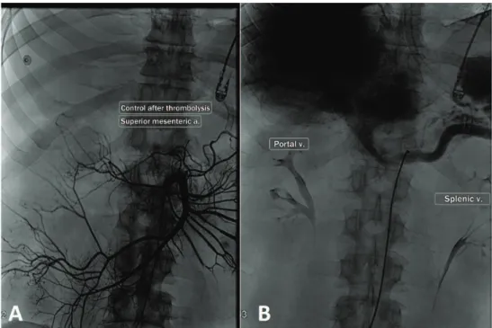
patient’s jejunal segment was removed. On the next day, recanalization of the portal system by thrombolysis was initiated with selective catheterization of the superior mesenteric artery and administration of recombinant tissue plasminogen activator (Alteplase), 10 mg in bolus and 40 mg every 24 hours for 3 days, followed by full heparinization via continuous infusion pump. A control SPG on the fourth day after admission (Figure 3) showed that blood low through the portal vein had been restored and hepatic perfusion had improved.
Postoperative follow-up was provided in the ICU, since the patient remained critical, sedated, on mechanical ventilation, hemodynamically unstable, and on vasoactive drugs for 6 days. He developed severe sepsis and was transferred to a hospital in São Paulo, where he remained in the ICU for a further 19 days, on low molecular weight heparin. He developed heparin-induced thrombocytopenia and, because of a delay in diagnosis, this was complicated by a further DVT episode. Heparin was substituted with fondaparinux and his platelet count and general condition improved. He remained in a standard ward for another 20 days and then he was discharged from hospital on oral anticoagulant.
During later investigations, it was found that there was a methylenetetrahydrofolate reductase mutation in the family, although homocysteine was not increased. A polymorphism of the plasminogen
activator inhibitor 1 (PAI-1) gene was also detected, but no diagnostic conclusions could be made. Other test results were normal or negative, such as mutation of factor V Leiden, antithrombin, S and C proteins, anticardiolipin, and lupus anticoagulant.
DISCUSSION
Mesenteric ischemia due to PVT with intestinal infarction is a severe and greatly feared complication that is associated with a mortality rate of 60%9 and
requires surgical management that may involve extensive intestinal resection. Signs of peritonitis indicate a need for exploratory laparotomy and resection of necrotic areas.8,10-12 From 20 to 50% of intestinal infarction
cases result in death.1,13,14 Portal vein thrombosis is a
rare but important cause of vascular acute abdomen.
The conluence of the splenic and superior mesenteric
veins posterior to the neck of the pancreas gives rise to the portal vein, which drains blood from the abdominal gastrointestinal tract and pancreas to the liver.5 Compensatory mechanisms are activated in
response to interruption of this low, including relexive
vasodilation of the hepatic artery and formation of collateral vessels, allowing blood to bypass the site of obstruction.15,16
Etiologies of PVT other than those related to cancer and cirrhosis17 include vascular malformations
and hypercoagulable states,1 such as deiciencies
Figure 3. Control splenoportography after thrombolysis by superior mesenteric artery catheterization (3A), showing patent portal
of antithrombin III or deiciencies of protein C and protein S, dysibrinogenemia, and the G20210A
prothrombin gene mutation.1,5,17,18 Around 60% of
patients with mesenteric thrombosis have a history of DVT.5,7 The patient in the case described here had
both personal and family history of DVT. This raised the suspicion of PVT with a hereditary etiology. Tests revealed a methylenetetrahydrofolate reductase mutation and polymorphism of the PAI-1 gene, but no conclusive diagnosis of an increased hypercoagulable state could be made, since homocysteine levels were normal. The number of cases of PVT that are considered truly “idiopathic” has been reduced by
identiication of underlying causes in 80% of patients
by rigorous investigation.14
The clinical presentation of PVT cases involves complications related to portal hypertension in 30% of cases,1 including ascites, appearance of gastric and
esophageal varices, and upper digestive hemorrhage.1,7
Thrombosis of the mesenteric vein is responsible for 5-15% of cases of mesenteric ischemia.7,13,19 Initially,
there is ischemia of the mucosa, which causes diffuse peritonitis as it progresses to transmural infarction.10
Ultrasonography is considered the irst line for
diagnosis of disorders in the portal vein system, although it was not used in this case because of the
acute abdomen presentation. It offers speciicity
and sensitivity greater than 80%, which improve if Doppler imaging is employed.2,20 Common indings
include, echogenic material adhering to the wall of the vessel causing partial obstruction of the lumen, collateral portal veins, increases in portal vein caliber
and cavernous transformation, absence of low through
the vessel on Doppler, and high frequency arterial
low caused by vasodilation of the hepatic artery.20
Full abdominal CT with contrast or magnetic resonance with contrast can also be used and the following
indings are relevant: failure of portal vein illing or
increased vein lumen.2 Since it is quicker and more
comfortable for the patient, the examination of choice for acute abdomen is abdominal CT with contrast.2
Splenoportography provides better images of size of
thrombus, site, and compromised blood low, offering
diagnostic sensitivity of 90%.19 The procedure is
conducted in two phases: irst contrast is injected into
the superior mesenteric artery and the arterial territory is examined, then a venous phase is conducted and any venous obstruction and intraluminal thrombi are recorded.11 In the case described here, the obstruction
was located at the conluence of the splenic and
superior mesenteric veins (Figure 2) and it was this
involvement that caused mesenteric ischemia, which is the principal complication of acute PVT.4
Treatment of acute PVT is on a case-by-case basis and is dependent on the cause of the thrombosis.12
For acute cases in patients free from cirrhosis and cancer, the American Association for the Study of Liver Diseases recommends full heparinization for 2 to 3 weeks as initial treatment, followed by vitamin K inhibitors to maintain a international normalized ratio of between 2 and 3. Before starting anticoagulation, patients should be assessed for portal hypertension, esophageal varices, and thrombocytopenia due to hypersplenism, in order to evaluate the risk of hemorrhagic complications.1,12 In one study, around
20% of prothrombotic patients had a recurrence of thrombosis.21
When anticoagulation fails or the superior mesenteric vein is involved, systemic treatment with a thrombolytic combined with low molecular weight heparin should be considered. In cases in which systemic anticoagulation is contraindicated, guided thrombolysis by catheter is indicated,12,22 either via direct access (transjugular,
trans-hepatic, or trans-splenic) or indirectly, injecting a thrombolytic agent into the superior mesenteric artery.22 Surgical thrombectomy is contraindicated
and is associated with a high rate of recurrence.3
In the case described here, the conduct chosen was guided thrombolysis with a catheter positioned in the superior mesenteric artery, combined with full heparinization. There is growing evidence to support the use of early thrombolytic treatment for patients with acute PVT.14 High rates of recanalization have
been observed with thrombolysis, when compared with conservative treatment with anticoagulation.14
When PVT is complicated by intestinal infarction, morbidity and mortality rates are high.14 When
presentation is with acute abdomen, resection is conducted as an emergency procedure and treatment of the underlying cause prevents new areas of necrosis from appearing.
There are no studies of when thrombolysis should be preferred to anticoagulation, but it has been shown
that the irst of these offers eficacy when treatment
with heparin is unsuccessful, and so it is reserved for patients with severe PVT who fail to respond to anticoagulation.1 In the case described here,
because of the extensive and severe thrombosis, it was decided to employ a combination of systemic heparinization and guided thrombolysis via catheter, which successfully reestablished circulation through the portal vein. A possible underlying prothrombotic
REFERENCES
1. Ferri PM, Ferreira AR, Fagundes EDT, et al. Trombose de veia porta em crianças e adolescentes: revisão de literatura. Rev Méd Minas Gerais. 2011;21:36-44.
2. Berzigotti A, Garcia-Criado A, Darnell A, Garcia-Pagán RC. Imaging in clinical decision-making for portal vein thrombosis. Nat Rev Gastroenterol Hepatol. 2014;11(5):308-16. PMid:24419395. http:// dx.doi.org/10.1038/nrgastro.2013.258.
3. Chawla Y, Duseja A, Dhiman K. The modern management of portal vein thrombosis. Aliment Pharmacol Ther. 2009;30(9):881-94. PMid:19678814. http://dx.doi.org/10.1111/j.1365-2036.2009.04116.x.
4. Alves RLJ, Macedo FA, Latorre MV, Rala de Paula BH, Barradas F, Tavares M. Trombose de veia porta: revisão de literatura e relato de caso. Cadernos UniFOA. 2012;18:101-8.
5. Guerreiro TEA. Trombose venosa esplâncnica: fatores de risco [dissertação]. Porto: Universidade do Porto; 2012.
6. Makdissi FF, Herman P, Machado MAC, Pugliese V, D’Albuquerque LAC, Saad WA. Trombose de veia porta após desconexão ázigo-portal e esplenectomia em pacientes esquistossomóticos. Qual a real importância? Arq Gastroenterol. 2009;46(1):50-6. PMid:19466310. http://dx.doi.org/10.1590/S0004-28032009000100014.
7. Menon NJ, Amin AM, Mohammed A, Hamilton G. Acute Mesenteric Ischaemia. Acta Chir Belg. 2005;105(4):344-54. PMid:16184714. http://dx.doi.org/10.1080/00015458.2005.11679734.
8. Lang SA, Loss M, Wohlgemuth WA, Schlitt HJ. Clinical management of Acute Portal/Mesenteric vein thrombosis. Viszeralmedizin. 2014;30(6):394-400. PMid:26285602. http:// dx.doi.org/10.1159/000369896.
9. Yoshida RA, Vieira PRB, Yoshida WB, Sobreira ML, Jaldin RG. Tratamento endovascular da isquemia mesentérica aguda iatrogênica. J Vasc Bras. 2013;12(2):151-4. http://dx.doi.org/10.1590/ S1677-54492013000200010.
10. Joh J-H, Kim D-I. Mesenteric and portal vein thrombosis: treated with early initiation of anticoagulation. Eur J Vasc Endovasc Surg. 2005;29(2):204-8. PMid:15649730. http://dx.doi.org/10.1016/j. ejvs.2004.10.005.
11. Bradbury MS, Kavanagh PV, Bechtold RE, et al. Mesenteric venous thrombosis: diagnosis and noninvasive imaging. Radiographics. 2002;22(3):527-41. PMid:12006685. http://dx.doi.org/10.1148/ radiographics.22.3.g02ma10527.
12. Schultheiß M, Bettinger D, Thimme R. Nonsurgical therapeutic options in portal vein thrombosis. Viszeralmedizin. 2014;30(6):388-92. PMid:26288606. http://dx.doi.org/10.1159/000369848.
13. Henao EA, Bohannon WT, Silva MB Jr. Treatment of portal venous thrombosis with selective superior mesenteric artery infusion of recombinant tissue plasminogen activator. J Vasc Surg. 2003;38(6):1411-5. PMid:14681650. http://dx.doi.org/10.1016/ S0741-5214(03)01052-8.
14. Webster GJM, Burroughs AK, Riordan SM. Portal vein thrombosis: new insights into aetiology and management. Aliment Pharmacol Ther. 2005;21(1):1-9. PMid:15644039. http://dx.doi. org/10.1111/j.1365-2036.2004.02301.x.
15. Chawla YK, Bodh V. Portal vein thrombosis. J Clin Exp Hepatol. 2015;5(1):22-40. PMid:25941431. http://dx.doi.org/10.1016/j. jceh.2014.12.008.
16. Schettino GCM, Fagundes EDT, Roquete MLV, Ferreira AL, Penna FJ. Trombose de veia porta em crianças e adolescentes. J Pediatr. 2006;82(3):171-8.
17. Trebicka J, Strassburg CP. Etiology and complications of portal vein thrombosis. Viszeralmedizin. 2014;30(6):375-80. PMid:26288604. http://dx.doi.org/10.1159/000369987.
18. Dentali F, Galli M, Gianni M, Ageno W. Inherited thrombophilic abnormalities and risk of portal vein thrombosis: a meta-analysis. Thromb Haemost. 2008;99(4):675-82. PMid:18392325. 19. Yanar F, Agcaoglu O, Gok AFK, et al. The management of mesenteric
vein thrombosis: a single institution’s experience. Ulus Travma Acil Cerrahi Derg. 2013;19(3):223-8. PMid:23720109. http://dx.doi. org/10.5505/tjtes.2013.47542.
20. Machado MM, Rosa ACF, Mota OM, et al. Aspectos ultra-sonográficos da trombose da veia porta. Radiol Bras. 2006;39(2):151-5. http:// dx.doi.org/10.1590/S0100-39842006000200015.
21. Spaander MCW, Hoekstra J, Hansen BE, Van Buuren HR, Leebeek FWG, Janssen HLA. Anticoagulant therapy in patients with non-cirrhotic portal vein thrombosis: effect on new thrombotic events and gastrointestinal bleeding. J Thromb Haemost. 2013;11(3):452-9. PMid:23289370. http://dx.doi.org/10.1111/jth.12121.
22. Chamarthy MR, Anderson ME, Pillai AK, Kalva SP. Thrombolysis and Transjugular Intrahepatic Portosystemic shunt creation for acute and subacute portal vein thrombosis. Tech Vasc Interv Radiol. 2016;19(1):42-51. PMid:26997088. http://dx.doi.org/10.1053/j. tvir.2016.01.005.
23. Condat B, Pessione F, Hillaire S, et al. Current outcome of portal vein thrombosis in adults: risk and benefit of anticoagulant therapy. Gastroenterology. 2001;120(2):490-7. PMid:11159889. http://dx.doi.org/10.1053/gast.2001.21209.
Correspondence
Milena de Fátima Queiroz Oliveira Rua Estatístico Teixeira de Freitas, 86/1309, Cond. Spazio Vittá - Farol CEP 57055-660 - Maceió (AL), Brazil Tel.: + 55 (82) 99949-2403 E-mail: [email protected]
Author information
GBBP - PhD in Vascular Surgery from Escola Paulista de Medicina (EPM), Universidade Federal de São Paulo (UNIFESP); Post-doctoral studies at Universidade Federal do Rio Grande do Sul (UFRGS) and internship at Leipzig University (Germany); Adjunct professor, Disciplina de Cirurgia Cardiovascular, Universidade Estadual de Ciências da Saúde de Alagoas (UNCISAL). DAP, MFQO e EAG - Medical student, Faculdade de Medicina, Universidade Federal de Alagoas (UFAL). JAS - Vascular surgeon, Hospital Memorial Arthur Ramos (HMAR).
Author contributions
Conception and design: DAP, MFQO, EAG Analysis and interpretation: JAS, DAP, MFQO Data collection: JAS, DAP, MFQO Writing the article: DAP, MFQO, EAB Critical revision of the article: GBBP Final approval of the article*: GBBP, DAP, MFQO, EAG, JAS Statistical analysis: N/A. Overall responsibility: GBBP