Feasibility, complications and oncologic results of a limited
inguinal lymph node dissection in the management of penile
cancer
_______________________________________________
Igor Tsaur
1, Carmen Biegel
1, Kilian Gust
1, Tanja Huesch
1, Hendrik Borgmann
1, Maximilian P.J.K.
Brandt
1, Martin Kurosch
1, Michael Reiter
1, Georg Bartsch
1, David Schilling
1, Axel Haferkamp
11 Department of Urology and Pediatric Urology, Goethe-University, Frankfurt am Main, Germany
ABSTRACT
ARTICLE
INFO
______________________________________________________________ ______________________
Purpose: In patients with penile cancer (PeCa) and increased risk of inguinal lympha-tic dissemination, inguinal lymphadenectomy offers a direct histological staging as the most reliable tool for assessment of the nodal metastasic status and a definitive oncologic treatment simultaneously. However, peri- and/or postoperative mutilating sequalae often occurn. We report on clinical outcome and complications of a limited inguinal lymph node (LN) dissection.
Materials and Methods: Clinical and histopathological data of all patients with PeCa who underwent limited inguinal lymphadenectomy (LIL) at our institution between 1986 and 2012 were comprehensively analyzed. Perioperative results were presented in relation to one-sided procedures, if appropriate, which were assessed without cross comparison with contralateral LILs.
Results: 29 consecutive patients with PeCa aged 60±10.3 years were included in the current study with 57 one-sided LIL performed. Mean operative time for one-sided LIL was 89.0±37.3 minutes with 8.1±3.7 LNs removed. A complication rate of 54.4% (n=31), including 16 minor and 15 major complications was found in a total of 57 procedures with leg oedema being the most prevalent morbidity (15.8%). 4 patients with clinically positive LNs developed inguinal lymphatic recurrence within 9 months after surgery.
Conclusions: Our technique of limited inguinal LN dissection provided an acceptable complication rate without aggravating morbidity. We experienced no recurrences in clinically LN negative patients, so that the approach might be a reasonable option in this scenario. In patients with enlarged LNs, radical inguinal lymphadenectomy still appears to represent the gold standard.
Key words:
Penile Neoplasms; Neoplasm Metastasis; Lymph Node Excision; Morbidity; Recurrence
Int Braz J Urol. 2015; 41: 486-95
_____________________
Submitted for publication: June 20, 2014
_____________________
Accepted after revision: September 22, 2014
INTRODUCTION
Inguinal lymphadenectomy (iLAD) is es-sential within the treatment algorithm of penile squamous cell carcinoma (PeCa). The inability of conventional imaging techniques to reliably detect the presence of micrometastases and the fact that
emerges considering whether to perform this pro-cedure in an individual patient. Being standard of care for patients with nodal invasion, iLAD is not justified for every patient with clinically negative nodes and may represent in up to 75% an over-treatment with risk of aggravating complications (1, 3, 4). Conversely, keeping a potentially curative approach of an iLAD in mind, around 75% of pa-tients with up to 2 positive inguinal LNs present a long term survival (1), while a significant pro-portion of patients submitted to surveillance will have a consecutive LN relapse and will face survi-val disadvantages, not having undergone surgery in first place (5, 6).
Identifying appropriate candidates with non-palpable nodes who are at high risk for occult regional lymphatic involvement and might benefit most likely from surgery is challenging but crucial for long-term survival (2). Moreover, taking into account that experience with dynamic sentinel LN biopsy (DSNB) as diagnostic procedure with favo-rable complications rate is still limited to several centers (7), reduction of morbidity of iLAD is an important prerequisite for changing physician`s cautious attitude to indicate surgical approach.
In the current study, we present our ex-perience with limited open iLAD (LIL) in PeCa patients treated at our institution with particular emphasis to assessment of morbidity as well onco-logic parameters.
MATERIALS AND METHODS
Clinical Characteristics
The study was conducted after receiving approval of the study protocol by the Ethics Com-mittee of the Goethe University (no. 162/13). Epi-demiological, clinical and histopathological data of all patients with PeCa consecutively treated with LIL at our institution between 1986 and 2012 were retrospectively collected from the patient charts and analyzed comparatively. Charlson comorbid-ity index was calculated as proposed by Charlson et al. (8). Patients were generally assessed for in-guinal and visceral metastases by physical exami-nation and ultrasound of the inguinal region and computed tomography of the abdomen and pelvis as well as chest X-ray. Tumor stage and grade was
determined according to the current TNM classifi-cation, which remained unchanged for PeCa from fourth to sixth edition between 1987 and 2009.
Risk groups for occult metastasis were de-fined according to pathologic stage of the primary tumor: stages ≤pT1 G1 being low risk group, stage pT1 G2 – intermediate risk and stages ≥pT1 G3 be-ing high risk group. In cases of intermediate or high risk for occult metastases without clinical evidence for inguinal LN dissemination, LILs were defined to be performed with prophylactic intent. If clinical or radiological signs for inguinal LN involvement were present, LILs were considered therapeutic. Surgical procedures for recurrent inguinal dissemi-nation after primary resection or significantly ul-cerated inguinal LN metastases were excluded from the study. Neoadjuvant chemotherapy or radiation was not performed in any case.
After hospital discharge, patients were fol-lowed in our outpatient clinic or cooperating outpa-tient units every three months for two years and bi-annually thereafter until five years after diagnosis. Subsequent follow-up controls took place annually.
Surgical procedure
flap. While fascia lata and great saphenous vein were preserved, vena circumflexa, ilium super-ficialis, vena epigastrica superficialis and venae pudendae externae were dissected. Afterwards, fi-brofatty tissue with superficial LNs was removed en bloc. Finally, deep LNs were dissected in fossa ovalis and femoral canal after exposing femo-ral vessels. Wound drains without suction were placed in the cranial and caudal part of the dis-sected field. Camper fascia was adapted at fascia lata and the wound closed. The leg was immedi-ately winded with short-stretch bandage from the sole of the foot till the inguinal crease.
Postoperative care
All patients received antibiotic therapy in-itiated at the latest before skin incision and main-tained at least until removal of drains. Generally,
β-lactam antibiotics were used unless cultures from the primary tumor or urine yielded results necessitating application of other antibiotics. Pa-tients were mobilized starting on postoperative day 1. Prior to discharge, short-stretch bandage was replaced by individually matched elastic com-pression stockings. Unfractionated or low molecu-lar weight heparin was prescribed after surgery and discontinued on postoperative day 6 at the
Figure 1 - (A) Preoperative view of the left groin with dissection borders and selected anatomic structures marked on the skin. (B) Cranial extension of the dissection field. Circle - external inguinal ring, arrow - lymphatic tissue being removed, white dotted line with white arrow - inguinal ligament. (C) Caudal dissection area with thick skin flaps preserved (circle). (D) Deep inguinal nodes (asterisk) are being dissected in the femoral canal.
earliest unless clinical signs of a significant leg oedema were present. Drains were removed after the daily secretion amount was below 30 mL.
Complications
Complications occurring during a 30-day period after surgery were defined as early and thereafter as late complications. Classification of the complication type and severity was performed according to Bevan-Thomas et al. (9) and the modified Clavien system (10).
Statistics
Clinical variables are presented as abso-lute numbers, mean ± standard deviation or per-centage. Fisher´s exact test was used for compara-tive assessment of the complication rate between prophylactic and therapeutic LIL.
RESULTS
Twenty-nine consecutive patients with PeCa aged 59.5±10.3 years were included in the current study with 57 one-sided LILs performed (Table-1). Body-Mass-Index (BMI) of the study co-hort was 28.6±3.6.
Radical circumcision was the definitive treatment of the primary lesion limited to the fore-skin in 1 (3.4%) patient. Circumcision combined with local tumor excision was performed in 7 (24.1%) men with superficial tumors below 4 cm dimension. In 17 (58.6%) patients with tumors larger than 4 cm and/or invasive disease, partial penectomy was carried out. Total penectomy was the treatment of choice in 4 men (13.8%) with large tumors and/or extensive proximal involvement of the penile shaft or scrotum. Clinical and histopathological character-istics of primary PeCa and LN status are presented in Table-2. At our institution, no patients with PeCa staged pTis or pTa underwent LIL due to low risk of lymphatic metastases and clinically negative ingui-nal LNs. On the other hand, all patients with pT4 tumors presented systemic metastases so that they have not received inguinal lymphadenectomy but palliative chemotherapy.
28 patients underwent bilateral LIL, while one patient rejected further surgical treatment
Table 1 - Demographic and clinical characteristics of the study cohort. Nr - patient number, age in years at cancer diagnosis, CCI - Charlson Comorbidity Index, Y/N - circumcision in the past before cancer diagnosis yes/no.
Nr Age Circumcision CCI
1 33 Y 2
2 71 N 3
3 58 N 4
4 41 Y 2
5 64 N 2
6 46 N 2
7 63 N 3
8 72 N 5
9 66 N 2
10 56 N 2
11 53 N 4
12 67 N 4
13 69 N 6
14 61 N 2
15 67 N 2
16 71 N 4
17 58 N 3
18 67 N 4
19 56 N 2
20 66 N 6
21 61 N 2
22 61 N 7
23 41 N 6
24 54 N 6
25 53 N 7
26 50 N 6
27 62 Y 2
28 60 N 7
Table 2 - Clinical and pathological characteristics of primary tumour and clinical LN status of 29 patients diagnosed with penile cancer undergoing LIL.
Total Grade 1 Grade 2 Grade 3 Tumour size (cm) cN-/cN+
pT1 (N (%)) 16 (55.2) 3 (10.3) 9 (31.0) 4 (13.8) 2.1±2.5 10/6
pT2 (N (%)) 11 (37.9) 2 (6.9) 8 (27.6) 1 (3.4) 2.5±1.4 7/4
pT3 (N (%)) 2 (6.9) 0 (0) 1 (3.4) 1 (3.4) 7.0±2.8 0/2
after undergone a one-sided LIL. 33 LILs (57.9%) were conducted with prophylactic intention and 24 (42.1%) therapeutic. 42 bilateral LILs (75.0%) were performed simultaneously, whereas 14 bilat-eral LILs (25.0%) were carried in a two step pro-cedure, starting with the clinically positive side, if present, and performing the contralateral LIL 4 to 13 weeks after surgical convalescence using the same dissection technique. Time from definitive primary tumor therapy to LIL was 1 to 53 weeks, while in 6 patients (20.7%) these interventions oc-curred simultaneously. Time of hospital stay was 14.2±6.1 days. Operative time for one-sided LIL was 89.0±37.3 minutes. 8.1±3.7 LNs were removed during unilateral LIL. From 242 LNs removed dur-ing prophylactic LIL, histopathological examina-tion revealed one LN metastasis. In contrast, 27 nodes were found to be metastatic out of 202 ex-tirpated during therapeutic LIL. None of the low risk patients presented histologically positive LN, while LN metastases were found in 3 intermediate risk patients (33.3%) and in 7 patients (41.2%) of the high risk group.
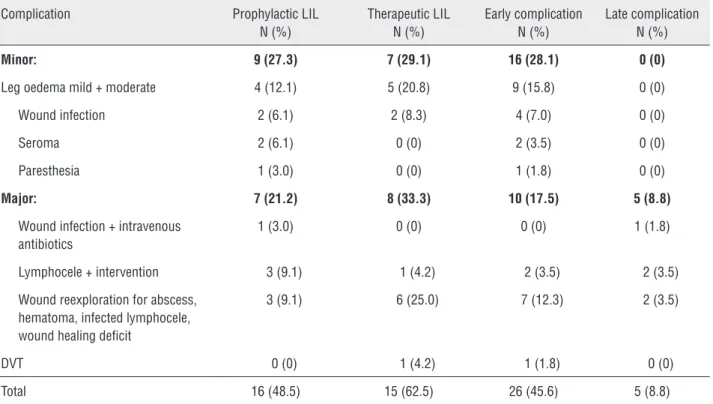
A total of 31 complications (54.4%) in-cluding 16 minor (28.1%) and 15 major (26.3%) were observed in 57 performed LILs. 9 patients (31.0%) experienced no postoperative complica-tions. Separate complication types and rates strat-ified to prophylactic and therapeutic LIL as well as early and late onset are described in Table-3 ac-cording to Bevan-Thomas (9). Classification of the complications according to Clavien (10) is depicted in Suppl. Table-1. Complication rates of prophy-lactic LIL (n=16, 48.5%) were decreased compared to therapeutic LIL (n=15, 62.5%) without reach-ing statistical significance (p˃0.05). There were no relevant intraoperative complications or mortal-ity associated with LIL. 4 patients (13.8%) who
received a therapeutic LIL and revealed LN me-tastases developed histologically proven inguinal lymphatic recurrence 3 to 9 months after LIL. Fol-low-up period of the study cohort was 49.5±42.6 months with 5 patients lost in the course of post-operative surveillance.
DISCUSSION
Penile cancer is a rare entity in Europe and North America with an incidence rate of less than 1 per 100,000 males (11). In many central Euro-pean countries, particularly in Germany, thera-peutic management of this cancer entity is not limited to high-volume referral centres but rather distributed to a large number of hospitals result-ing in relatively small case numbers per institu-tion. Strategies concerning the indication to iLAD as well as surgical technique vary significantly. Thus, different template extension and periopera-tive management on one hand and inconsistent methodology of complication definition, grading, reporting and way of data acquisition (prospec-tively/retrospectively) on the other (12) contribute to a great variability of iLAD complication rates reported in literature. We comprehensively ana-lyzed the outcome of our technique with horizon-tal skin incision, limited dissection field including thick skin flaps, preservation of fascia lata and great saphenous vein without transposition of the sartorius muscle and compared our results with those of other institutions.
Table 3 - 31 clinically relevant complications associated with 57 LILs. In brackets - complication rates. Complication rates of prophylactic and therapeutic LIL presented in relation to respectively performed LILs, early and late complication rates related to all LILs.
Complication Prophylactic LIL N (%)
Therapeutic LIL N (%)
Early complication N (%)
Late complication N (%)
Minor: 9 (27.3) 7 (29.1) 16 (28.1) 0 (0)
Leg oedema mild + moderate 4 (12.1) 5 (20.8) 9 (15.8) 0 (0)
Wound infection 2 (6.1) 2 (8.3) 4 (7.0) 0 (0)
Seroma 2 (6.1) 0 (0) 2 (3.5) 0 (0)
Paresthesia 1 (3.0) 0 (0) 1 (1.8) 0 (0)
Major: 7 (21.2) 8 (33.3) 10 (17.5) 5 (8.8)
Wound infection + intravenous antibiotics
1 (3.0) 0 (0) 0 (0) 1 (1.8)
Lymphocele + intervention 3 (9.1) 1 (4.2) 2 (3.5) 2 (3.5)
Wound reexploration for abscess, hematoma, infected lymphocele, wound healing deficit
3 (9.1) 6 (25.0) 7 (12.3) 2 (3.5)
DVT 0 (0) 1 (4.2) 1 (1.8) 0 (0)
Total 16 (48.5) 15 (62.5) 26 (45.6) 5 (8.8)
DVT = deep venous thrombosis.
Suppl. Table 1 - Type, rate per procedure (in brackets) and severity of 31 complications related to 57 LILs.
Complication Grade 1 N(%)
Grade 2 N(%)
Grade 3a (N%)
Grade 3b (N%)
Total (N%)
Wound infection 0 (0) 5 (8.8) 0 (0) 8 (14) 13 (22.8)
Leg oedema 9 (15.8) 0 (0) 0 (0) 0 (0) 9 (15.8)
Seroma/lymphocele 2 (3.5) 0 (0) 4 (7) 0 (0) 6 (10.5)
Hematoma 0 (0) 0 (0) 0 (0) 1 (1.8) 1 (1.8)
Paresthesia 1 (1.8) 0 (0) 0 (0) 0 (0) 1 (1.8)
DVT 0 (0) 1 (1.8) 0 (0) 0 (0) 1 (1.8)
Total 12 (21.1) 6 (10.5) 4 (7.0) 9 (15.8) 31 (54.4)
DVT = deep venous thrombosis.
morbidity rates. The most common cited compli-cations included wound infection (10-20%), lym-phocele/seroma (19-45%), particularly mutilating skin edge necrosis (14-65%), and lymphoedema (2-100%) (1). Johnson et al. (16) reported that
rate of 53% in 32 patients with RIL. A recently published large series from the Netherlands by Stuiver et al. (19) including 237 RILs reported 195 complications (82.3%). In contrast, Koifman et al. (20) observed in a large series of 170 patients with 340 RILs without muscle transposition an overall complication rate of only 10.3%. To our knowl-edge this is the currently lowest reported compli-cation rate for this technique. The high incidence of PeCa in Brazil with nearly 300 newly diagnosed patients per year (15), leading to the high number of patients within a 10 years study period and a consequently expanded expertise of the group in RIL surgical techniques as well as an optimized postoperative patient management contributed to these outstanding results. However, only a few centres experience such a high volume of patients with PeCa resulting conceivably in a higher perio-perative complication rate.
Hence, several modifications of this radical technique mainly aiming to reduce the dissection area have been proposed to alleviate immediate and long-term sequelae and to improve the quality of life, while maintaining the oncologic benefit of the procedure. The most commonly used technique is the modified inguinal lymphadenectomy (MIL) proposed by Catalona (21). This technique is mainly characterized by a shorter skin incision of 6-7 cm, reduced dissection field which is predominantly fo-cusing on deep inguinal nodes in fossa ovalis with omitting of the regions lateral to femoral artery and caudal to fossa ovalis, as well as maintenance of the saphenous vein and no transposition of sarto-rius muscle (1). In case of histologically proven me-tastases during this procedure, RIL is recommended to be performed (1). In a series with 6 patients with clinically negative LNs, Catalona reported one lym-phocele and mild lymphoedema in most cases (21). In a small cohort of 12 patients, Parra et al. (22) observed no major complications, skin flaps necro-sis or leg oedema. Similarly, no significant sequelae were indicated by Jacobellis et al. (23) in bilateral MILs of 8 patients with PeCa and 2 with penile leio-myosarcoma. In concert with these results, Bouchot et al. (4) describes only 12 minor complications in 118 MILs (10.2%).
Our approach with a limited extent of dis-section compared to the standard RIL was
con-ducted aiming the reduction of perioperative sequalae and simultaneously preserving the onco-logic safety. Particularly, the dissection of the great saphenous vein, fascia lata, wide exposure of the femoral vessels and consequent transposition of the sartorius muscle was avoided in order to limit the dissection of the deep lymphatic vessels and deterioration of lymph flow. With this technique, we experienced an overall complication rate of 54.4%, which ranges expectably between that of the most series with RIL and MIL as proposed by Catalona (21) Thereby, leg oedema represented the most prevalent morbidity (overall 15.8%). Surgical re-exploration was required in 9 patients (15.8%). The spectrum of complications was comparable to other publications on RIL, except that relevant skin edge necrosis was not observed in our cohort using a horizontal skin incision, most likely based on the fact that skin vascularisation is horizon-tal at the level of the subcutaneous adipose tissue (14), resulting in a high incidence of skin flap ne-crosis if a vertical dissection is used, interrupting the blood supply.
of the factors most strongly associated with the occurrence of moderate to severe wound compli-cations in a multivariate analysis in this study. The selection of the one vs. two step surgery still seems to be on the discretion of the surgeon. Obvi-ously, prospective randomized trials are required to comparatively assess both strategies in terms of general health cost, complication rates as well as if clinical variables as Charlson comorbidity index or BMI might assist individually the selection of the most appropriate approach. Further research is also needed to shed more light on the influence of the histopathologic result of the one-side surgery on extension of the contralateral dissection.
Local inguinal tumour recurrence is the main concern of surgical approaches with a lim-ited dissection field. Surprisingly, only a few studies reported about this critical issue (2). Thus, Lopes et al. (24) criticized Catalona´s procedure with avoiding the dissection of the LNs lateral to the femoral artery as unreliable due to local recurrence in 2 out of 13 patients and the fact that no patient presented metastases in the medial quadrant LNs. In concert with these results, 2 out of 18 patients experienced local recurrence within two years after MIL published by D´Ancona et al. (25). In contrast, Colberg et al. (26) observed no local recurrence in nine patients despite the his-tological finding of metastases in three of them. Nevertheless, omitting of the dissection laterally to the femoral artery as well as superior zones has to be critically reviewed. A recently published study by Leijte et al. on the penile lymphatic drainage (27) provided evidence for location of sentinel and higher-tier nodes in superior and central inguinal zones. Furthermore, the number of removed nodes should be taken into account as tenuous data on the groin lymphatic anatomy suggest the presence of 10-15 superficial and 0-5 deep inguinal nodes (20, 28, 29).
The average number of removed LNs per side in our study was 8.1, which is slightly lower compared to most recent studies (19, 20) using RIL with 9 (median) and 10.9 (mean) nodes, respective-ly. Unfortunately, 4 patients with therapeutic LIL in our study experienced local inguinal recurrence within 9 months after surgery despite our approach with dissection of all five anatomical sectors of the
inguinal LNs as described by Daseler et al. (30). Lo-cal recurrence might be based on insufficient dis-section of the deep lymphatics on the femoral ves-sels due to their reduced exposure and preservation of fascia lata using our approach of LIL.
Recently, video-endoscopy (31) with fur-ther advancement of a single-port access (32) and even robotic-assisted techniques (33) have been proposed for iLAD aiming to further decrease peri-operative sequalae. These series yielded promising preliminary results. However, until a definitive as-sessment of the reliability and oncologic safety of these approaches will be possible after presenta-tion of studies with larger sample size and longer follow-up in the future, open iLAD still remains a state-of-the-art procedure if surgical approach is indicated. Also, the role of DSNB, currently ap-plied only in a few centres worldwide, should be further elucidated.
The current study is limited by its restricted sample size and retrospective nature, which might have contributed to underestimated complication rates (12). Taking into account a low incidence of PeCa in Europe, cooperative research in this area might be crucial to achieve more robust evidence with higher patient numbers and a shorter recruit-ment period. Nevertheless, we believe that our re-sults reflect the “real life” in central Europe, where procedures are performed out of high-volume re-ferral centres and in regions with a low incidence of the disease.
In conclusion, our technique of a limited inguinal dissection provided an acceptable com-plication rate without aggravating morbidity. We experienced no recurrences in clinically negative patients, so that LIL might be a reasonable option for this cohort and incorporation of frozen sec-tion analysis into this approach with extending the dissection field to radical template in case of positivity might further reduce the risk of local re-currence. In patients with clinically enlarged LNs, more extended resection is required and RIL still is the gold standard.
CONFLICT OF INTEREST
REFERENCES
1. Horenblas S. Lymphadenectomy for squamous cell carcinoma of the penis. Part 2: the role and technique of lymph node dissection. BJU Int. 2001;88:473-83.
2. Protzel C, Alcaraz A, Horenblas S, Pizzocaro G, Zlotta A, Hakenberg OW. Lymphadenectomy in the surgical management of penile cancer. Eur Urol. 2009;55:1075-88. 3. Naumann CM, Filippow N, Seif C, van der Horst C, Roelver L,
Braun PM, et al. Penile carcinoma (pT1 G2): surveillance or inguinal lymph node dissection? Onkologie. 2005;28:135-8. 4. Bouchot O, Rigaud J, Maillet F, Hetet JF, Karam G. Morbidity
of inguinal lymphadenectomy for invasive penile carcinoma. Eur Urol. 2004;45:7615; discussion 765-6.
5. Kroon BK, Horenblas S, Lont AP, Tanis PJ, Gallee MP, Nieweg OE. Patients with penile carcinoma benefit from immediate resection of clinically occult lymph node metastases. J Urol. 2005;173:816-9.
6. Leijte JA, Kirrander P, Antonini N, Windahl T, Horenblas S. Recurrence patterns of squamous cell carcinoma of the penis: recommendations for follow-up based on a two-centre analysis of 700 patients. Eur Urol. 2008;54:161-8. 7. Leijte JA, Kroon BK, Valdés Olmos RA, Nieweg OE, Horenblas
S. Reliability and safety of current dynamic sentinel node biopsy for penile carcinoma. Eur Urol. 2007;52:170-7. 8. Charlson ME, Pompei P, Ales KL, MacKenzie CR. A new
method of classifying prognostic comorbidity in longitudinal studies: development and validation. J Chronic Dis. 1987;40:373-83.
9. Bevan-Thomas R, Slaton JW, Pettaway CA. Contemporary morbidity from lymphadenectomy for penile squamous cell carcinoma: the M.D. Anderson Cancer Center Experience. J Urol. 2002;167:1638-42.
10. Dindo D, Demartines N, Clavien PA. Classification of surgical complications: a new proposal with evaluation in a cohort of 6336 patients and results of a survey. Ann Surg. 2004;240:205-13.
11. Barnholtz-Sloan JS, Maldonado JL, Pow-sang J, Giuliano AR. Incidence trends in primary malignant penile cancer. Urol Oncol. 2007;25:361-7. Erratum in: Urol Oncol. 2008;26:112. Guiliano, Anna R [corrected to Giuliano, Anna R].
12. Campbell PG, Malone J, Yadla S, Chitale R, Nasser R, Maltenfort MG, et al. Comparison of ICD-9-based, retrospective, and prospective assessments of perioperative complications: assessment of accuracy in reporting. J Neurosurg Spine. 2011;14:16-22.
13. Daseler EH. Radical excision of the inguinal and iliac lymph glands. Univ Hosp Bull. 1949;15:70-4.
14. Baronofsky ID. Technique of inguinal node dissection. Surgery. 1948;24:555-67.
15. Favorito LA, Nardi AC, Ronalsa M, Zequi SC, Sampaio FJ, Glina S. Epidemiologic study on penile cancer in Brazil. Int Braz J Urol. 2008;34:587-91;discussion 591-3.
16. Johnson DE, Lo RK. Complications of groin dissection in penile cancer. Experience with 101 lymphadenectomies. Urology. 1984;24:312-4.
17. Kamat MR, Kulkarni JN, Tongaonkar HB. Carcinoma of the penis: the Indian experience. J Surg Oncol. 1993;52:50-5. 18. Horenblas S, van Tinteren H, Delemarre JF, Moonen LM,
Lustig V, van Waardenburg EW. Squamous cell carcinoma of the penis. III. Treatment of regional lymph nodes. J Urol. 1993;149:492-7.
19. Stuiver MM, Djajadiningrat RS, Graafland NM, Vincent AD, Lucas C, Horenblas S. Early wound complications after inguinal lymphadenectomy in penile cancer: a historical cohort study and risk-factor analysis. Eur Urol. 2013;64:486-92. 20. Koifman L, Hampl D, Koifman N, Vides AJ, Ornellas
AA. Radical open inguinal lymphadenectomy for penile carcinoma: surgical technique, early complications and late outcomes. J Urol. 2013;190:2086-92.
21. Catalona WJ. Modified inguinal lymphadenectomy for carcinoma of the penis with preservation of saphenous veins: technique and preliminary results. J Urol. 1988;140:306-10. 22. Parra RO. Accurate staging of carcinoma of the penis in men
with nonpalpable inguinal lymph nodes by modified inguinal lymphadenectomy. J Urol. 1996;155:560-3.
23. Jacobellis U. Modified radical inguinal lymphadenectomy for carcinoma of the penis: technique and results. J Urol. 2003;169:1349-52.
24. Lopes A, Rossi BM, Fonseca FP, Morini S. Unreliability of modified inguinal lymphadenectomy for clinical staging of penile carcinoma. Cancer. 1996;15;77:2099-102.
25. d’Ancona CA, de Lucena RG, Querne FA, Martins MH, Denardi F, Netto NR Jr. Long-term followup of penile carcinoma treated with penectomy and bilateral modified inguinal lymphadenectomy. J Urol. 2004;172:498-501; discussion 501.
26. Colberg JW, Andriole GL, Catalona WJ. Long-term follow-up of men undergoing modified inguinal lymphadenectomy for carcinoma of the penis. Br J Urol. 1997;79:54-7.
27. Leijte JA, Valdés Olmos RA, Nieweg OE, Horenblas S. Anatomical mapping of lymphatic drainage in penile carcinoma with SPECT-CT: implications for the extent of inguinal lymph node dissection. Eur Urol. 2008;54:885-90. 28. Hudson CN, Shulver H, Lowe DC. The surgery of
‘inguino-femoral’ lymph nodes: is it adequate or excessive? Int J Gynecol Cancer. 2004;14:841-5.
29. Spratt J. Groin dissection. J Surg Oncol. 2000;73:243-62. 30. Daseler EH, Anson BJ, Reimann AF. Radical excision of
31. Tobias-Machado M, Tavares A, Ornellas AA, Molina WR Jr, Juliano RV, Wroclawski ER. Video endoscopic inguinal lymphadenectomy: a new minimally invasive procedure for radical management of inguinal nodes in patients with penile squamous cell carcinoma. J Urol. 2007;177:953-7; discussion 958.
32. Tobias-Machado M, Correa WF, Reis LO, Starling ES, de Castro Neves O, Juliano RV, et al. Single-site video endoscopic inguinal lymphadenectomy: initial report. J Endourol. 2011;25:607-10.
33. Matin SF, Cormier JN, Ward JF, Pisters LL, Wood CG, Dinney CP, et al. Phase 1 prospective evaluation of the oncological adequacy of robotic assisted video-endoscopic inguinal lymphadenectomy in patients with penile carcinoma. BJU Int. 2013;111:1068-74.