Antilithiatic Activity of phlorotannin rich extract of
Sarghassum Wightii on Calcium Oxalate Urolithiais – In
Vitro and In Vivo Evaluation
_______________________________________________
D. Sujatha
1, Kiranpal Singh
1, Mursalin Vohra
1, K. Vijay Kumar
1, S. Sunitha
11 Department of Pharmacology and Toxicology, National Institute of Pharmaceutical Education and
Research, Hyderabad, Andhrapradesh, India
ABSTRACT
ARTICLE
INFO
______________________________________________________________ ______________________
Purpose: Urolithiasis is a common urological disorder responsible for serious human affliction and cost to the society with a high recurrence rate. The aim of the present study was to systematically evaluate the phlorotannin rich extract of Sargassum wi-ghtii using suitable in vitro and in vivo models to provide scientific evidence for its antilithiatic activity.
Materials and Methods: To explore the effect of Sargassum wightii on calcium oxalate crystallization, in vitro assays like crystal nucleation, aggregation and crystal growth were performed. Calcium oxalate urolithiasis was induced in male Sprague dawley rats using a combination of gentamicin and calculi producing diet (5% ammonium oxalate and rat pellet feed). The biochemical parameters like calcium, oxalate, magnesium, phosphate, sodium and potassium were evaluated in urine, serum and kidney homo-genates. Histopathological studies were also done to confirm the biochemical findings. Results: The yield of Sargassum wightii extract was found to be 74.5 gm/kg and con-firmed by quantitative analysis. In vitro experiments with Sargassum wightii showed concentration dependent inhibition of calcium oxalate nucleation, aggregation and growth supported by SEM analysis. In the in vivo model, Sargassum wightii reduced both calcium and oxalate supersaturation in urine, serum and deposition in the kidney. The biochemical results were supported by histopathological studies.
Conclusion: The findings of the present study suggest that Sargassum wightii has the ability to prevent nucleation, aggregation and growth of calcium oxalate crystals. Sargassum wightii has better preventive effect on calcium oxalate stone formation indicating its strong potential to develop as a therapeutic option to prevent recur-rence of urolithiasis.
Key words:
Calcium Oxalate; Calculi; Phaeophyta
Int Braz J Urol. 2015; 41: 511-20
_____________________
Submitted for publication: July 18, 2014
_____________________
Accepted after revision: October 18, 2014
INTRODUCTION
The risk of developing urolithiasis in adults appears to be higher in the western hemisphere (5-9% in Europe, 12% in Canada, and 13-15% in the USA) (1), than in the eastern hemisphere (1-5%), although the highest risks have been reported in
some Asian countries such as Saudi Arabia (20.1%) with lifetime recurrence rates of up to 50% (2).
long accepted simple explanation of formation of calcium oxalate (CaOx) stones is supersaturation (3). Deviating from the hypothesis new insights suggest a primary plaque formation in the inters-titial space of the renal papilla. This plaque acts as support commonly referred as nidus made of ei-ther calcium phosphate crystals or organic matrix derived from damaged membranes. This membra-ne damage is mainly because of various apoptotic pathways as ROS, oxidative stress, altered pH and co-morbid conditions damaging the kidney.
Hence, it has been hypothesized that com-pounds that either alter supersaturation or prevent membrane damage can be a useful aid to treat and prevent recurrence of urolithiasis. Phlorotannins, important secondary metabolites obtained from many marine sources are structurally similar to tannins, can easily complex these divalent ions and accordingly reduce supersaturation of urine. On the other hand, phlorotannins are claimed and proved to be good antioxidants from earlier re-ports (4). Hence, they may also reduce the renal tubular damage and prevent stone formation.
Sargassum wightii is an important marine species (Family: Phaeophyceae), widely distribu-ted in tropical and temperate oceans. S. wightii shows presence of good amount of flavonoids, alkaloids, phenolics, phlorotannins and steroids with various pharmacological activities like anti-bacterial and antioxidant activity (5-8). Still many pharmaceutical and therapeutical applications of S. wightii are untapped. Hence, the present stu-dy has been initiated with an objective to obtain phlorotannin rich extract of S. wightii (PTSW) and to evaluate whether PTSW has any preventive or curative affect against calcium oxalate stones using suitable in vitro crystallization methods and animal model.
MATERIALS AND METHODS
Collection of S. Wightii and Extraction
The brown algae S. wightii was collected in November from sea shore of MANDAPAM re-gion Rameshwaram coast. The brown algae was authenticated by Dr. B. Seetharam, Professor, Sri Venkateswara Ayurvedic Medical College Tirupa-thi, Andhra Pradesh, India and a voucher
speci-men (M-001) was deposited in the departspeci-ment of pharmacology and toxicology of National Insti-tute of Pharmaceutical Education and Research, Hyderabad, India.
Air dried S.wightii was extracted to obtain phlorotannin rich extract as explained by Young et al. with some modifications (9). Briefly, air dried S.wightii was kept for maceration at room tem-perature with 70% methanol (v/v) for 24 hrs un-der nitrogen environment. Methanolic extract was then collected by using rota evaporator (Rotavac, Heidolf, Germany) at 40ºC and fractioned thrice with distilled water and n-hexane for 24 hr (1:1). All the aqueous portions were pooled and acetyla-ted with ethyl acetate in pyridine environment. The acetylated aqueous extract was then dialyzed against distilled water using dialyzing membrane (3000 kd cutoff). The obtained phlorotannin rich S.wightii extract (PTSW) was collected and stored at 2-8ºC.
Quantification of PTSW
For qualitative estimation of phlorotan-nins, TLC was carried out on 10×20 cm silica gel plate as per the procedure of Jeeva et al. (10). The chloroform and methanol (9:1) served as mobi-le phase. Folin-Ciocalteu reagent was used as spraying agent to detect the phenolic compoun-ds. Quantification of phlorotannins in PTSW was done according to modified Folin-Ciocalteu me-thod, using phloroglucinol as standard (11). Total phlorotannin content was expressed as gram equi-valents of phloroglucinol.
In vitro crystallization methods
Antilithiatic activity of S. wightii Animals
Male Sprague Dawley (SD) rats (150-200g,) were obtained from Teena laboratories and housed under conditions of optimum light, temperature and humidity (12 h light–dark cycle, 22±2˚C and relative humidity of 45 to 55%), with food and water provided ad libitum. The animal experimen-tal protocols were approved by the Institutional Animal Ethics Committee (IAEC No: NIP/10/2013/ PC/66). Acute toxicity study for S. wightii (PTSW) was performed as per OECD guideline no 425 to determine the dose for antilithiatic study.
Experimental design
Hyperoxaluria and calcium oxalate depo-sition was induced using gentamicin and calculi producing diet (CPD) (15). The standard rat pellet feed was powdered and mixed with ammonium oxalate (5%), then made into pellets used as CPD.
Male SD rats were randomly grouped in to 7 groups (n=8). The group Normal (N) recei-ved only vehicle (distilled water, 2 mL/kg/p.o.). The remaining six groups received gentamicin (40 mg/kg/s.c., 1-8th day) and CPD from 5-15th day. The preventive-control (PC) and curative-control (CC) groups received only vehicle (distilled water, 2 mL/kg/orally) from day 1 to 15 and day 16 to 30 respectively. The preventive groups (P1 and P2) received the PTSW (200 and 400 mg/kg/p.o. respectively) from day 1 to 15, whereas curative groups (C1 and C2) received the PTSW (200 and 400 mg/kg/p.o. respectively) from day 16 to 30. As the extract was administered from 1st day, P1 and P2 assess ability of PTSW to prevent stone forma-tion for which the PC group serves as control. The purpose of groups C1 and C2 was to assess the ability of the extract to treat already developed renal calculi for which CC group serves as control.
Parameters monitored
Biochemical parameters were measured for preventive groups on the day 15 and for cura-tive groups on the day 30. At regular intervals, as mentioned above the animals were individually housed in metabolic cages for collection of urine samples for 24 hr. The urine volume was measured and then centrifuged at 3000 rpm for 5 min to
collect the supernatant for estimation of urinary parameters like sodium, potassium, calcium, oxa-late, magnesium and phosphate.
Blood was collected from the retro-orbi-tal region at the same time points and allowed to clot at room temperature and serum was separa-ted by centrifugation at 3000 rpm for 5 minutes. The serum was used to analyze the serum level of sodium, potassium, calcium, oxalate, magnesium and phosphate.
Animals were sacrificed at the end of the experimental period day 15 (PC, P1 and P2) and day 30 (CC, C1 and C2) by decapitation. The kidneys were carefully excised, weighed and one kidney of each animal was put in 10% formalin for histolo-gical studies. The other kidney was sliced into two equal halves. One half was homogenized in 10% HCl and another in 10 % Tris buffer. The superna-tant from HCl homogenate was used for estimation of calcium and oxalate and Tris buffer homogenate for estimation of magnesium and phosphate. All the biochemical parameters mentioned were measured using commercial analytical kits (Accurex Pvt Ltd) except oxalate. The urine, serum and kidney levels of oxalate were estimated manually using procedu-re of Hodgkinson and Williams (16).
Histopathological evaluation
For microscopic evaluation, kidneys were fixed in 10% formalin. Tissue sections of 5µm thi-ckness were stained with hematoxylin-eosin. A minimum of 10 fields for each kidney slide were examined for necrosis, calcium oxalate crystals deposits, stone size, membrane damage and other histopathological parameters. Tissue slices were photographed using research microscope (Nikon Eclipse Ti-U, USA) at 10X magnification.
Statistical analysis
RESULTS
PTSW yield and quantification
The yield of methanolic extract of S. wi-ghtii was found to be 74.5 gm/kg of dry material. The yield of phlorotannins rich extract (PTSW) was found to be 20g / 100 g of methanolic ex-tract of S. wightii. Total phlorotannin content of PTSW was determined from the calibration cur-ves of phloroglucinol (y=0.002x-0.022, R²=0.989) by Folin-Ciocalteu method. The total phlorotan-nin content of PTSW was found to be 74.5µg/mg phloroglucinol equivalents.
Nucleation assay
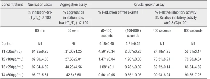
The simultaneous incubation along with the different concentrations of PTSW significantly re-duced the nucleation of calcium oxalate at 30 and 60 min significantly (p<0.05), when compared to control setup. The % inhibition of nucleation, by PTSW was found to be dose dependent (Table-1).
Crystal aggregation assay
The simultaneous incubation of COM seed along with different concentrations of PTSW (t1 -50µg, t2-100µg, t3-250µg and t4-500µg) had shown
a significant reduction in aggregation of COM crys-tals. Results showed a significant inhibitory effect of PTSW in all the different concentration when compared to the control COM slurry (p<0.05), but the effect was dose independent. The % aggrega-tion inhibiaggrega-tion rate was calculated and found to be dose independent effect as shown in Table-1.
Crystal growth assay
A significant deposition of 6.18% of oxa-late was observed at 400 s, whereas an additional 5.71 % was deposited by 800s indicating signifi-cant growth of COM crystal in control samples. When COM crystals were incubated with different concentrations of PTSW, it was observed that the oxalate deposition was significantly reduced in dose and time dependent manner as shown in Ta-ble-1.The SEM analysis of calcium oxalate growth assay at 0 and 60 min showed the reduction in size and the shape of COM crystals in the incu-bation mixtures having different concentrations of PTSW when compared with control (Figure-1), which clearly indicate ability of PTSW to redu-ce crystal growth supporting the results of crystal growth assay.
Table 1 - Effect of PTSW on in vitro crystal nucleation, aggregation and growth assay.
Concentrations Nucleation assay Aggregation assay Crystal growth assay
% inhibition={(1-(TSi/TSc)} X 100
% aggregation inhibition rate, Ir=(1-TSi/TSc) X 100
% Reduction of free oxalate % Relative inhibitory activity {% Relative inhibitory activity
=((C-S)/C)×100}
60 min 60 ⇒ in (0–400)
seconds
(400-800 ) seconds
400 seconds 800 seconds
Control Nil Nil 6.18±0.45 5.71±0.32 Nil Nil
T1 (50µg/mL) 91.95±6.25 31.65±1.25 4.50*±0.34 2.38*±0.23 27.18±1.25 58.31±3.14
T2 (100µg/mL) 92.90±4.56 27.66±2.01 1.47*±0.04 1.20*±0.06 76.21±8.21 78.98±6.54
T3 (250µg/mL) 97.04±6.89 48.28±4.56 1.08*±0.1 0.78*±0.09 82.52±9.14 86.34±4.89
T4 (500µg/mL) 98.97±5.61 42.6±3.58 0.56*±0.05 0.55*±0.05 90.93±6.24 90.36±7.28
All values were mean ± SD of 3 observations. Where; *=p<0.05, when compared with control and
TSi = Turbidity slope in the presence of the test sample;
TSc = Turbidity slope of the control;
C= Rate of reduction of free oxalate without test sample;
Acute toxicity study
PTSW was found to be safe up to 2 g/kg, p.o. as no mortality was observed in 48 hrs follo-wed by 14 days observation except increase in urination in the fi rst 6 hrs. The dose for pharma-cological evaluation was fi xed at 200 and 400 mg/ kg p.o as per standard guidelines.
Effect of PTSW on biochemical parameters With administration of CPD and gentami-cin, signifi cant (p<0.05) increase in urinary excre-tion of calcium and oxalate was observed in PC and CC groups when compared with normal group (N) confi rming that CPD and gentamicin treatment has caused supersaturation of urine with calcium and oxalate. After treatment with PTSW a signifi -cant (p<0.05) decrease in calcium and oxalate was observed in P1, P2, C1 and C2 groups when com-pared with their respective control groups, indica-ting the ability of PTSW to reduce super saturation of urine caused by CPD and gentamicin adminis-tration. PC and CC has not shown any signifi cant increase in magnesium and phosphorus excretion when compared with normal animals (N), but with PTSW treatment a signifi cant (p<0.05) decrease in urinary excretion of magnesium and phosphorus was observed in P1, P2, C1 and C2 groups when
compared with their respective control groups. Similar signifi cant (p<0.05) increase was observed in serum concentrations of calcium, oxalate and magnesium, in PC and CC, when compared with normal group (N). Whereas on treatment with PTSW for 15 days, a signifi cant (p<0.05) decrease in serum concentrations of cal-cium and oxalate alone was found P1, P2, C1 and C2 groups when compared with their respective control groups, without any effect on magnesium levels (Table-2).
While on treatment with gentamicin and CPD for 15 days, a signifi cant (p<0.05) deposition of calcium, oxalate, magnesium and phosphorous in kidney was observed in PC and CC groups when compared to the normal group (N). Administration of PTSW in two different doses for 15 days, signi-fi cantly (p<0.05) decreased deposition of calcium, oxalate and magnesium in P1, P2, C1 and C2 groups when compared to their respective control groups. A signifi cant decrease in phosphate depo-sition was also observed but only with higher con-centration of PTSW i.e., only in P2 and C2 groups as shown in Table-2.
Though urinary, serum and kidney deposi-tion of sodium and potassium were also measured, they were not documented as no signifi cant chan-Figure 1 - SEM Images of Crystal growth assay taken at 0 and 60 min.
a, b = Control (0 and 60 min respectively); c, d = t1-50µg/mL (0 and 60 min respectively); e, f = t2- 100µg/mL (0 and 60 min respectively);g, h = t3- 250 µg/mL (0 and 60
IBJU
|
ANTILITHIA
TIC A
CTIVITY OF SARGHASSUM WIGHTII
51
6
Calcium (m Mol/L)
Oxalate (µg/dL)
Magnesium (m Mol/L)
Phosphorus (m Mol/L)
Calcium (m Mol/L)
Oxalate (µg/dL)
Magnesium (m Mol/L)
Calcium (mg/g wet
tissue)
Oxalate (mg/g wet
tissue)
Magnesium (mg/g wet
tissue)
Phosphorous (mg/g wet tissue)
N 0.618±0.15 80.84±11.4 4.52±0.78 18.33±2.51 14.17±1.2 8.8±4.4 0.23±0.03 1.59±0.37 0.98±0.03 11.21±1.0 2.26 ± 0.76
PC 10.6±6.11a 105.8±12.2a 3.92±0.98ns 14.27±3.26ns 23.31±0.5a 23.18±2.0a 2.30±0.6a 5.85±0.6a 6.43±0.7a 28.99±9.01a 8.88 ± 2.7a
P1 3.76±0.79b 24.2±5.2b 1.20±0.51b 6.5±2.4b 14.77±1.2b 13.76±1.6b 1.97±0.4ns 2.59±1.2b 2.52±0.9b 12.16±6.2b 6.77 ± 2.6ns
P2 3.32±0.91b 32.2±4.9b 2.03±0.49b 3.27±0.9b 14.55±0.71b 15.36±3.6b 2.01±0.5ns 2.17±0.9b 1.46±0.2b 14.8±3.2b 3.83 ± 1.1b
CC 16.01±3.5a 155.67±9.3a 5.86±2.2ns 15.4±2.04ns 29.48±0.7a 24.67±1.8a 2.15±0.42a 5.05±0.5a 6.04±0.7 21.61±5.2a 9.05 ±0.7a
C1 6.24±1.77b 35.2±8.5b 5.65±0.41ns 2.5±0.76b 10.15±0.6ns 17.17±6.4b 2.08±0.22ns 1.3±0.3b 1.27±0.27b 11.79±1.1b 8.04 ±0.3ns
C2 4.79±2.28b 28.4±2.3 5.29±1.01ns 6.50±1.08b 10.65±0.8ns 12.88±3.7b 2.06±0.45ns 1.3±0.8b 1.12±0.07b 15.43±4.1b 2.85 ±0.3b
ges were observed between the groups and serum levels of phosphorous have also not been included because of their non significant changes.
Histopathology
The sections photographed highlights the junction of medulla and cortex region in kid-ney as both in humans and rats CaOx deposition mainly occurs in this region. Gentamicin and CPD administration has caused severe glomerular da-mage, tubular dilation, membrane damage and deposition of CaOx crystals as highlighted in the photomicrographs of PC and CC.
Treatment with PTSW at both the doses clearly ameliorated the nephrotoxic damage
cau-sed by gentamicin and CaOx deposition by CPD as noted in the sections of P1, P2, C1 and C2.These results not only support the biochemical estima-tions but also testify that PTSW has antilithiatic property (Figure-2).
DISCUSSION
In clinical scenario, irrespective of the che-mical nature of kidney stones they are treated by various surgical procedures like extracorporeal shock wave lithotripsy, percutaneous nephrolitho-tomy etc.,. Beyond doubt the surgical procedures cause immediate relief from the kidney stones but cannot overcome their recurrence, a very common
Figure 2 - Photomicrographs of kidney sections at 100 X magnification where N,PC,P1,P2,CC,C1 and C2 corresponds to groups.
problem associated with kidney stones and consi-dered unmet clinical need in the area of urology.
Phlorotannins, important secondary meta-bolites are obtained from many marine sources. These being structurally similar to tannins can ea-sily complex the divalent ions and might reduce supersaturation of urine (17).On the other hand, phlorotannins like tannins/polyphenols obtained from plant sources are claimed and proved to be good antioxidants from earlier reports (18). Hence, the present study focuses on the systematic evalu-ation of PTSW for its benefit as antilithiatic agent. Extraction of phlorotannins is not always easy and uniform to isolate phlorotannins due to the susceptible chemical structure of phlorotan-nins with interphloroglucinol linkages which can be oxidized easily (19). Hence, ascorbic acid and acetylation of phlorotannins was used during the extraction procedure which might be responsible for increase in the yield of phlorotannins.
The rationale behind the selection of CaCl2 and NaOx solution for in vitro studies is their swift solubility and they rapidly complex to form CaOx crystals uniformly, which can be used as probes for in vitro studies as mentioned in many former reports (17). Results of nucleation assay indica-te that PTSW complexes the free Ca and Ox ions available in the incubation mixture, thus causing dose dependent inhibition of nucleation. After nucleation, agglomeration of particles is a critical step in urinary stone formation, as larger crystals are less likely to pass spontaneously in the urinary tract. If the extract keeps CaOx particles dispersed in solution for longer time they are more easily eliminated. Thus, from aggregation assay results it can be affirmed that phlorotannins can main-tain divalent ions in dispersed condition for lon-ger durations such that they can be eliminated out through urine.
Antilithiatic substances can also prevent the growth of COM by inhibiting oxalate depo-sition on the COM crystals surface present in in-cubation mixture (20). PTSW has shown similar dose dependent ability to inhibit the growth of CaOx crystals. The growth assay was followed by SEM analysis of crystals which clearly testified ability of PTSW to reduce crystal growth. PTSW incubated samples showed comparatively less
number and smaller size smooth surfaced crys-tals indicating PTSW ability of converting COM to CaOx dehydrate crystals, which may adhere less to renal membranes and can be easily excre-ted along with urine.
In the in vivo studies, from the results of urinary biochemical parameters, we can explain that PTSW clearly reduces supersaturation of urine with divalent cations with much specific effect on calcium and oxalate. Sodium and po-tassium excretion were not altered, but those ions are neither induced nor regulated by gentamicin and CPD diet, which was confirmed once again by our studies. Moreover, in urolithiasis oxalic salts are soluble when formed with magnesium but when complexes with calcium forms inso-luble calcium oxalate thus causing crystalline precipitation of renal calculi of calcium oxalate type (21). Accordingly, hyperoxaluria is far more significant risk factor in the pathogenesis of re-nal stones than hypercalciuria and changes in urinary oxalate levels are relatively much more important than those of magnesium, phosphorus, sodium and potassium (22). As, PTSW lowered the levels of both oxalate and calcium in urine it can be suggested that PTSW reduces both hype-roxaluria and hypercalciuria.
After serum studies, it was clear that super-saturation of urine with certain urinary salts such as calcium and oxalate was comparative with their serum levels. CPD and gentamicin clearly increased serum levels of calcium and oxalate which can be attributed to the enzymatic disturbances caused by gentamicin and increased absorption of both cal-cium and oxalate by feeding with CPD as mentio-ned earlier (23). PTSW ability to reduce hyperoxa-luria and hypercalciuria can thus also be attributed to its ability to reduce absorption of calcium and oxalate from dietary sources and also might recti-fy the enzymatic disturbances leading to idiopathic hyperoxaluria, evident by decreased serum levels of calcium and oxalate. Further mechanistic studies are warranted to explain the exact effect of PTSW on absorption of divalent ions.
But PTSW at both the doses clearly dimini-shed the deposition of calcium and oxalate in kid-ney. These results were further supported by his-tological findings, where preventive control and curative control groups showed marked glomeru-lar damage, widened gaps in tubuglomeru-lar duct, depo-sition of honey colored CaOx crystals, inflamma-tory damage and collecting duct dilatation. The different concentration of PTSW has shown the marked reduction in glomerular damage, mem-brane damage and infiltration of inflammatory cells. CaOx deposition was significantly reduced as shown in both preventive treatment groups and curative treatment groups. These findings support the in vitro results and biochemical changes cau-sed by PTSW, suggesting the antilithiatic activity of PTSW.
CONCLUSIONS
The findings of the present study highlight the ability of PTSW to prevent nucleation, aggre-gation and growth of calcium oxalate crystals as proved in in vitro studies. PTSW has also shown re-markable decrease in supersaturation of urine and serum with calcium, oxalate and magnesium. All these conditions put together will create an envi-ronment to prevent formation of kidney stone ra-ther than dissolving them. Though PTSW has sho-wn both the properties, i.e.; ability to prevent stone formation and dissolved already formed stones, the preventive effect was more predominant. This stu-dy encourages isolation of active constituents of PTSW responsible for antilithiatic activity.
CONFLICT OF INTEREST
None declared.
REFERENCES
1. Soucie JM, Thun MJ, Coates RJ, McClellan W, Austin H. Demographic and geographic variability of kidney stones in the United States. Kidney Int.1994;46:893-9.
2. Hughes P; Caring for Australians with Renal Impairment (CARI). The CARI guidelines. Kidney stones epidemiology. Nephrology (Carlton). 2007;12:S26-30.
3. Khan A, Bashir S, Khan SR, Gilani AH. Antiurolithic activity of Origanum vulgare is mediated through multiple pathways. BMC Complement Altern Med. 2011;11:96.
4. Pareta S K, Patra KC, Harwansh R: In-vitro calcium oxalate crystallization inhibition by Achyranthes indica linn. Hydroalcoholic extract: An approach to antilithiasis. International Journal of Pharma & Bio Sciences. 2011;2:432-7.
5. Josephine A, Nithya K, Amudha G, Veena CK, Preetha SP, Varalakshmi P. Role of sulphated polysaccharides from Sargassum Wightii in Cyclosporine A-induced oxidative liver injury in rats. BMC Pharmacol. 2008;8:4.
6. Vallinayagam K, Arumugam R, Kannan R, Thirumaran G, Anantharaman P: Antibacterial activity of some selected seaweeds from pudumadam coastal regions. Global J Pharmacol. 2009;3:50-2.
7. Syad AN, Shunmugiah KP, Kasi PD. Antioxidant and anti-cholinesterase activity of Sargassum wightii. Pharm Biol. 2013;51:1401-10.
8. Jang KH, Lee BH, Choi BW, Lee HS, Shin J. Chromenes from the brown alga Sargassum siliquastrum. J Nat Prod. 2005;68:716-23.
9. Young MH, Jong SB, Jin WH, Nam HL. Isolation of a New Phlorotannin, Fucodiphlorethol G, from a Brown Alga Ecklonia cava. Bull. Korean Chem. Soc. 2007;28:1595-7. 10. Jeeva S, Antonisamy JM, Domettila C, Anantham B, Mahesh
M. Preliminary phytochemical studies on some selected seaweeds from Gulf of Mannar, India. Asian Pacific Journal of Tropical Biomedicine. 2012;2:S30-3.
11. Waterman PG, Mole S: Analysis of Phenolic Plant Metabolites. Blackwell Scientific Publications, Oxford. 1994;pp.85. 12. Hennequin C, Lalanne V, Daudon M, Lacour B, Drueke T.
A new approach to studying inhibitors of calcium oxalate crystal growth. Urol Res. 1993;21:101-8.
13. Atmani F, Khan SR. Role of urinary bikunin in the inhibition of calcium oxalate crystallization. J Am Soc Nephrol. 1999;1:S385-8.
14. Nakagawa Y, Abram V, Parks JH, Lau HS, Kawooya JK, Coe FL. Urine glycoprotein crystal growth inhibitors. Evidence for a molecular abnormality in calcium oxalate nephrolithiasis. J Clin Invest. 1985;76:1455-62.
15. Kumar S, Sigmon D, Miller T, Carpenter B, Khan S, Malhotra R, et al. A new model of nephrolithiasis involving tubular dysfunction/injury. J Urol. 1991;146:1384-9.
16. Hodgkinson A, Williams A. An improved colorimetric procedure for urine oxalate. Clin Chim Acta. 1972;36:127-32.
18. Li Y, Qian ZJ, Ryu B, Lee SH, Kim MM, Kim SK. Chemical components and its antioxidant properties in vitro: an edible marine brown alga, Ecklonia cava. Bioorg Med Chem. 2009;17: 1963-73.
19. Peng S, Jay-Allemand C. Use of antioxidants in extraction of tannins from walnut plants. J Chem Ecol. 1991;17:887-96. 20. Tayal S, Duggal S, Bandyopadhyay P, Aggarwal A, Tandon
S, Tandon C. Cytoprotective role of the aqueous extract of Terminalia chebula on renal epithelial cells. Int Braz J Urol. 2012;38:204-13; discussion 213-4.
21. Pak CY, Adams-Huet B, Poindexter JR, Pearle MS, Peterson RD, Moe OW. Rapid Communication: relative effect of urinary calcium and oxalate on saturation of calcium oxalate. Kidney Int. 2004;66:2032-7.
22. Robertson WG, Hughes H. Importance of mild hyperoxaluria in the pathogenesis of urolithiasis--new evidence from studies in the Arabian peninsula. Scanning Microsc. 1993;7:391-401.
23. Jonassen JA, Cao LC, Honeyman T, Scheid CR. Mechanisms mediating oxalate-induced alterations in renal cell functions. Crit Rev Eukaryot Gene Expr. 2003; 13:55-72.
_______________________ Correspondence address:
D. Sujatha, MD Assistant Professor, Department of Pharmacology and Toxicology, National Institute of Pharmaceutical Education and