Rev Bras Ter Intensiva. 2015;27(4):402-405
Severe hypercalcemia as a form of acute
lymphoblastic leukemia presentation in children
CASE REPORT
INTRODUCTION
Hypercalcemia is an uncommon metabolic disorder in children. he diferential diagnosis is complex and varies with age at presentation. Metabolic, nutritional, drug-induced, genetic, inlammatory and neoplastic factors may also be involved.(1)
Although common in adults, malignancy-associated hypercalcemia (MAH) is a rare complication at pediatric age and occurs in 0.4 to 1.3% of cancers, of which acute lymphoblastic leukemia is the most common in this age group.(2,3)
Treatment of MAH consists in the treatment of the underlying malignancy. In severe and persistent hypercalcemia, the initial approach is hyperhydration.(4) As a part of standard treatment, prednisolone is efective in cases of moderate severity.(4) Calcitonin is often reported as a treatment for pediatric MAH but has a modest hypocalcemic efect and is not marketed in Portugal. Bisphosphonates have been extensively studied and are efective in adult MAH. However, due to the rarity of the disease in children and the potential adverse efects with respect to osteogenesis, studies of eicacy and safety in this age group are Andreia Luís Martins1, Marta Moniz1, Pedro
Sampaio Nunes1, Clara Abadesso1, Helena Cristina Loureiro1, Ximo Duarte2, Helena Isabel Almeida1
1. Pediatric Intensive Care Unit, Hospital Prof. Doutor Fernando Fonseca, EPE - Amadora, Portugal.
2. Department of Child and Adolescent Oncology, Instituto Português de Oncologia Lisboa, Francisco Gentil, EPE - Lisbon, Portugal.
Hypercalcemia is a rare metabolic disorder in children and is potentially fatal. It has a wide diferential diagnosis, including cancer. Here, we report the case of a previously healthy 3-year-old who was admitted to the emergency room with fatigue, hyporeactivity, fever and limping gait that had evolved over 5 days and that was progressively worsening. On examination the patient was unconscious (Glasgow coma score: 8). Laboratory tests indicated severe hypercalcemia (total calcium 21.39mg/dL, ionized calcium 2.93mmol/L) and microcytic anemia. Hyperhydration was initiated, and the child was transferred to the pediatric intensive care unit. Continuous
Conflicts of interest: None.
Submitted on September 17, 2015 Accepted on November 19, 2015
Corresponding author:
Andreia Martins
Departamento de Pediatria do Hospital Professor Doutor Fernando Fonseca, EPE; IC 19, 2720-276 - Amadora, Portugal
E-mail: [email protected]
Responsible editor: Jefferson Piva
Hipercalcemia grave como forma de apresentação de leucemia
linfoblástica aguda em crianças
ABSTRACT
Keywords: Hypercalcemia; Precursor T-cell lymphoblastic leukemia-lymphoma; Hemodiailtration; Case reports
venovenous hemodiailtration with calcium-free solution was instituted, which brought progressive normalization of serum calcium and an improved state of consciousness. Zoledronate was administered, and metabolic and infectious causes and poisoning were excluded. he bone marrow smear revealed a diagnosis of acute lymphoblastic leukemia. Hypercalcemia associated with malignancy in children is rare and occurs as a form of cancer presentation or recurrence. Continuous venovenous hemodiailtration should be considered in situations where there is imminent risk to life.
Severe hypercalcemia as a form of acute lymphoblastic leukemia 403
Rev Bras Ter Intensiva. 2015;27(4):402-405
limited.(4) Nonetheless, small case series have conirmed its efectiveness.(5) Severe symptomatic hypercalcemia requires emergency correction with continuous venovenous hemodiailtration.
CASE REPORT
A 3-year-old male child weighting 16kg with unremarkable past medical history presented with tiredness that had evolved over 1 week. Five days before admission, he started fever, left coxalgia and limping gait in the context of recent trauma. Due to symptom maintenance, the child was re-evaluated 3 days before admission. Imaging and laboratory studies did not suggest osteoarticular infection, and the child was given symptomatic treatment. Since the clinical picture persisted and was, accompanied by prostration, hyporeactivity and refusal to eat, he returned to the emergency room.
On admission, the patient was unconscious (Glasgow coma score: 8) with the maintenance of osteotendinous relexes. He was hemodynamically stable and did not present any other alterations, such as rash, blood dyscrasia, lymphadenopathy, hepatomegaly or splenomegaly.
Laboratory evaluation revealed compensated metabolic alkalosis (pH of 7.41, partial pressure of
carbon dioxide [PaCO2] of 48.7mmHg, bicarbonate
[HCO3] of 32.5mmol/L and base excess of 9.6) and
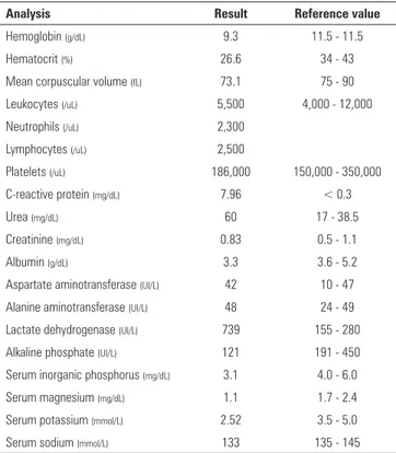
severe hypercalcemia (total calcium 21.8mg/dL, ionized calcium 2.93mmol/L). Other evaluations are shown in table 1. Craniocephalic computed tomography and renal, abdominal and hip joint ultrasound showed no signiicant changes.
Given the clinical and laboratory severity of hypercalcemia, on suspicion of osteoarticular infection, intravenous hyperhydration was initiated (2,500mL/
m2/day), and antibiotics were given (lucloxacillin
and gentamicin). he child was transferred to the pediatric intensive care unit. Continuous venovenous hemodiailtration was initiated after a 6.5F hemodialysis central venous catheter was placed in the right femoral vein. An HF20 ilter was used and priming was performed with 5,000 UI of heparin in 1L of 0.9% sodium chloride. Continuous venovenous hemodiailtration was programmed in accordance with the pediatric protocol (25 - 40mL/kg/h = 1/3 dialysis luid + 2/3 luid replacement (2/3 preilter + 1/3 post-ilter)). Ultrailtrate was calculated according to the desired luid balance.(6) A replacement and calcium-free dialysis solution was used (Prism0Cal®, Gambro - Lund, Sweden). Regional anticoagulation was
Table 1 - Evaluation performed on admission
Analysis Result Reference value
Hemoglobin (g/dL) 9.3 11.5 - 11.5
Hematocrit (%) 26.6 34 - 43
Mean corpuscular volume (fL) 73.1 75 - 90
Leukocytes (/uL) 5,500 4,000 - 12,000
Neutrophils (/uL) 2,300
Lymphocytes (/uL) 2,500
Platelets (/uL) 186,000 150,000 - 350,000
C-reactive protein (mg/dL) 7.96 < 0.3
Urea (mg/dL) 60 17 - 38.5
Creatinine (mg/dL) 0.83 0.5 - 1.1
Albumin (g/dL) 3.3 3.6 - 5.2
Aspartate aminotransferase (UI/L) 42 10 - 47
Alanine aminotransferase (UI/L) 48 24 - 49
Lactate dehydrogenase (UI/L) 739 155 - 280
Alkaline phosphate (UI/L) 121 191 - 450
Serum inorganic phosphorus (mg/dL) 3.1 4.0 - 6.0
Serum magnesium (mg/dL) 1.1 1.7 - 2.4
Serum potassium (mmol/L) 2.52 3.5 - 5.0
Serum sodium (mmol/L) 133 135 - 145
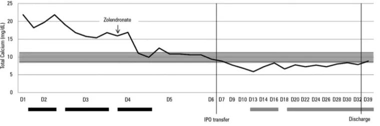
performed with machine-perfused unfractionated heparin, the dose of which was adjusted according to the patient’s activated partial thromboplastin time. he technique was maintained for 72 hours and took place without complications. As a therapeutic supplement, intravenous zoledronate (0.025mg/kg) was administered on the third day of hospitalization. here was a progressive decrease in total and ionized calcium levels and an improved state of consciousness (Figure 1).
he investigation revealed low (unmeasurable) serum intact parathyroid hormone (PTH) and excluded metabolic and infectious causes and vitamin or drug poisoning (Table 2). Skeletal radiography excluded osteolytic lesions.
During hospitalization, progressive pancytopenia (hemoglobin 7.7g/dL, leukocytes 2200/uL, platelets 67000/uL) was identiied and bone marrow examination was performed, which conirmed precursor B-cell acute lymphoblastic leukemia.
404 Martins AL, Moniz M, Nunes PS, Abadesso C, Loureiro HC, Duarte X, et al.
Rev Bras Ter Intensiva. 2015;27(4):402-405
Table 2 - Laboratory tests performed
Analysis Result Reference value
PTH intact < 20
PTHrp (pmol/L) 1.2 < 2.0
1.25 (OH)2D (pmol/L) 2 39 - 193
25 (OH)D (ng/dL) 12.7 30 - 100
Retinol (ng/dL) 21 30 - 70
ACE (U/L) 20.10 12 - 68
EBV-VCA IgM/IgG Negative/positive
Parvovirus B19 IgM/IgG Negative/positive
CMV IgM/IgG Negative/positive
Mycoplasma pneumoniae IgM/IgG Negative/negative
HIV 1/2 Negative
FT3 (pg/mL) 2.31 4.0 - 7.10
FT4 (ng/dL) 0.56 0.9 -1.70
TSH (mUI/L) 0.496 0.8 - 7.5
Total proteins (g/dL) 5.6 6 - 8
Albumin (g/dL) 2.53 3.75 - 5.01
Uric acid (mg/dL) 9.4 3.0 - 5.5
PTH - parathyroid hormone; PTHrp - parathyroid hormone-related protein; 1.25 (OH)2D - 1.25-dihydroxy-vitamin D; 25(OH)D - Vitamin D in the form of 25-hydroxyvitamin D; ACE - angiotensin converting enzyme; EBV-VCA - viral capsid antigen of the Epstein-Barr virus; CMV - cytomegalovirus; HIV - human immunodeficiency virus; FT3 - free T3 hormone; FT4 - free T4 hormone; TSH - thyroid stimulating hormone.
5.9mg/dL at day 10 post-zoledronate), which required intravenous correction. A bone marrow smear performed 4 weeks after the beginning of induction therapy conirmed complete remission both morphologically and by molecular biology.
Currently, approximately 10 months after starting treatment, the child remains in remission.
DISCUSSION
Hypercalcemia is a potentially fatal disorder, regarding its neurological and cardiac complications. he treatment includes hyperhydration, bisphosphonates and treatment of the underlying disease.(1) Occasionally, rapid correction of the disturbance becomes crucial, particularly in the setting of loss of consciousness or when the hypercalcemia is refractory to conventional measures. In such situations, the use of continuous venovenous hemodiailtration has been identiied as an efective treatment.(7,8) Its successful use in severe hypercalcemia has been reported in adults,(8-11) but the use of the technique in pediatrics has rarely been described in the literature.(7,12) In this case report, due to severe hypercalcemia on admission, the use of dialysis solution with calcium-free replacement was chosen. Regular analytical calcemia controls were performed in order to avoid a sudden decrease and below-normal values. he renal replacement therapy settings were set in order to provide a gradual decrease in serum calcium, thereby avoiding complications such as circuit clotting. As the patient did not present spontaneous diuresis, it was decided to program losses to ensure a neutral luid balance. Following clinical and laboratory stabilization, zoledronate was introduced to maintain normocalcemia, as the efect of continuous venovenous hemodiailtration is temporary.(11) Continuous monitoring of serum calcium levels was assured due to the risk of hypocalcemia observed in this case.(13)
he etiological investigation suggested an independent PTH mechanism. Metabolic and infectious causes and vitamin or drug poisoning were excluded. Progressive pancytopenia led to the suspicion of MAH, which ultimately led to the inal diagnosis.
Severe hypercalcemia as a form of acute lymphoblastic leukemia 405
Rev Bras Ter Intensiva. 2015;27(4):402-405
he pathogenesis of MAH includes the stimulation of bone resorption, mediated by proteins and cytokines produced by the tumor cells or by the tumoral microenvironment. Two distinct mechanisms are described, which include hypercalcemia by local osteolytic lesions (bone metastasis) and humoral hypercalcemia by the activation of RANK-RANKL (receptor activator of nuclear factor κB and its ligand). Parathyroid hormone-related protein (PTHrP) is the most frequently involved mediator, but other mediators, such as interleukin (IL)-1, IL-6, tumor necrosis factor alpha (TNF-α), transforming growth factor beta (TGF-β), prostaglandins and even calcitriol and ectopic PTH production may be involved.(4)
In acute lymphoblastic leukemia, an association with hypercalcemia in patients with t (17;19) has been reported, suggesting the possible induction of PTHrP.(3,14) In this
case, this cytogenetic abnormality was not observed, and high levels of PTHrP were not detected, thus excluding this mechanism as the primum movens of hypercalcemia.
CONCLUSION
he described case shows an infrequent complication, not only at pediatric age, but also in children with oncological diseases, suggesting that this metabolic emergency unveils of the underlying disease. Continuous venovenous hemodiailtration with calcium-free solution as a irst-line treatment in cases of severe and symptomatic hypercalcemia was found to be efective in the rapid induction of normocalcemia and neurological improvement, buying valuable time until maintenance treatment focused on the etiology can exert a sustained efect.
A hipercalcemia é um distúrbio metabólico raro em pediatria, potencialmente fatal, apresentando um vasto diagnóstico diferencial, incluindo neoplasias. Relatamos aqui o caso de uma criança de 3 anos, previamente saudável, admitida no serviço de urgência por fadiga, hiporreatividade, febre e claudicação da marcha com 5 dias de evolução, de agravamento progressivo. À observação, apresentava-se inconsciente (escore de coma Glasgow: 8). Laboratorialmente, apresentava hipercalcemia grave (cálcio total 21,39mg/dL, ionizado 2,93mmol/L) e anemia microcítica. Iniciou hiper-hidratação e foi transferido para a unidade de cuidados intensivos pediátricos. Instituiu-se
hemodiailtração venovenosa contínua com soluto livre de cálcio, ocorrendo a progressiva normalização da calcemia, com melhoria do estado de consciência. Administrou-se zolendronato. Excluíram-se causas metabólicas, infecciosas e intoxicação. O mielograma permitiu o diagnóstico de leucemia linfoblástica aguda. A hipercalcemia associada à malignidade em pediatria é rara, ocorrendo como forma de apresentação da neoplasia ou na recorrência desta. Em situações com risco de vida iminente, deve se considerar hemodiailtração venovenosa contínua.
RESUMO
Descritores: Hipercalcemia; Leucemia-linfoma linfoblástico de células T precursoras; Hemodiailtração; Relatos de casos
REFERENCES
1. Lietman SA, Germain-Lee EL, Levine MA. Hypercalcemia in children and adolescents. Curr Opin Pediatr. 2010;22(4):508-15.
2. McKay C, Furman WL. Hypercalcemia complicating childhood
malignancies. Cancer. 1993;72(1):256-60.
3. Inukai T, Hirose K, Inaba T, Kurosawa H, Hama A, Inada H, et al. Hypercalcemia in childhood acute lymphoblastic leucemia: frequent implication of parathyroid hormone-related peptide and E2A-HLF from translocation 17;19. Leukemia. 2007;21(2):288-96.
4. Sargent JT, Smith OP. Haematological emergencies managing
hypercalcaemia in adults and children with haematological disorders. Br J Haematol. 2010;149(4):465-77.
5. Kerdudo C, Aerts I, Fattet S, Chevret L, Pacquement H, Doz F, et al. Hypercalcemia and childhood cancer: a 7-year experience. J Pediatr Hematol Oncol. 2005;27(1):23-7.
6. Ruza F. Tratado de cuidados intensivos pediatricos. 3a ed. Madri: Norma-Capitel Ediciones; 2003.
7. Pradhan M, Leonard MB. Calcium-free hemodialysis for hypercalcemia of malignancy in a newborn. Pediatr Nephrol. 2003;18(5):474-6.
8. Wang CC, Chen YC, Shiang JC, Lin SH, Chu P, Wu CC. Hypercalcemic crisis successfully treated with prompt calcium-free hemodialysis. Am J Emerg Med. 2009;27(9):1174.e1-3.
9. Au S, Dunham M, Godinez T. Treatment of medically refractory hypercalcemic crisis. Int J Artif Organs. 2012;35(7):538-41.
10. Kindgen-Milles D, Kram R, Kleinekofort W, Morgera S. Treatment of severe hypercalcemia using continuous renal replacement therapy with regional citrate anticoagulation. ASAIO J. 2008;54(4):442-4.
11. Camus C, Charasse C, Jouannic-Montier I, Seguin P, Tulzo YL, Bouget J, et al. Calcium free hemodialysis: experience in the treatment of 33 patients with severe hypercalcemia. Intensive Care Med. 1996;22(2):116-21. 12. Bahoush G, Miri-Aliabad G. Severe hypercalcemia: a rare and unusual
presentation of childhood acute lymphoblastic leukemia. Int J Hematol Oncol Stem Cell Res. 2014;8(2):38-40.
13. Kreutle V, Blum C, Meier C, Past M, Müller B, Schütz P, et al. Bisphosphonate induced hypocalcemia - report of six cases and review of the literature. Swiss Med Wkly. 2014;144:w13979.