Inluence of diferent degrees of head elevation on
respiratory mechanics in mechanically ventilated
patients
INTRODUCTION
he positioning of a patient in bed can directly afect respiratory function in mechanically ventilated (MV) patients.(1,2) he posture imposed on MV patients may facilitate diaphragmatic performance, but it may also increase the mechanical load against the respiratory system airlow.(3,4)
he current recommendation is that the head of MV patients should be maintained between 30° and 45° because of the high risk of bronchoaspiration and because this position can reduce the risk of mechanical ventilation-associated pneumonia.(5,6) In addition to promoting a reduction in the risk of developing pneumonia, some postural positions can increase the possibility of more homogeneous alveolar ventilation and possibly reduce the risk of lung injury caused by mechanical ventilation similar to that in patients undergoing ventilation in the prone position.(7)
Bruno Prata Martinez1,2, Thaís Improta Marques3, Daniel Reis Santos3, Vanessa Salgado Silva4, Balbino Rivail Nepomuceno Júnior1, Giovani Assunção de Azevedo Alves1,5, Mansueto Gomes Neto4, Luiz Alberto Forgiarini Junior6
1. Hospital Aliança - Salvador (BA), Brazil. 2. Universidade do Estado da Bahia - Salvador (BA), Brazil.
3. Hospital Santo Antônio, Obras Sociais Irmã Dulce - Salvador (BA), Brazil.
4. Universidade Federal da Bahia - Salvador (BA), Brazil.
5. Postgraduate Program, Universidade Cidade de São Paulo - São Paulo (SP), Brazil. 6. Centro Universitário Metodista - IPA - Porto Alegre (RS), Brazil.
Objective: he positioning of a patient in bed may directly afect their respiratory mechanics. he objective of this study was to evaluate the respiratory mechanics of mechanically ventilated patients positioned with diferent head angles hospitalized in an intensive care unit.
Methods: his was a prospective physiological study in which static and dynamic compliance, resistive airway pressure, and peripheral oxygen saturation were measured with the head at four diferent positions (0° = P1, 30° = P2, 45° = P3, and 60° = P4). Repeated-measures analysis of variance (ANOVA) with a Bonferroni post-test and Friedman analysis were used to compare the values obtained at the diferent positions.
Results: A comparison of the 35 evaluated patients revealed that the
Conflicts of interest: None.
Submitted on July 21, 2015 Accepted on September 21, 2015
Corresponding author:
Bruno Prata Martinez
Departamento de Ciências da Vida I da Universidade do Estado da Bahia Rua Silveira Martins, 2.555 - Cabula Zip code: 41150-000 - Salvador (BA), Brazil E-mail: [email protected]
Responsible editor: Carmen Valente Barbas
Inluência de diferentes graus de elevação da cabeceira na
mecânica respiratória de pacientes ventilados mecanicamente
ABSTRACT
Keywords: Respiratory mechanics; Inpatients; Patient positioning; Intensive care units
resistive airway pressure values in the 0° position were higher than those obtained when patients were positioned at greater angles. he elastic pressure analysis revealed that the 60° position produced the highest value relative to the other positions. Regarding static compliance, a reduction in values was observed from the 0° position to the 60° position. he dynamic compliance analysis revealed that the 30° angle produced the greatest value compared to the other positions. he peripheral oxygen saturation showed little variation, with the highest value obtained at the 0° position.
Conclusion: he highest dynamic compliance value was observed at the 30° position, and the highest oxygenation value was observed at the 0° position.
Although the efects of positioning the head at 30° and 45° on the reduction of mechanical ventilation-associated pneumonia are known, no studies have evaluated the diference in mean values obtained for mechanical ventilation at diferent head angles in this population. herefore, the objective of this study was to evaluate the respiratory mechanics of MV patients admitted to the intensive care unit (ICU) who were positioned with diferent head angles (0°, 30°, 45°, and 60°).
METHODS
his was a prospective physiological study conducted in the ICU of the Hospital Santo Antônio, Obras Sociais Irmã Dulce, in the city of Salvador (BA), between October 2009 and January 2010. he study included adult patients of both genders who were over 18 years of age, in the ICU for more than 24 hours, undergoing invasive MV, sedated, not interacting with the mechanical ventilator, which was visualized by graphical analysis, and hemodynamically stable, characterized by the absence or low doses of vasoactive or inotropic drugs. Patients with recent fractures (chest wall, spine, and hip) and those with a clinical diagnosis of pulmonary ibrosis or acute respiratory distress syndrome were excluded. Patients who showed changes in mean arterial pressure greater than 20% relative the baseline value, a systolic blood pressure < 90mmHg in invasive blood pressure measurements, and peripheral oxygen saturation < 90% during mechanical measurements were also excluded. he present study was approved by the Research Ethics Committee of the Hospital Santo Antônio (protocol number 46/09). Individuals who were responsible for the patients were informed about the study and signed an informed consent form authorizing participation.
he measured values of respiratory mechanics were obtained from a TBIRD VELA mechanical ventilator (Viasys Respiratory Care, United States) and included respiratory system static (Cst, rs) and dynamic (Cdyn, rs) compliance and resistive airway pressure. Hemodynamic data such as mean arterial pressure, systolic blood pressure, heart rate, and peripheral oxygen saturation were obtained from a multiparameter monitor (DIXTAL, Manaus, Brazil).
he patients included in the study were evaluated at four diferent positions (0° = P1, 30° = P2, 45° = P3, and 60° = P4), which were randomly allocated,
and randomization of the positions was conducted in a point by point manner. For greater accuracy, a goniometer was used to verify the head angle adopted for each position.
Before the evaluation of respiratory mechanics, a single alveolar recruitment maneuver was performed for pulmonary homogenization, with patients in a pressure controlled ventilation mode with a 100% inspired oxygen fraction and an increased positive end expiratory pressure (PEEP) of 2cmH2O every minute until a value of 20cmH2O was reached. his condition was maintained for two minutes and followed by reduction of 2cmH2O per minute until the initial PEEP level was achieved.(8) After 30 minutes, the patients were placed in a controlled volume ventilation mode for evaluation of respiratory mechanics with the following parameters: tidal volume of 6 - 8mL/kg in relation to the ideal weight, 40 L/min low, square wave low, a respiratory rate of 15 breaths per minute, and an inspiratory pause time of 0.5 seconds.(9) hese parameters were maintained for approximately two minutes in each position, and the peak and plateau pressure values and the mean PEEP were recorded. he screen was paused to record the peak and plateau pressures; the highest value was considered the peak, and the pressure value closest to the 0.5-second pause time and with a low equal to zero was recorded as the plateau.
Static compliance was calculated by dividing the tidal volume by the respiratory system elastic pressure or driving pressure (plateau pressure subtracted from the mean PEEP value). For dynamic compliance, the tidal volume was divided by the peak pressure subtracted from the mean PEEP value. Resistive airway pressure was calculated as the diference between the peak and plateau pressures.
RESULTS
During the data collection period, 35 patients were included in the study, of whom 27 (77.7%) had a primary diagnosis of pneumonia and eight (22.3%) were undergoing a postoperative period after abdominal surgery. he mean age was 58.1 ± 15.6 years, and 66.6% of the patients were male. No complications, such as peripheral oxygen saturation below 90% or hemodynamic changes, were reported during the procedures. Table 1 and igure 1 show the static and dynamic compliance, resistive airway pressure, and alveolar distension pressure values.
(p = 0.001). Peripheral oxygen saturation did not difer signiicantly when the 0o and 60 o positions were compared (p = 0.465).
DISCUSSION
A change in the angle of the head afects the respiratory mechanics of MV patients. In this study, the largest resistive pressure value was found at the 0o position, and the largest value of elastic pressure was found at the 60o position. For the Cdyn, rs, the highest value occurred in the 30o position.
In an intervention study involving early mobilization of intubated abdominal surgery patients, Zairopoulos et al.(10) observed that high thoracic positions, such as sitting upright for 20 minutes, led to an improvement in transthoracic pressure, with consequent improvement in the Cst, rs. his gain enabled a reduction in the driving pressure required for the generation of a similar lung volume. his knowledge is crucial and must be employed in ventilatory lung protection strategies. Such diferences may be relevant to clinical practice because variations in driving pressure, for example, may be associated with lower mortality in patients with and without acute respiratory distress syndrome, which has been demonstrated in recent meta-analyses.(11,12)
In the present study, the lowest driving pressure was observed at the 30o position, but the values were higher than 15cmH2O, which is not consistent with current recommendations for ventilatory strategy in MV patients.(13) One possible explanation for this inding is that when the study was conducted, that recommendation did not exist, and patients were ventilated with tidal volumes of 6 to 8mL/kg because they had not been diagnosed with acute respiratory distress syndrome.
Although the literature reports improved respiratory system compliance in the sitting position compared to the dorsal and lateral decubitus positions,(8) our study revealed a reduction of these values at greater angles, possibly due to the higher transthoracic pressure. However, it is not possible to state that this inding results from an increase in intra-abdominal pressure, as this variable was not evaluated; however, in all of our measurements, the legs were parallel to the ground to prevent further tilting with higher head positions.
To distinguish this possible chest wall change from a pulmonary change, it would be necessary to measure transpulmonary pressure, which would require the use of an esophageal balloon to estimate the pleural pressure
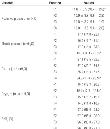
Table 1 - Evaluation of respiratory mechanics variables at different body positions
Variable Position Values
Resistive pressure (cmH2O)
P1 11.6 ± 3.5 (10.4 - 12.8)* P2 10.9 ± 3.8 (9.6 - 12.2) P3 10.8 ± 3.2 (9.6 - 11.9) P4 10.8 ± 3.5 (9.6 - 12.0)
Elastic pressure (cmH2O)
P1 17.4 (14.0 - 22.1)
P2 16.6 (13.7 - 21.4)
P3 17.3 (14.8 - 23.8)
P4 19.3 (16.1 - 25.3)*
Cst, rs (mL/cmH2O)
P1 27.1 (19.5 - 32.3)
P2 27.0 (20.7 - 34.8)
P3 25.2 (18.4 - 31.5)
P4 24.5 (17.4 - 29.9)*
Cdyn, rs (mL/cm H2O)
P1 15.4 (12.3 - 20.2)
P2 16.4 (12.7 - 19.5)*
P3 15.6 (13.7 - 19.1)
P4 14.6 (11.8 - 18.7)
SpO2 (%)
P1 97.0 (96.0 - 98.0)
P2 97.0 (96.0 - 98.0)
P3 96.0 (96.0 - 97.0)
P4 96.0 (96.0 - 97.0)
Cst, rs - static compliance; Cdyn, rs - dynamic compliance; SpO2 - pulse oximetry. Results are expressed as the mean ± standard deviation (95% confidence interval) or median (25%-75%). * p < 0.001.
A comparison of resistive pressure values revealed that the 0o position values were higher than those recorded for greater angles (Table 1). he elastic pressure analysis revealed that the 60o position produced the highest value of all positions (p = 0.001).
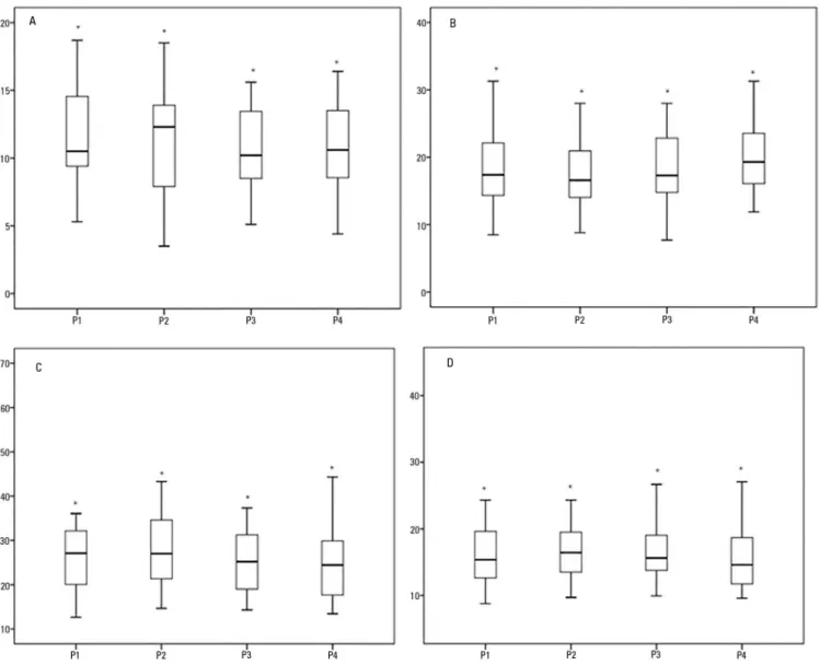
Figure 1 - Analysis of resistive airway pressure (A), respiratory system elastic pressure (B), respiratory system static compliance (C), and respiratory system dynamic compliance (D) in the four positions (P1 = 0º, P2 = 30º, P3 = 45º and P4 = 60º). * p value < 0.05.
value. hus, the mechanical variations obtained at diferent positions could be related to variations in pleural pressure (thoracic) or alveolar pressure changes (pulmonary).(9)
Similarly, airway resistance was inluenced by body position, with the highest value found with the head at 0o. his result was not expected because it had been thought that the lowest resistance would be found in the pulmonary areas of greatest respiratory system compliance. However, the opposite result was observed because greater resistance was found at the position of greatest compliance, which indicates that no inverse linear relationship exists between these two variables.(14,15)
In the evaluation of the Cdyn, rs, the highest value was observed at the 30o position, which may be explained by
the close relationship between reduced resistive pressure and increased elastic pressure. A possible explanation for this inding is the reduction in resistance due to an increase in functional residual capacity (FRC), as well as a reduction in the intrathoracic blood volume.(14,16) In addition to likely facilitating ventilation, this angle reduces the risk of mechanical ventilation-associated pneumonia.(5,6)
facilitating a reduction in mechanical load that opposes the entry of air are therefore fundamental for greater stabilization of the air in the alveoli.(19-21)
he quasi-static method used to measure respiratory mechanics with occlusion at the end of inspiration was chosen due to the ease of bedside application in critically ill patients, but this method cannot diferentiate between the chest wall and lung components.(22) For this purpose, the use of invasive methods that can quantify pleural pressure is necessary.(23)
he increased peripheral saturation at the 0o position can be explained by the movement of blood to areas with a better ventilation/perfusion ratio that have proportionately greater vascularity at the posterior region of the lung, which remains dependent in the supine position at 0o.(22) However, the 0.6% diference was not statistically signiicant.
In the case of obese patients, increased intra-abdominal pressure and general anesthesia can also cause changes in respiratory mechanics. An increase in intra-abdominal pressure increases chest wall elastance, reduces compliance, and promotes cranial displacement of the diaphragm. hese factors may explain the higher esophageal pressure values in overweight/obese individuals. In obese patients, increased intra-abdominal pressure is the major determinant of lung volume reduction and premature closure of the small airways, especially when associated with anesthesia, which increases the reduction in functional capacity.(24-27)
In the present study, the alveolar recruitment maneuver was performed to homogenize the lung before applying the diferent head inclinations, thereby ensuring that the behavior of the variables of interest exhibited less bias
due to possible gain or loss of alveolar unit recruitment between position changes.
his study had some limitations, such as the lack of non-invasive, intra-abdominal pressure measurements and the absence of pleural pressure measurements, which may also be a confounding variable in respiratory mechanics. In addition, mortality scores, cumulative luid balance, use of vasopressor and inotropic drugs, use of renal replacement therapy, total mechanical ventilation time, hospital survival, and mean tidal volume were not measured. However, because this is the irst study to evaluate diferent head angles with respect to respiratory mechanics, additional studies are needed to evaluate the efect of these variables. Another limitation was that the evaluated population had a very heterogeneous proile, which, although mostly consisting of patients with pneumonia, also included patients in the postoperative period after abdominal surgery. As stated previously, the driving pressure and tidal volume values were higher than the current recommendation, which is also a possible limitation of this study. Further studies evaluating the elastic components of the respiratory system that can afect these changes, such as intra-abdominal, chest wall, and pulmonary pressure and the ventilation versus infusion relationship, are needed.
CONCLUSION
Head angle afected the respiratory mechanics of mechanically ventilated patients. he highest dynamic compliance value was observed at the 30o position relative to the other angles, and the driving pressure was increased at head angles of 45o and 60o.
Objetivo: O posicionamento do paciente no leito pode afetar diretamente a mecânica respiratória. Este estudo teve como objetivo avaliar a mecânica respiratória em diferentes angulações da cabeceira em pacientes internados na unidade de terapia intensiva sob ventilação mecânica.
Métodos: Trata-se de um estudo prospectivo isiológico, no qual foram mensuradas a complacência estática e dinâmica; a pressão resistiva das vias aéreas e saturação periférica de oxigênio nas diferentes posições adotadas (0° = P1, 30° = P2, 45° = P3 e 60° = P4). Para comparação dos valores obtidos nas diferentes posições, foi utilizada a Análise de Variância de medidas repetidas (ANOVA) com pós-teste de Bonferroni e análise de Friedman.
Resultados: Quando comparamos os 35 pacientes avaliados, os valores da pressão resistiva das vias aéreas na posição a 0°
foram superiores em relação às angulações mais elevadas. Já na análise da pressão elástica, a posição a 60° apresentou o maior valor em relação às outras posições. Em a relação à complacência estática, houve redução dos valores da posição 0° para a posição 60°. Quando analisada a complacência dinâmica, observou-se que a angulação de 30° apresentou o maior valor, quando comparada às demais posições. A saturação periférica de oxigênio apresentou pequena variação, sendo o maior valor obtido na posição 0°.
Conclusão: A complacência dinâmica apresentou maior valor na posição a 30° em relação às outras angulações, sendo que a posição de maior oxigenação foi a 0°.
RESUMO
REFERENCES
1. Reinius H, Jonsson L, Gustafsson S, Sundbom M, Duvernoy O, Pelosi P, et al. Prevention of atelectasis in morbidly obese patients during general anesthesia and paralysis: a computerized tomography study. Anesthesiology. 2009;111(5):979-87.
2. Mulier JP, Dillemans B, Van Cauwenberge S. Impact of the patient’s body position on the intraabdominal workspace during laparoscopic surgery. Surg Endosc. 2010;24(6):1398-402.
3. Gea J. La especie humana: un largo camino para el sistema respiratorio. Arch Bronconeumol. 2008;44(5):263-70.
4. França EE, Ferrari F, Fernandes P, Cavalcanti R, Duarte A, Martinez BP, et al. Fisioterapia em pacientes críticos adultos: recomendações do Departamento de Fisioterapia da Associação de Medicina Intensiva Brasileira. Rev Bras Ter Intensiva. 2012;24(1):6-22.
5. Drakulovic MB, Torres A, Bauer TT, Nicolas JM, Nogué S, Ferrer M. Supine body position as a risk factor for nosocomial pneumonia in mechanically patients: a randomised trial. Lancet. 1999;354(9193):1851-8.
6. Grap MJ, Munro CL, Hummel RS 3rd, Elswick RK Jr, McKinney JL, Sessler CN. Effect of backrest elevation on the development of ventilator-associated pneumonia. Am J Crit Care. 2005;14(4):325-32; quiz 333. 7. Guérin C, Reignier J, Richard JC, Beuret P, Gacouin A, Boulain T, Mercier
E, Badet M, Mercat A, Baudin O, Clavel M, Chatellier D, Jaber S, Rosselli S, Mancebo J, Sirodot M, Hilbert G, Bengler C, Richecoeur J, Gainnier M, Bayle F, Bourdin G, Leray V, Girard R, Baboi L, Ayzac L; PROSEVA Study Group. Prone positioning in severe acute respiratory distress syndrome. N Eng J Med. 2013;368(23):2159-68.
8. Porto EF, Castro AA, Leite JR, Miranda SV, Lancauth A, Kumpel C. Análise comparativa da complacência do sistema respiratório em três diferentes posições no leito (lateral, sentada e dorsal) em pacientes submetidos à ventilação mecânica invasiva prolongada. Rev Bras Ter Intensiva. 2008;20(3):213-9.
9. Hess DR. Respiratory mechanics in mechanically ventilated patients. Respir Care. 2014;59(11):1773-94.
10. Zafiropoulos B, Alison JA, McCarren B. Physiological responses to the early mobilisation of the intubated, ventilated abdominal surgery patient. Aust J Physiother. 2004;50(2):95-100.
11. Amato MB, Meade MO, Slutsky AS, Brochard L, Costa EL, Schoenfeld DA, et al. Driving pressure and survival in the acute respiratory distress syndrome. N Engl J Med. 2015;372(8):747-55.
12. Serpa Neto A, Simonis FD, Barbas CS, Biehl M, Determann RM, Elmer J, et al. Association between tidal volume size, duration of ventilation, and sedation needs in patients without acute respiratory distress syndrome: an individual patient data meta-analysis. Intensive Care Med. 2014;40(7):950-7.
13. Barbas CS, Ísola AM, Farias AM, Cavalcanti AB, Gama AM, Duarte AC, et al. Recomendações brasileiras de ventilação mecânica 2013. Parte I. Rev Bras Ter Intensiva. 2014;26(2):89-121.
14. Saddy F. Avaliação da mecânica respiratória na síndrome do desconforto respiratório agudo. Pulmão RJ. 2011;20(1):31-6.
15. Scanlan CL, Stoller JK, Wilkins RL. Fundamentos da terapia respiratória de Egan. 7a ed. São Paulo: Manole; 2000.
16. Behrakis PK, Baydur A, Jaeger MJ, Milic-Emili J. Lung mechanics in sitting and horizontal body positions. Chest. 1983;83(4):643-6.
17. Barbas CS, de Matos GF, Pincelli MP, da Rosa Borges E, Antunes T, de Barros JM, et al. Mechanical ventilation in acute respiratory failure: recruitment and high positive end-expiratory pressure are necessary. Curr Opin Crit Care. 2005;11(1):18-28. Review.
18. Seeley EJ, McAuley DF, Eisner M, Miletin M, Zhuo H, Matthay MA, et al. Decreased respiratory system compliance on the sixth day of mechanical ventilation is a predictor of death in patients with established acute lung injury. Respir Res. 2011;12:52.
19. Pelosi P, Brazzi L, Gattinoni L. Prone position in acute respiratory distress syndrome. Eur Respir J. 2002;20(4):1017-28. Review.
20. Blanch L, Mancebo J, Perez M, Martinez M, Mas A, Betbese AJ, et al. Short-term effects of prone position in critically ill patients with acute respiratory distress syndrome. Intensive Care Med. 1997;23(10):1033-9. 21. West JB. Fisiologia respiratória: princípios básicos. 9a ed. Porto Alegre:
Artmed; 2013.
22. Faustino EA. Mecânica pulmonary de pacientes em suporte ventilatório na unidade de terapia intensiva. Conceitos e monitorização. Rev Bras Ter Intensiva. 2007;19(2):161-9.
23. Fernandes CR. A importância da pressão pleural na avaliação da mecânica respiratória. Rev Bras Anestesiol. 2006;56(3):287-303.
24. Delgado PM, Lunardi AC. Complicações respiratórias pós-operatórias em cirurgia bariátrica: revisão da literatura. Fisioter Pesqui. 2011;18(4):388-92. 25. Pelosi P, Quintel M, Malbrain ML. Effect of intra-abdominal pressure on
respiratory mechanics. Acta Clin Belg Suppl. 2007;(1):78-88.
26. Owens RL, Campana LM, Hess L, Eckert DJ, Loring SH, Malhotra A. Sitting and supine esophageal pressures in overweight and obese subjects. Obesity (Silver Spring). 2012;20(12):2354-60.
27. Talmor D, Sarge T, O’Donnell CR, Ritz R, Malhotra A, Lisbon A, et al. Esophageal and transpulmonary pressures in acute respiratory failure. Crit Care Med. 2006;34(5):1389-94.
ERRATUM
In the article Inluence of diferent degrees of head elevation on respiratory mechanics in mechanically ventilated
patients, DOI number: 10.5935/0103-507X.20150059, published in Revista Brasileira de Terapia Intensiva