www.rbo.org.br/
issn/$–see front matter © 2013 Sociedade Brasileira de Ortopedia e Traumatologia. Published by Elsevier Editora Ltda. All rights reserved. doi: 10.1016/j.rboe.2012.05.006
*Corresponding author at: Santa Casa de Misericórdia de São Paulo, Departamento de Ortopedia e Traumatologia, Pavilhão Fernandinho Simonsen, Rua Dr. Cesário Mota Júnior, 112, Vila Buarque, São Paulo, SP, Brazil, CEP 01220-020, Phone/Fax (0xx11) 3222-6866. Phone: 00 3519 6622 1650.
E-mail: [email protected] A RT I C L E I N F O
Article history:
Received on February 24, 2012 Approved on May 28, 2012
Keywords: Osteoarthritis
Arthroplasty, replacement Evaluation studies Shoulder joint
A B S T R A C T
Objective: In this study we aim at statistically evaluating the results of the surgical treatment of the osteoarthrosis of the shoulder (OAS) with partial shoulder arthroplasty (PSA) and at correlating them with the several variables involved. Methods: In this study we evaluated 36 shoulders of 31 patients with OAS who underwent treatment with PSA in the Grupo de Ombro e Cotovelo (Group of Shoulders and Elbows) of the Department of Traumatology and Orthopedics of the Faculdade de Ciências Médicas da Santa Casa de São Paulo – Pavillion Fernandinho Simonsen between January, 1989 and November, 2010. Patients who underwent PSA and who had a post-operative follow-up of at least 12 months were included in the study. Results: After the surgery the range of elevation, external rotation, internal rotation and the UCLA scale improved (with average differences of 35o, 27o, 4o and 17 points, respectively), with a significant level of 5% (p < 0.05). For the same level of significance, the relation between a satisfactory UCLA and two variables was found: patients with maximum age of 60 years old at the moment of the surgery and patients that underwent tenotomy of the long head of biceps. Conclusion: Patients under 60 who underwent surgery and patients who underwent tenotomy of the long head of biceps achieved better results.
© 2013 Sociedade Brasileira de Ortopedia e Traumatologia. Published by Elsevier Editora Ltda. All rights reserved.
Original Article
Evaluation of the results from partial arthroplasty for treating
shoulder osteoarthrosis
Alberto Naoki Miyazaki,
1Marcelo Fregoneze,
2Pedro Doneux Santos,
3Luciana Andrade da Silva,
3Guilherme do Val Sella,
3Rodrigo Zampieri,
4Eduardo Régis de Alencar Bona Miranda,
4Sergio Luiz Checchia
51PhD. Assistant Professor in the Department of Orthopedics and Traumatology, School of Medical Sciences, and Head of the Shoulder and
Elbow Group, Santa Casa de São Paulo, São Paulo, SP, Brazil.
2Assistant Professor in the Department of Orthopedics and Traumatology, School of Medical Sciences, and Attending Physician in the
houlder and Elbow Group, Santa Casa de São Paulo, São Paulo, SP, Brazil.
3 Attending Physician in the Shoulder and Elbow Group, Santa Casa de São Paulo, São Paulo, SP, Brazil.
4Trainee Physician in the Shoulder and Elbow Group, Santa Casa de São Paulo, São Paulo, SP, Brazil.
5PhD. Adjunct Professor and Head of Clinical Medicine, Department of Orthopedics and Traumatology, School of Medical Sciences, and
cademic Coordinator of the Shoulder and Elbow Group, Santa Casa de São Paulo, São Paulo, SP, Brazil.
Introduction
Shoulder osteoarthrosis is a painful and often incapacitating condition that occurs less frequently than in other joints such as the hip and knee.1 It may be primary or secondary to a series
of events such as trauma, instability or avascular necrosis of the humeral head. Independent of its etiology, it leads to a clinical condition of pain, diminished range of motion and functional limitation of the arm affected.1
Total shoulder arthroplasty has been widely accepted as a successful treatment for severe shoulder osteoarthrosis since the start of the 1970s.2 According to Cofield, Neer,
Morrisson, Hawkins et al, the results from this procedure have been extremely positive.3-8 Glenoid components made
of cemented polyethylene were introduced with the aim of enabling anatomical reconstruction of the shoulder and thereby providing pain relief, while increasing the range of motion of the shoulder. However, loosening of this component is the main cause of lack of success in total shoulder arthroplasty, as proven in the study by Hill and Norris (2001), for example.9
The difficulties in the implantation technique for the glenoid component and difficulties with bone stock, in which the glenoid cavity does not tolerate a polyethylene component because of excessive wear, or in cases of younger individuals (with the likelihood of requiring revision arthroplasty procedures), have led some authors to recommend partial shoulder arthroplasty instead of total shoulder arthroplasty, for treating shoulder osteoarthrosis.
According to Levine et al.,1 partial shoulder arthroplasty
provides pain relief and improvements in function, range of motion and capacity to perform activities of daily living, for shoulders presenting osteoarthrosis. In a recent study, Saltzman demonstrated that partial shoulder arthroplasty with concentric milling of the glenoid cavity, in patients under the age of 55 years, led to improvements in pain and shoulder function.10 The study by Bonnevialle, published
in 2011, demonstrated that partial shoulder arthroplasty is a reliable procedure in shoulders with osteoarthrosis and dysplastic morphology, thus leading to satisfactory clinical results.11
Along general lines, indications for total shoulder arthroplasty are reserved for cases in which the patients are older, are less demanding with regard to physical activity and have adequate bone stock for implantation of a glenoid component,12,13 always with an intact rotator cuff. Indications
for partial shoulder arthroplasty are reserved for cases of younger patients with higher physical demands or cases presenting glenoid abnormalities in which implantation of a component becomes impossible14 (Fig. 1). There is still no
consensus in the literature with regard to using or not using a glenoid component in cases of shoulder osteoarthrosis, and thus, surgeons have the task of choosing between performing partial and total shoulder arthroplasty.15
The present study had the aim of evaluating the results obtained by the Shoulder and Elbow Group of Santa Casa de Misericórdia de São Paulo from treating shoulder osteoarthrosis with partial shoulder arthroplasty.
Materials and methods
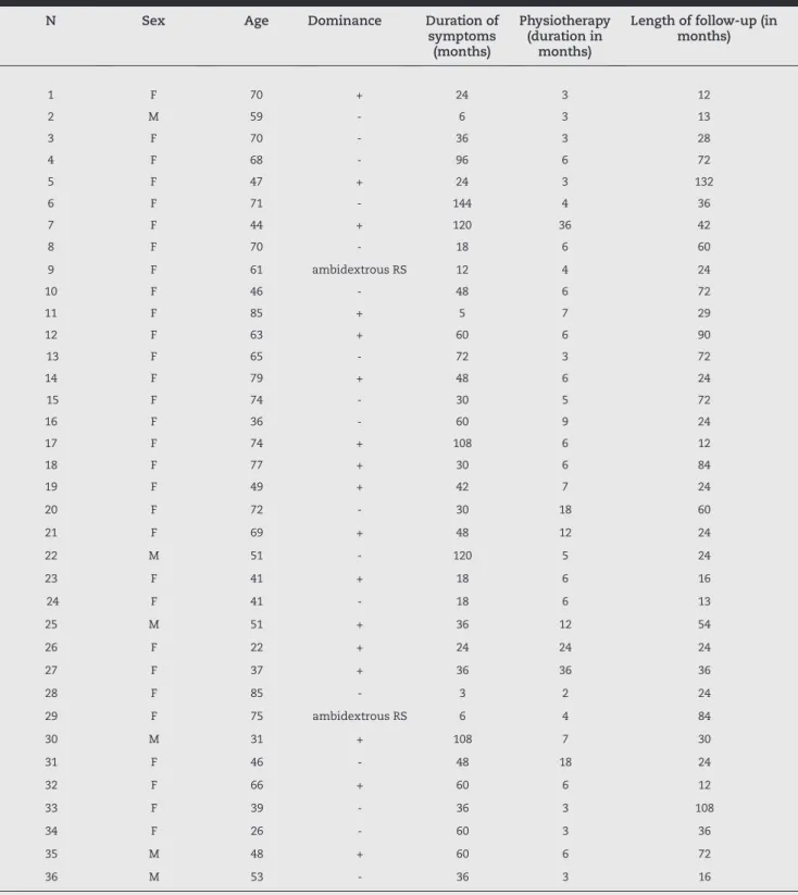
Thirty-six shoulders from 31 patients (five with bilateral disease) who were treated for shoulder osteoarthrosis using partial shoulder arthroplasty were retrospectively assessed (Table 1). The operations were performed by the Shoulder and Elbow Group, Department of Orthopedics and Traumatology, School of Medical Sciences, Santa Casa de São Paulo, between January 1989 and November 2010. This study was approved by the Research Ethics Committee.
The patients included in this study underwent partial shoulder arthroplasty to treat shoulder osteoarthrosis, with a minimum follow-up duration of one year.
The following patients were excluded: those who underwent partial shoulder arthroplasty due to any humeral head disease without the presence of arthrosis (due to fractures, for example) or due to arthropathy of the rotator cuff; individuals with arthrosis classified as B2 or C according to Walch et al.16 (Fig. 2); those who underwent treatment with
surface prostheses; and those with length of follow-up less than one year.
The postoperative follow-up ranged from 12 to 132 months (mean of 43.8 months) and these patients’ ages ranged from 22 to 85 years (mean of 57.2 years). Twenty-six patients were female, of whom four were bilateral cases (83.3% of the total number of shoulders were female), and five patients were male, of whom one was a bilateral case (16.7% of the total number of shoulders were male). In 17 situations (47.2%), the shoulder of the dominant arm was operated, while in another 17 (47.2%), the shoulder of the non-dominant arm was operated. Two patients (5.6%) who were ambidextrous were operated on the right shoulder (Table 1).
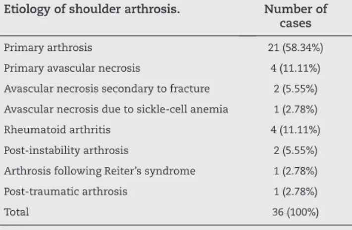
In relation to etiology, 21 cases of arthrosis were primary (58.34%) and seven resulted from necrosis (19.44%), of which four were idiopathic necrosis (11.11%), two were necrosis secondary to fractures (5.55%) and one was necrosis secondary to sickle-cell anemia (2.78%). Four cases were arthrosis secondary to rheumatoid arthritis (11.11%), one was secondary to Reiter’s syndrome (2.78%), two were secondary to instability (5.55%) and one was secondary to trauma (wound caused by white arms) (2.78%) (Table 2).
Fig. 1 - Image of a glenoid cavity (arrow) with insufficient bone stock, in which it would be possible to insert an implant.
The arthrosis were classified in accordance with Walch et al.16 (Fig. 2) as A1 (four shoulders), A2 (31 shoulders) and B1
(one shoulder) (Table 3).
Regarding cementation, five prostheses were not cemented and the other 31 were cemented. In five cases, a technique of
interposition of the glenoid cavity was performed, in which in four cases the individual’s own joint capsule was used and in one case a graft from a tissue bank (calcaneal tendon) was used.
In order to measure the degree of joint mobility, we used the AAOS method17 (American Academy of Orthopedic Surgeons).
N Sex Age Dominance Duration of
symptoms (months)
Physiotherapy (duration in
months)
Length of follow-up (in months)
1 F 70 + 24 3 12
2 M 59 - 6 3 13
3 F 70 - 36 3 28
4 F 68 - 96 6 72
5 F 47 + 24 3 132
6 F 71 - 144 4 36
7 F 44 + 120 36 42
8 F 70 - 18 6 60
9 F 61 ambidextrous RS 12 4 24
10 F 46 - 48 6 72
11 F 85 + 5 7 29
12 F 63 + 60 6 90
13 F 65 - 72 3 72
14 F 79 + 48 6 24
15 F 74 - 30 5 72
16 F 36 - 60 9 24
17 F 74 + 108 6 12
18 F 77 + 30 6 84
19 F 49 + 42 7 24
20 F 72 - 30 18 60
21 F 69 + 48 12 24
22 M 51 - 120 5 24
23 F 41 + 18 6 16
24 F 41 - 18 6 13
25 M 51 + 36 12 54
26 F 22 + 24 24 24
27 F 37 + 36 36 36
28 F 85 - 3 2 24
29 F 75 ambidextrous RS 6 4 84
30 M 31 + 108 7 30
31 F 46 - 48 18 24
32 F 66 + 60 6 12
33 F 39 - 36 3 108
34 F 26 - 60 3 36
35 M 48 + 60 6 72
36 M 53 - 36 3 16
+: dominant side operated; F: female; M: male; N: number; RS: right shoulder.
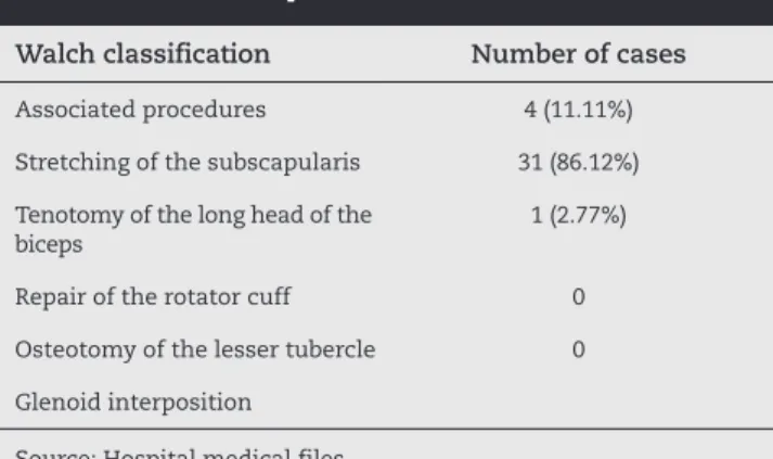
The associated procedures during the transoperative period are shown in Table 5.
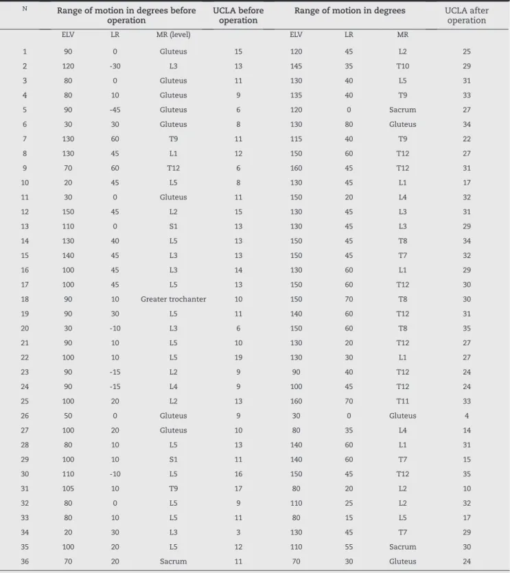
To evaluate the results, we used the UCLA method (University of California at Los Angeles),18 with statistical
comparisons of the following variables: age, gender, etiology, side operated, dominance, bilaterality, length of time with symptoms and ranges of elevation, lateral rotation and medial rotation (before and after the operation). The partial UCLA variables relating to pain, function, active anterior flexion, active anterior flexion strength and satisfaction were also gathered, along with the preoperative and postoperative UCLA scores (Table 4).
With regard to radiographs, it was ascertained whether the arthrosis was concentric or eccentric. The state of the rotator cuff was evaluated, along with whether the individual had undergone osteotomy of the lesser tubercle, stretching of the subscapularis and cementation of the prosthesis. It was also noted whether the individual had undergone procedures of tenotomy of the tendon of the long head of the biceps (all the individuals who underwent tenotomy also underwent tenodesis), repair of the rotator cuff or interposition of the glenoid cavity.
Following this, a descriptive analysis was conducted on the abovementioned variables, and we verified the hypotheses relating to whether the differences in mean ranges of motion and UCLA scores improved after the surgery or not. Each of the variables was divided into two groups in order to facilitate the analysis, because of the small sample size. These groups were then analyzed using the Mann-Whitney test.
The UCLA variable was divided into two categories: unsatisfactory (from 0 to 27) and satisfactory (from 28 to 35): the range of elevation was divided into unsatisfactory (less than 90°) and satisfactory (at least 90°); and the range of lateral rotation was divided into unsatisfactory (less than 20°) and satisfactory (greater than or equal to 20°).
To construct the confidence intervals and to perform Student’s t test, respectively, a confidence interval of 95% and a significance level of 5% were used. The data analysis was done using the Minitab® software, version 16.
Results
We found that 21 shoulders (58.3%) had a satisfactory final result and 15 shoulders (41.7%) had an unsatisfactory result.
Regarding the ranges of elevation, lateral rotation and medial rotation and the UCLA score, all of these increased after the surgery (mean differences of 35°, 27°, 4° and 17 points, respectively). At the significance level of 5%, it was observed that there were gains in all these variables (Table 4).
According to the Mann-Whitney test for the variables of gender, age group, side operated, duration of postoperative physiotherapy, length of follow-up, dominance and bilaterality, there were no differences in the means for the differences in range of elevation, lateral rotation or medial rotation. There were also no differences in UCLA and postoperative UCLA (p < 0.05).
In relation to the duration of symptoms, there was no difference in the means for the ranges of elevation and medial Etiology of shoulder arthrosis. Number of
cases
Primary arthrosis 21 (58.34%)
Primary avascular necrosis 4 (11.11%) Avascular necrosis secondary to fracture 2 (5.55%) Avascular necrosis due to sickle-cell anemia 1 (2.78%)
Rheumatoid arthritis 4 (11.11%)
Post-instability arthrosis 2 (5.55%)
Arthrosis following Reiter’s syndrome 1 (2.78%)
Post-traumatic arthrosis 1 (2.78%)
Total 36 (100%)
Source: Hospital medical files.
Table 2 - Etiology of shoulder arthrosis.
Walch classification Number of cases
A1 4 (11.11%)
A2 31 (86.12%)
B1 1 (2.77%)
B2 0
C 0
Source: Hospital medical files. Anteversion of the second measurement.
Table 3 - Walch classification for shoulder arthrosis.
Fig. 2 -Walch classification in relation to the different types of glenoid morphology in cases of glenohumeral arthrosis. A1 = centered head with minimal erosion. A2 = centered head with greater erosion. B1 = posteriorly subluxated head with sclerosis and posterior osteophytes. B2 = posteriorly subluxated head with biconcave appearance of the glenoid. C = glenoid retroversion greater than 25°, independent of its erosion.
N Range of motion in degrees before
operation
UCLA before operation
Range of motion in degrees UCLA after operation
ELV LR MR (level) ELV LR MR
1 90 0 Gluteus 15 120 45 L2 25
2 120 -30 L3 13 145 35 T10 29
3 80 0 Gluteus 11 130 40 L5 31
4 80 10 Gluteus 9 135 40 T9 33
5 90 -45 Gluteus 6 120 0 Sacrum 27
6 30 30 Gluteus 8 130 80 Gluteus 34
7 130 60 T9 11 115 40 T9 22
8 130 45 L1 12 150 60 T12 27
9 70 60 T12 6 160 45 T12 31
10 20 45 L5 8 130 45 L1 17
11 30 0 Gluteus 11 150 20 L4 32
12 150 45 L2 15 130 45 L3 31
13 110 0 S1 13 130 45 L3 29
14 130 40 L5 13 150 45 T8 34
15 140 45 L3 13 150 45 T7 32
16 100 45 L3 14 130 60 L1 29
17 100 45 L5 13 150 60 T12 30
18 90 10 Greater trochanter 10 150 70 T8 30
19 90 30 L5 11 140 60 T12 31
20 30 -10 L3 6 150 60 T8 35
21 90 10 L5 10 130 20 T12 27
22 100 10 L5 19 130 30 L1 27
23 90 -15 L2 9 90 40 T12 24
24 90 -15 L4 9 100 45 T12 24
25 100 20 L2 13 160 70 T11 33
26 50 0 Gluteus 9 30 0 Gluteus 4
27 100 20 Gluteus 10 80 35 L4 14
28 80 10 L5 13 140 60 L1 31
29 100 10 S1 11 140 60 T7 15
30 110 -10 L5 16 150 45 T12 35
31 105 10 T9 17 80 20 L2 10
32 80 0 L5 9 110 25 L2 32
33 80 10 L5 11 80 15 L5 17
34 20 30 L3 3 130 45 T7 29
35 100 20 L5 12 110 55 Sacrum 30
36 70 20 Sacrum 11 70 30 Gluteus 24
Source: Hospital medical files; ELV: elevation; L: lumbar vertebra followed by its number; LR: lateral rotation; MR: medial rotation; N: number; T: thoracic vertebra followed by its number.
rotation, or for the postoperative UCLA score. In relation to the range of lateral rotation, the difference was greater when the duration of symptoms was up to three years. The mean for the difference in lateral rotation in patients with symptoms of duration up to three years was 35° and with duration greater than three years, 20°.
We also tested whether there was any difference in relation to the range of postoperative lateral rotation in groups that had and had not undergone stretching of the subscapularis, but the difference between the means was not significant.
We also took into consideration the possible complications of infection, loosening, fractures and nerve lesions, but none of these occurred in any of our patients.
At the significance level of 5% (p < 0.05), no relationship was found between the UCLA score and the following variables: gender (p = 1.000), side operated (p = 0.864), dominance (p = 1.000), bilaterality (p = 0.309), concentric arthrosis (p = 0.417), eccentric arthrosis (p = 0.417), state of the rotator cuff (p = 1.000), presence of osteotomy of the lesser tubercle (p = 0.705), presence of cementation (p = 0.630), presence of repairs to the rotator cuff (p = 1.000), presence of interposition (p = 1.000), symptom duration of up to three years (p = 0.155), physiotherapy duration of up to six months (p = 1.000), length of follow-up of up to two years (p = 0.364), lateral rotation of at least 20° (p = 0.064), elevation of at least 90° (p = 0.063) and having undergone stretching of the subscapularis (p = 0.082).
For the same significance level of 5% (p < 0.05), relationships were found between satisfactory UCLA score and two variables: patients with maximum age of 60 years at the time of the surgery (p = 0.016); and patients who underwent tenotomy of the long head of the biceps (p = 0.046).
Discussion
In the literature, arthroplasty for treating shoulder osteoarthro-sis has presented excellent results in most patients, regardless of whether this is partial or total arthroplasty. Nevertheless, controversy still continues in relation to coverage of the glenoid cavity or not.19
Neer2 first described partial shoulder arthroplasty for
treating shoulder arthrosis in 1974. In his series of 46 patients,
20 had excellent results, 20 had satisfactory results and six had unsatisfactory results. The results were encouraging, but many patients reported that their strength was slow to return and that they had difficulty in doing activities above head height. With the aim of improving these patients’ function, total shoulder arthroplasty was developed, in which the glenoid component was cemented, and the results were considered to be favorable.1 High incidence of radiolucent lines
was observed on the cement-bone interface,3-5,7,20-23 and this
reached 100% in patients with rheumatoid arthritis.24 Even
though several subsequent studies have demonstrated that the great majority of these lines do not progress to symptomatic loosening,3-5,20,22,24,25 many authors have recommended partial
shoulder arthroplasty for treating shoulder osteoarthrosis, in order to minimize the chance of needing glenoid component revision because of its loosening,26-28 which is the main
complication of this type of prosthesis.
Many studies6,19,29-31 comparing the results from total
and partial shoulder arthroplasty for treating shoulder osteoarthrosis have shown slightly better results from using total shoulder arthroplasty, in relation to long-term pain relief. However, in terms of patients’ strength, function, range of motion and general satisfaction, the results remain unclear.19
Among our results, we observed that there was a notable improvement in UCLA score through using partial shoulder arthroplasty for treating shoulder osteoarthrosis.
In 2002, a study by Godenèche et al.32 demonstrated good
and excellent results in 77% of the individuals who underwent shoulder arthroplasty to treat shoulder osteoarthrosis, without any statistically significant difference in the results between total and partial prostheses, but with greater pain relief among individuals who underwent tenotomy of the tendon of the long head of the biceps, which is compatible with our study. Levine et al.1 demonstrated in 1997 that partial shoulder arthroplasty
may be an effective treatment for shoulder osteoarthrosis, albeit in selected cases in which the result was dependent on the preoperative state of the glenoid cavity. In our study, we were unable to find any relationship between the preoperative state of the glenoid and the final result. On the other hand, we did not indicate partial shoulder arthroplasty for glenoid cases classified by Walch et al.16 as B2 or C.
With regard to correlating the surgical results with the individuals’ ages, the study by Saltzman et al.10 demonstrated
that partial shoulder arthroplasty in patients with glenohumeral arthrosis who were under 55 years of age led to improvements in pain and shoulder function. Likewise, the study by Bartelt et al.29 demonstrated improvement of range
of motion in patients under the age of 55 years with shoulder osteoarthrosis who underwent partial shoulder arthroplasty. In our study, we had a result similar to these, in which we observed better results in the patients operated at ages of less than 60 years (Fig. 3 A-D).
We can also cite the study by Burkhead and Hutton (1995),33
who described the technique of glenoid interposition of the fascia lata or the anterior joint capsule combined with arthroplastic replacement of the humeral head, with good results. We applied this technique in five cases, but we were unable to demonstrate any relationship between this and the UCLA score.
Walch classification Number of cases
Associated procedures 4 (11.11%)
Stretching of the subscapularis 31 (86.12%) Tenotomy of the long head of the
biceps
1 (2.77%)
Repair of the rotator cuff 0
Osteotomy of the lesser tubercle 0 Glenoid interposition
Source: Hospital medical files.
Given the known possible complications from revision of total shoulder arthroplasty and the possible difficulties in implanting the glenoid component (Fig. 1), we demonstrated in our study that partial shoulder arthroplasty is a viable option for treating shoulder osteoarthrosis in cases in which there is a contraindication against implantation of a glenoid component. The results were shown to be better in patients under the age of 60 years who underwent surgery, and also in those who underwent tenotomy of the tendon of the long head of the biceps.
Conclusion
We found that the patients with shoulder osteoarthrosis who underwent partial shoulder arthroplasty achieved better results if they underwent the operation under the age of 60 years and if tenotomy with tenodesis of the long head of the biceps was performed as an associated procedure, independent of age.
Conflicts of interest
The authors declare that there was no conflict of interests in conducting this study.
R E F E R E N C E S
1. Levine WN, Djurasovic M, Glasson JM, Pollock RG, Flatow EL, Bigliani LU. Hemiarthroplasty for glenohumeral osteoarthritis: results correlated to degree of glenoid wear. J Shoulder Elbow Surg. 1997;6(5):449-54.
2. Neer CS, 2nd. Replacement arthroplasty for glenohumeral osteoarthritis. J Bone Joint Surg Am. 1974;56(1):1-13. 3. Amstutz HC, Thomas BJ, Kabo JM, Jinnah RH, Dorey FJ. The
Dana total shoulder arthroplasty. J Bone Joint Surg Am. 1988;70(8):1174-82.
4. Barrett WP, Franklin JL, Jackins SE, Wyss CR, Matsen FA, 3rd. Total shoulder arthroplasty. J Bone Joint Surg Am. 1987;69(6):865-72.
5. Barrett WP, Thornhill TS, Thomas WH, Gebhart EM, Sledge CB. Nonconstrained total shoulder arthroplasty in patients with polyarticular rheumatoid arthritis. J Arthroplasty. 1989;4(1):91-6.
6. Cofield RH. Total shoulder arthroplasty with the Neer prosthesis. J Bone Joint Surg Am. 1984;66(6):899-906.
7. Hawkins RJ, Bell RH, Jallay B. Total shoulder arthroplasty. Clin Orthop Relat Res. 1989;(242):188-94.
8. Neer CS 2nd, Morrison DS. Glenoid bone-grafting in total shoulder arthroplasty. J Bone Joint Surg Am. 1988;70(8):1154-62.
9. Hill JM, Norris TR. Long-term results of total shoulder arthroplasty following bone-grafting of the glenoid. J Bone Joint Surg Am. 2001;83(6):877-83.
10. Saltzman MD, Chamberlain AM, Mercer DM, Warme WJ, Bertelsen AL, Matsen FA 3rd. Shoulder hemiarthroplasty with concentric glenoid reaming in patients 55 years old or less. J Shoulder Elbow Surg. 2011;20(4):609-15.
11. Bonnevialle N, Mansat P, Mansat M, Bonnevialle P.
Hemiarthroplasty for osteoarthritis in shoulder with dysplastic morphology. J Shoulder Elbow Surg. 2011;20(3):378-84.
12. Bell SN, Gschwend N. Clinical experience with total
arthroplasty and hemiarthroplasty of the shoulder using the Neer prosthesis. Int Orthop. 1986;10(4):217-22.
13. Rodosky MW, Bigliani LU. Indications for glenoid resurfacing in shoulder arthroplasty. J Shoulder Elbow Surg. 1996;5(3):231-48. 14. Boileau P, Sinnerton RJ, Chuinard C, Walch G. Arthroplasty of
the shoulder. J Bone Joint Surg Br. 2006;88(5):562-75. 15. Pfahler M, Jena F, Neyton L, Sirveaux F, Mole D.
Hemiarthroplasty versus total shoulder prosthesis: results of cemented glenoid components. J Shoulder Elbow Surg. 2006;15(2):154-63.
16. Walch G, Badet R, Boulahia A, Khoury A. Morphologic study of the glenoid in primary glenohumeral osteoarthritis. J Arthroplasty. 1999;14(6):756-60.
17. Codsi M, Mccarron J, Brams JJ. The shoulder. Clinical evaluation of shoulder problems. 4th ed. Philadelphia: Saunders Elsevier; 2009.
18. Ellman H, Kay SP. Arthroscopic subacromial decompression for chronic impingement. Two- to five-year results. J Bone Joint Surg Br. 1991;73(3):395-8.
19. Edwards TB, Kadakia NR, Boulahia A, Kempf JF, Boileau P, Némoz C, et al. A comparison of hemiarthroplasty and total shoulder arthroplasty in the treatment of primary glenohumeral osteoarthritis: results of a multicenter study. J Shoulder Elbow Surg. 2003;12(3):207-13.
20. Fenlin Jr. JM, Ramsey ML, Allardyce TJ, Frieman BG. Modular total shoulder replacement. Design rationale, indications, and results. Clin Orthop Relat Res. 1994;(307):37-46.
21. Franklin JL, Barrett WP, Jackins SE, Matsen FA 3rd. Glenoid loosening in total shoulder arthroplasty. Association with rotator cuff deficiency. J Arthroplasty. 1988;3(1):39-46.
22. Weiss AP, Adams MA, Moore JR, Weiland AJ. Unconstrained shoulder arthroplasty. A five-year average follow-up study. Clin Orthop Relat Res. 1990;(257):86-90.
23. Wirth MA, Rockwood CA Jr. Complications of shoulder arthroplasty. Clin Orthop Relat Res. 1994;(307):47-69. 24. Kelly IG, Foster RS, Fisher WD. Neer total shoulder
replacement in rheumatoid arthritis. J Bone Joint Surg Br. 1987;69(5):723-6.
25. Bonutti PM, Hawkins RJ, Saddemi S. Arthroscopic assessment of glenoid component loosening after total shoulder arthroplasty. Arthroscopy. 1993;9(3):272-6.
26. Boyd AD Jr, Thornhill TS. Surgical treatment of osteoarthritis of the shoulder. Rheum Dis Clin North Am. 1988;14(3):591-611. 27. Brostrom LA, Kronberg M, Wallensten R. Should the glenoid
be replaced in shoulder arthroplasty with an unconstrained Dana or St. Georg prosthesis? Ann Chir Gynaecol.
1992;81(1):54-7.
28. Clayton ML, Ferlic DC, Jeffers PD. Prosthetic arthroplasties of the shoulder. Clin Orthop Relat Res. 1982;(164):184-91. 29. Bartelt R, Sperling JW, Schleck CD, Cofield RH. Shoulder
arthroplasty in patients aged fifty-five years or younger with osteoarthritis. J Shoulder Elbow Surg. 2011;20(1):123-30. 30. Thomas WH, Scott RD, Sledge CB, Thornhill TS. Total shoulder
arthroplasty versus hemiarthroplasty. Indications for glenoid resurfacing. J Arthroplasty. 1990;5(4):329-36.
31. Gartsman GM, Roddey TS, Hammerman SM. Shoulder arthroplasty with or without resurfacing of the glenoid in patients who have osteoarthritis. J Bone Joint Surg Am. 2000;82(1):26-34.
32. Godeneche A, Boileau P, Favard L, Le Huec JC, Lévigne C, Nové-Josserand L, et al. Prosthetic replacement in the treatment of osteoarthritis of the shoulder: early results of 268 cases. J Shoulder Elbow Surg. 2002;11(1):11-8.