1 – Trainee Physician, specializing in Shoulder and Elbow Surgery at the Orthopedics and Traumatology Clinic, Public Servants’ Hospital of the State of São Paulo (HSPE), São Paulo, SP, Brazil.
2 – Attending Physician in the Shoulder and Elbow Group, Orthopedics and Traumatology Clinic, Public Servants’ Hospital of the State of São Paulo (HSPE), São Paulo, SP, Brazil (HSPE), São Paulo, SP, Brazil.
3 – Head of the Shoulder and Elbow Group, Orthopedics and Traumatology Clinic, Public Servants’ Hospital of the State of São Paulo (HSPE), São Paulo, SP, Brazil. Work performed in the “Paulo Francisco Morato de Oliveira” Public Servants’ Hospital of the State of São Paulo (HSPE), São Paulo, SP.
Correspondence: Rua Borges Lagoa 1755, sala 180, 1º andar, Vila Clementino, 04038-034 São Paulo, SP. Fax: (11) 5573-3087. E-mail: [email protected] Work received for publication: November 7, 2011; accepted for publication: February 14, 2012.
POSTEROSUPERIOR SURGICAL ACCESS ROUTE FOR TREATMENT
OF ACROMIOCLAVICULAR DISLOCATIONS:
RESULTS FROM 84 SURGICAL CASES
Danilo Canesin Dal Molin1, Fabiano Rebouças Ribeiro2, Rômulo Brasil Filho2, Cantídio Salvador Filardi Junior3, Antonio Carlos Tenor Junior2, Willian Nandi Stipp1, Rodrigo Souto Borges Petros1
ABSTRACT
Objective: To evaluate the results from surgical treatment of 84 cases of acute acromioclavicular dislocation, using a posterosuperior access route. Methods: Eighty-four cases of acute acromioclavicular dislocation (grade III in the Allman-Tossy classification) operated between November 2002 and May 2010 were evaluated. The patients’ mean age was 34 years. The diagnoses were made using clinical and radiographic evaluations. The patients were operated by the same surgical team, within three weeks of the date of the trauma, using a posterosuperior approach to the shoulder to access the top of the base of the coracoid process for placement of two anchors, which were used in reducing the dislocation. The minimum follow-up was 12 months. The postoperative
clinical-radiographic evaluation was done using the modified Karlsson criteria and the University of California at Los Angeles (UCLA) score. Results: 92.8% of the 84 patients treated presented good or excellent results, and 7.2% presented fair or poor results, using the UCLA assessment score. According to the modified Karlsson criteria, 76.2% were assessed as grade A, 17.9% as grade B and 5.9% as grade C. Conclusion: The posterosuperior access route to the shoulder is a new option for accessing the coracoid process and treating acromioclavicular dislocation, with clinical and radiographic results equivalent to those in the literature.
Keywords – Acromioclavicular Joint/anatomy & histolo-gy; Acromioclavicular Joint/injuries; Acromioclavicular Joint/surgery
INTRODUCTION
Acromioclavicular dislocation is a common lesion
in orthopedic practice(1). The commonest trauma
me-chanism is a fall on the shoulder, with force applied directly on the acromion with the shoulder
adduc-ted(2). It occurs most commonly among young adults
because it is associated with impact sports and high--speed vehicle accidents(3,4).
The classification system of Tossy et al(5)
divi-des acromioclavicular dislocations into three types,
among which types I and II (mild and moderate) should be treated conservatively and type III (severe, with dislocation greater than 1 cm or greater than the thickness of the clavicle) should be treated surgically.
The classification system of Rockwood et al(6)
modi-fied type III and added types IV, V and VI to Tossy’s classification. The treatment for Rockwood’s types I and II is initially non-surgical(2,7). The treatment for
type III remains a matter for discussion, since it de-pends on the patient’s age, activities and deformities,
The authors declare that there was no conflict of interest in conducting this work
either loss from follow-up (12) or associated lesions in the same limb (six), which made the assessment methods impossible.
The diagnosis of acromioclavicular dislocation was made by means of physical and radiographic examinations on the patient. The patients included in the study presented less than 21 days of evolution from the trauma and had lesions of Tossy grade III.
The patients’ ages ranged from 17 to 61 years, with a mean of 33.9 years. Among the 84 patients evalu-ated, 68 (80.9%) were male and 16 (19.1%) were female. The dominant side was affected in 53 pa-tients (63%) and the non-dominant side in 31 papa-tients (37%). The mean length of follow-up was 54 months, with a minimum of 12 months and maximum of eight years and five months.
The patients were assessed using the modified cri-teria of Karlsson et al(20) (Chart 1) and according to
the score of the University of California in Los
An-geles (UCLA)(21). Radiographic examinations were
performed using the bilateral anteroposterior view on the acromioclavicular joint, anteroposterior view on the chest and axillary view on the shoulder, bilaterally.
Chart 1 – Modified Karlsson criteria for clinical-radiographic evaluation of acromioclavicular dislocations.
Grade A: without pain, normal movements and without muscle atrophy; anatomical restoration of the joint or displacement less than 5 mm on radiography.
Grade B: slight pain, slight limitation of movements and slight muscle atrophy; with displacement greater than 5mm on radiography. Grade C: with pain, with muscle atrophy and with significant limitation of movements; with displacement greater than 5 mm on radiography.
while types IV, V and VI require surgical treatment .
More than 75 techniques have been described for sur-gically treating acromioclavicular dislocation, but none of them has been established as the gold standard(9).
The techniques include fixation of the acromioclavicular joint using wires or a plate(10,11), transfer of the
coraco-acromial ligament(12), fixation of the coracoacromial
interval using a screw, suturing anchors or lashing with suturing wires(13,14) and reconstruction of the
coracocla-vicular and acromioclacoracocla-vicular ligaments(15).
The modifications to the techniques for fixing the clavicle to the coracoid process go from use of screws or subcoracoid lashings to use of anchors and material
of Endobutton®
type (flip-button®
), which can be used as open, minimally invasive or arthroscopic procedu-res(16-18). Anchors have the advantage that they can be
placed more proximally to the anatomical insertion site of the coracoclavicular ligaments(19).
The posterosuperior shoulder access route for trea-ting acromioclavicular dislocations was presented at the
34th Brazilian Congress of Orthopedics and
Traumato-logy (2002) and was awarded the “Prof. Orlando Pinto de Souza” award for creativity. It was developed by the Shoulder and Elbow Group of the Public Servants’ Hos-pital of the State of São Paulo (HSPE/IAMSPE) with the aims of enabling easy access to the top of the base of the coracoid process, maintaining a more anatomical reduction of the acromioclavicular joint and preserving the anterior portion of the deltoid muscle, thereby allo-wing better functional recovery for the shoulder.
The aim of the present study was to evaluate the clinical and radiographic results from patients who underwent treatment for acute acromioclavicular dislocation by means of a posterosuperior shoulder access route.
METHODS
Eighty-four cases of acute acromioclavicular dislo-cation (grade III of Tossy’s classifidislo-cation) were trea-ted surgically between November 2002 and May 2010 using the posterosuperior access route. The operations were performed by the Shoulder and Elbow Group of the Public Servants’ Hospital of the State of São Paulo (HSPE/IAMSPE).
Over this period, 102 patients underwent surgery to treat acute acromioclavicular dislocations. Eighteen patients were excluded from this study because of
SURGICAL TECHNIQUE
After administration of general anesthesia and regional blockade, the patient was positioned semi--seated on the surgical table. Asepsis was performed on the affected shoulder and respective limb, while leaving the latter free for movement. A sabre incision was made in the skin and subcutaneous tissue in the anteroposterior direction, starting 1 cm anteriorly to the acromioclavicular joint and going to 3 cm posteriorly.
565
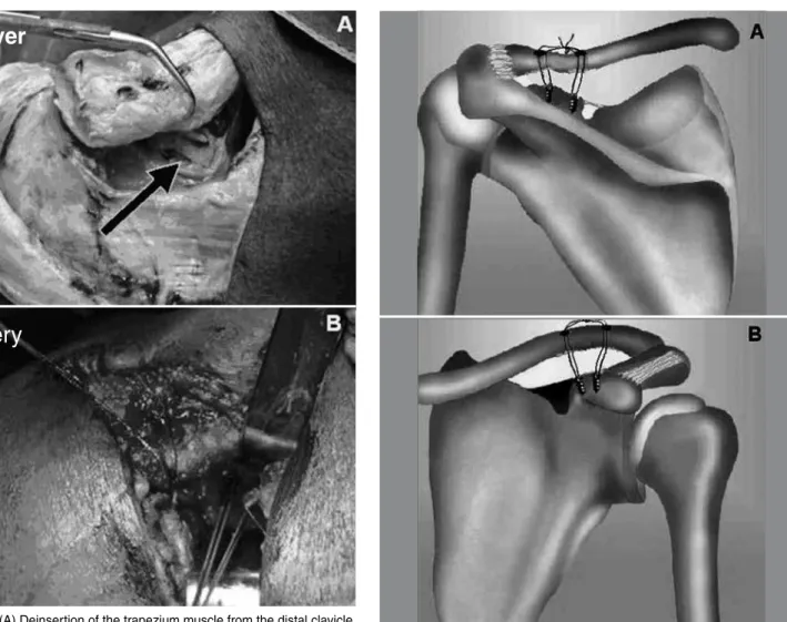
Figure 1 – (A) Deinsertion of the trapezium muscle from the distal clavicle, which was subsequently pushed away so that the top of the base of the coracoid process (arrow) could be viewed, in a cadaver. (B) Anchors
posi-tioned in the coracoid process during the surgery. Figure 2 – the holes made in the clavicle: (A) posterior view; and (B) anterior view.Schematic diagram showing the positioning of the anchors and
a Velpeau sling for 40 days and was instructed to per-form active flexion of the elbow starting in the imme-diate postoperative period. Pendulum exercises were performed starting in the third week after the operation.
RESULTS
Out of the 84 cases evaluated, 52 (61.9%) were considered to have excellent results according to the UCLA score, 26 (30.9%) had good results and six (7.2%) had fair results. None of the patients presented poor results. Among the 26 patients with good results, 18 had a score of 33, thus losing two points regarding the pain criterion (Figure 4).
Thirteen patients (15.4%) presented loss of reduction during the postoperative period. In three cases (3.5%), the loss occurred within the first six weeks after the operation. All the patients who presented fair results according to the UCLA score showed losses of joint
RESULTS FROM 84 SURGICAL CASES
high-resistance threads were then fixed to the top of the base of the coracoid process. In the clavicle, two holes were made in the superoinferior direction, at the level of the conoid tubercle and roughly along the line of the clavicle. The lateral orifice was used as the entry point, from inferiorly to superiorly, for the threads from the anchor that was fixed most la-terally in the coracoid process. The threads from the anchor that was fixed most medially in the coracoid process were passed through the medial orifice of the clavicle. To pass the anchor threads through the ori-fices that were made in the clavicle, a doubled-over fine steel wire was used. The acromioclavicular joint was then reduced and the threads were tied off above the clavicle (Figure 2). The trapezoid muscle was reinserted into the clavicle and deltoid-trapezoidal fascia. Control radiographs were produced after the operation (Figure 3).
During the postoperative evolution, the patient used
C
adaver
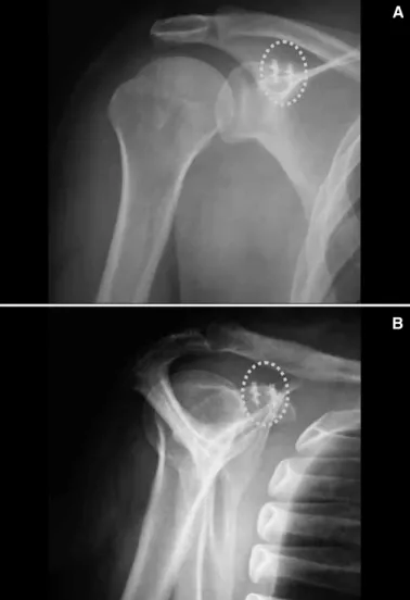
Figure 3 – Postoperative radiograph of the shoulder in anteroposterior view (A) and lateral view (B), demonstrating the anchors in the coracoid process and the reduction of the acromioclavicular joint.
Figure 4 – UCLA score results for the 84 patients operated through the posterosuperior access route to treat acromioclavicular dislocations.
Figure 5 – Modified Karlsson score results for the 84 patients operated by means of the posterosuperior access route to treat acromioclavicular dislocations.
DISCUSSION
The aim in surgically treating acute acromioclavi-cular dislocations is to achieve anatomical reduction so as to enable healing of the soft tissues and recovery of the previous functioning of the joint. The large number of procedures described in the literature demonstrates the lack of consensus regarding the ideal method(16).
The techniques for reconstructing the coracoclavi-cular ligaments through transferring the coracoacro-mial ligament or using biological or synthetic grafts present the complication of loss of reduction. This has been found in up to 24% of the cases treated using the Weaver-Dunn method(15,22). The lower resistance of the
coracoacromial ligament and the change in direction of the force vector in relation to the coracoclavicular ligaments are the main causes of this complication(23).
Despite the advantage of technical simplicity, acro-mioclavicular fixation by means of metal wires may present the complications of migration of the wires, infection, arthrosis and loss of reduction(24,25).
There are several techniques for fixation of the
clavicle to the coracoid process(9). Bosworth screws
present the disadvantage of requiring a new procedure to remove them, and they present high rates of osteoly-sis in the clavicle(9). High-resistance threads that are
tied off in the subcoracoid area may cause local bone erosion and maintain the anterior subluxation of the clavicle, and this has been shown to cause pain, arthri-tis and diminished strength(26). Fixation of the clavicle
to the coracoid process using suture anchors facilitates the operative technique, although the anterior access route still requires dissection of the anterior portion of the deltoid muscle and maintains residual subluxation of the clavicle, since it does not reestablish the force
A
B
reduction, but they did not present functional limitation and there was no need for any new intervention.
According to the modified Karlsson criteria, 64 patients (76.2%) presented grade A, 15 (17.9%) grade B and five (5.9%) grade C (Figure 5).
Excellent Good
567
vector of the coracoclavicular ligaments.
The posterosuperior shoulder access route provides direct access to the top of the base of the coracoid pro-cess without damage to the anterior deltoid muscle. This may keep the joint reduction more anatomical and reestablish joint stability because of the positio-ning of the anchors in the anatomical position of the ruptured coracoclavicular ligaments.
The results obtained were analyzed using the UCLA scale because this is the method most often used in the literature for analyzing the results from surgical treatment of acromioclavicular dislocations. The Karlsson criteria were used because they are spe-cific for diseases that involve the acromioclavicular joint, but they were modified so that the functional limitations and loss of reduction would be better quantified in the evaluation.
Algarín et al(27) evaluated 42 patients who were
treated using a minimally invasive technique for cora-coclavicular fixation using a cortical screw and acro-mioclavicular fixation using Kirschner wires. They found that 57% of their results were excellent and
29% were good, using the UCLA score, and their values were close to what we obtained in our study.
Lädermann et al(28) conducted a clinical,
radiogra-phic and isokinetic evaluation on 37 patients who un-derwent coracoclavicular cerclage and acromioclavicu-lar stabilization using non-absorbable sutures, to treat acromioclavicular dislocations. Seven patients (18.9%) presented loss of reduction, and this was correlated with less satisfactory results, just like in our study.
The posterosuperior shoulder access route for pla-cement of suture anchors at the base of the coracoid process was shown to be easy to learn and reprodu-cible, and the results presented were similar to those from techniques described in the literature, with a low rate of postoperative complications.
CONCLUSION
The posterosuperior shoulder access route is a new option for accessing the coracoid process and treating acromioclavicular dislocations, with clinical and ra-diographic results equivalent to those in the literature.
REFERENCES
1. Bannister GC, Wallace WA, Stableforth PG, Hutson MA. The management of acute acromioclavicular dislocation. A randomised prospective controlled trial. J Bone Joint Surg Br. 1989;71(5):848-50.
2. Lemos MJ. The evaluation and treatment of the injured cromioclavicular joint in athletes. Am J Sports Med. 1998;26(1):137-44.
3. Bishop JY, Kaeding C. Treatment of the acute traumatic acromioclavicular separation. Sports Med Arthrosc. 2006;14(4):237-45.
4. Mazzocca AD, Arciero RA, Bicos J. Evaluation and treatment of acromiocla-vicular joint injuries. Am J Sports Med. 2007;35(2):316-29.
5. Tossy JD, Mead NC, Sigmond HM. Acromioclavicular separations: useful and practical classification for treatment. Clin Orthop Relat Res. 1963;28:111-9. 6. Rockwood CA Jr, Williams GR Jr, Young DC. Disorders of the acromioclavicular
joint. Rockwood CA Jr, Matsen FA 3rd, editors. The shoulder. Philadelphia: Saunders: 1998. p. 485-553.
7. Mouhsine E, Garofalo R, Crevoisier X, Farron A. Grade I and II acromioclavicular dislo-cations: results of conservative treatment. J Shoulder Elbow Surg. 2003;12(6):599-602. 8. Nissen CW, Chatterjee A. Type III acromioclavicular separation: results of a
re-cent survey on its management. Am J Orthop (Belle Mead NJ). 2007;36(2):89-93. 9. Johansen JA, Grutter PW, McFarland EG, Petersen SA. Acromioclavicular
joint injuries: indications for treatment and treatment options. J Shoulder Elbow Surg.2011;20(Suppl 2):S70-82.
10. Phemister PB. The treatment of dislocation of the acromioclavicular joint by open reduction and threaded wire fixation. J Bone Joint Surg 1942; 24:166-68. 11. Sim E, Schwarz N, Höcker K, Berzlanovich A. Repair of complete acromiocla-vicular separations using the acromioclaacromiocla-vicular-hook plate. Clin Orthop Relat Res. 1995;(314):134-42.
12. Weaver JK, Dunn HK. Treatment of acromioclavicular injuries, especially com-plete acromioclavicular separation. J Bone Joint Surg Am. 1972;54(6):1187-94. 13. Bosworth BM. Acromioclavicular separation: new method of repair. Surg Gynecol
Obstet. 1941;73:866-71.
14. Breslow MJ, Jazrawi LM, Bernstein AD, Kummer FJ, Rokito AS. Treatment of acromioclavicular joint separation: suture or suture anchors? J Shoulder Elbow Surg. 2002;11(3):225-9.
15. Carofino BC, Mazzocca AD. The anatomic coracoclavicular ligament re-construction: surgical technique and indications. J Shoulder Elbow Surg. 2010;19(Suppl 2):37-46.
16. Simoni M, Brandão BL, Tumolo LH. Tratamento da luxação acromioclavicular com amarrilhos coracoclaviculares por meio de duas miniincisões. Rev Bras Ortop. 2005;40(8):483-9.
17. DeBerardino TM, Pensak MJ, Ferreira J, Mazzocca AD. Arthroscopic stabili-zation of acromioclavicular joint dislocation using the AC graftrope system. J Shoulder Elbow Surg. 2010;19(Suppl 2):47-52.
18. Vieira LAG, Visco A, Fernandes LFD, Cordero NGG. Tratamento artroscópico da luxação acromio-clavicular pelo método “Tight Rope” (Arthrex®). Rev Bras Ortop. 2009;44(1):52-6.
19. Wellmann M, Zantop T, Weimann A, Raschke MJ, Petersen W. Biomechani-cal evaluation of minimally invasive repairs for complete cromioclavicular joint dislocation. Am J Sports Med. 2007;35(6):955-61.
20. Karlsson J, Arnarson H, Sigurjónsson K. Acromioclavicular dislocations treated by coracoacromial ligament transfer. Arch Orthop Trauma Surg. 1986;106(1):8-11. 21. Ellman H, Hanker G, Bayer M. Repair of the rotator cuff. End-result study of
factors influencing reconstruction. J Bone Joint Surg Am. 1986;68(8):1136-44. 22. Rokito AS, Oh YH, Zuckerman JD. Modified Weaver-Dunn procedure for
acro-mioclavicular joint dislocations. Orthopedics. 2004;27(1):21-8.
23. Motamedi AR, Blevins FT, Willis MC, McNally TP, Shahinpoor M. Biomechanics of the coracoclavicular ligament complex and augmentations used in its repair and reconstruction. Am J Sports Med. 2000;28(3):380-4.
24. Lyons FA, Rockwood CA Jr. Migration of pins used in operations on the shoul-der. J Bone Joint Surg Am. 1990;72(8):1262-7.
25. Norrell H Jr, Llewellyn RC. Migration of a threaded steinmann pin from an acromioclavicular joint into the spinal canal. A case report. J Bone Joint Surg Am. 1965;47:1024-6.
26. Baker JE, Nicandri GT, Young DC, Owen JR, Wayne JS. A cadaveric study examining acromioclavicular joint congruity after different methods of coraco-clavicular loop repair. J Shoulder Elbow Surg. 2003;12(6):595-8.
27. Algarín JR, Salcedo JD, Rodríguez JO, Bello AG, Sancho FB. [Grade III ac-romioclavicular dislocation treated with a minimally invasive approach]. Acta Ortop Mex. 2010;24(5):317-23.