Acute hemodynamic, respiratory and metabolic
alterations after blood contact with a volume
priming and extracorporeal life support circuit: an
experimental study
Alterações hemodinâmicas respiratórias e metabólicas agudas
após o contato do sangue com o circuito extracorpóreo da
ECMO: estudo experimental
INTRODUCTION
Extracorporeal membrane oxygenation (ECMO) has been used to support patients with severe respiratory and/or cardiovascular failure.(1) Despite increasing clinical experience and success,(2-5) there are currently few reports in the literature about the physiology and about acute patients’
experienced with ECMO use.(6)
In animal studies, the use of polypropylene ECMO is associated with an acute elevation of plasmatic interleukins and with intestinal and
pulmonary edema.(7) Accordingly, gut barrier dysfunction associated with
polypropylene ECMO use can be an important contributor to
ECMO-related systemic inlammation.(8) In human beings, respiratory support
using polymethylpentene ECMO membranes is also associated with early, new, radiographic pulmonary iniltrates, a phenomenon known as
white-out.(9) he development of bioactive and biopassive coated extracorporeal
Marcelo Park, Eduardo Leite Vieira Costa, Alexandre Toledo Maciel, Adriana Sayuri Hirota, Edzangela Vasconcelos and Luciano Cesar Pontes Azevedo on behalf of Hospital das Clínicas de São Paulo and Hospital Sírio Libanês ECMO group*
ABSTRACT
Objective: To investigate the hemodynamic, respiratory and metabolic impact of blood contact with a priming volume and extracorporeal membrane oxygenation circuit, before the initiation of oxygenation and ventilation
Methods: Five animals were instru-mented and submitted to extracorpo-real membrane oxygenation. Data were collected at the baseline and 30 minutes after starting extracorporeal circulation, without membrane ventilatory (swee-per) low.
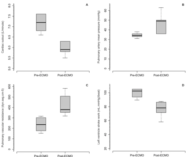
Results: After starting extracorporeal membrane oxygenation, there was a non-signiicant elevation in pulmonary vascular resistance from 235 (178,303) to 379 (353,508) dyn.seg.(cm5)-1 (P=0.065),
associated with an elevation in the alveolar arterial oxygen gradient from
235 (178,303) to 379 (353,508) mmHg (P=0.063). We also observed a reduction in the left ventricle stroke work from 102 (94,105) to 78 (71,87) (mL.mmHg)/beat (P=0.064), in addition to a reduction in cardiac output from 7.2 (6.8,7.6) to 5.9 (5.8,6.3) L/min (P=0.188). he right ventricle stroke work was counterbalanced between the pulmonary vascular resistance increment and the cardiac output reduction, maintaining a similar value.
Conclusions: We presented an experimental model that is feasible and safe. Blood contact with the priming volume and extracorporeal membrane oxygenation circuit resulted in non-signiicant systemic or metabolic changes.
Keywords: Acute respiratory
distress syndrome; Respiration, artiicial; Extracorporeal membrane oxygenation; Multiple organ failure; Swine
This study was conducted at the Instituto de Ensino e Educação do Hospital Sírio Libanês - São Paulo (SP), Brazil.
Conflicts of interest: The authors received a donation of PLS systems from Maquet Cardiopulmonary from Brazil, to conduct an experimental research study and for patient support.
Submitted on March 30, 2012 Accepted on June 15, 2012
Corresponding author:
Marcelo Park Rua Francisco Preto, 46
circuits reduced inlammatory and pro-coagulant pathway activation in a simulated model.(10)
Routine priming using calcium salts can prevent
severe hypocalcemia in neonates;(11) however, the
efects of priming with normal saline have not been characterized. hus, the aim of this study was to describe the acute hemodynamic, respiratory and metabolic efects of blood-extracorporeal circuit contact and priming with normal saline in an animal model.
METHODS
his study received approval from the Institutional Animal Research Ethics Committee and was performed according to National Institutes of Health guidelines for the use of experimental animals. he instrumentation and surgical preparation were performed as previously described.(12-14)
Instrumentation and surgical preparation
he room temperature was set at 24ºC. Five domestic female Agroceres pigs (80±3 kg) were anesthetized with thionembutal (10 mg/kg, Tiopental, Abbott, Brazil) and pancuronium bromide (0.1 mg/kg, Pavulon, AKZO Nobel, Brazil) and were connected to a mechanical ventilator (Evita XL Dräger, Dräger, Luebeck, Germany) with the following parameters: tidal volume 8 mL/kg; end-expiratory pressure 5 cm H2O; FIO2 initially set at 100% and subsequently adjusted to maintain arterial saturation between 94% and 96%; and respiratory rate
titrated to maintain PaCO2 between 35 and 45 mm
Hg or end-tidal CO2 (NICO, Dixtal Biomedica Ind.
Com, Sao Paulo, Brazil) between 30 and 40 mm Hg. he electrocardiogram, heart rate, oxygen saturation, and pressures of the animals were monitored with a multiparametric monitor (Ininity Delta XL, Dräger, Luebeck, Germany). Anesthesia was maintained during the study with midazolam (1- 5 mg.kg-1.h-1) and fentanyl (5-10 mcg.kg-1.h-1, Fentanyl; Janssen-Cilag, Brazil), and muscular relaxation was maintained with pancuronium
bromide (0.2 mg.kg-1.h-1). he adequate depth of
anesthesia during the surgical period was evaluated with the maintenance of physiological variables (heart rate and arterial pressure) and the absence of relexes (corneal and hind limb lexion response), as well as unresponsiveness to stimuli during manipulation. Supplementary boluses of 3 - 5 mcg/kg fentanyl and 0.1 – 0.5 mg/kg midazolam were administered as necessary.
he left external jugular vein was cannulated (guided by ultrasonography) to introduce a pulmonary
artery catheter, and the right external jugular vein was cannulated to introduce a 25-cm ECMO devolution cannula (Edwards Lifesciences, Irvine, CA, USA). he right femoral vein was punctured for the insertion of a 55-cm ECMO drainage cannula (Edwards Lifesciences, Irvine, CA, USA), which was positioned close to the right atrium with the aid of trans-hepatic ultrasonographic visualization. Only the guidewires were kept in place after the irst baseline measurements during the stabilization period; the guidewires were then replaced by the cannulas. After the insertion of the guidewires, an infusion of 1000 IU per hour of heparin was started. A central venous catheter and an invasive arterial blood pressure catheter were placed in the left femoral vein and artery, respectively.
hrough a midline laparotomy, a cistostomy was performed, and a bladder catheter was inserted. During the surgical interventions, 15 mL.kg-1.h-1 of lactated Ringer’s was continuously infused, and boluses of 250 mL were administered to maintain a systemic mean arterial blood pressure (ABPm) of 65 mm Hg or greater, a central
venous pressure (CVP) of 8 mm Hg or greater, and SvO2
greater than 65% until the end of instrumentation.
Stabilization and support of the animals
After the end of the instrumentation, the animals were allowed to stabilize for 1 h. At the beginning of the stabilization period, a continuous infusion of 3 mL.kg-1.h-1 of lactated Ringer’s was started and maintained throughout the entire experiment.
When the animals became hypotensive (ABPm<65 mm Hg), a bolus of 500 mL of lactated Ringer’s was infused. If the ABPm failed to rise above 65 mmHg after the bolus, an infusion of norepinephrine 0.1 mcg.kg-1.min-1 (Norepine, Opem Pharmaceuticals, São Paulo, Brazil) was started and titrated to an ABPm≥65 and <80 mmHg.
ECMO priming, starting, and maintenance
he ECMO system (Permanent life support system - PLS, Jostra – Quadrox D, Maquet Cardiopulmonary, Hirrlingen, Germany) was primed with a 37ºC normal saline solution and connected to a centrifugal pump (Rotalow, Jostra, Maquet Cardiopulmonary, Hirrlingen, Germany). With the circuit illed, 1000 IU of heparin were injected in the circulating luid.
he PLS uses a polymethylpentene membrane; the tubes are coated with a bioactive and biopassive system (Bioline, Maquet Cardiopulmonary, Hirrlingen,
Germany).(10) Two Luer locks were connected,
to allow for the measurement of pressures and for the collection of blood samples. Pressure lines were connected to the ports in the drainage tube (before the centrifugal pump), before and after the ECMO membrane; pressure measurements were performed in real time with a multiparametric monitor (Dx 2020, Dixtal Biomedical Ind. Com, Sao Paulo, Brazil).
After the baseline measurements, the extracorporeal circulation was initiated with blood low of 1.5 L/ minute and with the gas (sweeper) low turned of. After a stabilization period of 30 min, a second baseline was measured with blood, but no gas low, through the membrane, i.e., without gas exchange, with the aim of assessing the isolated efects of blood contact with the ECMO circuit.
Study procedures
he following data were collected: heart rate (HR); mean arterial blood pressure (ABPm); central venous pressure (CVP); mean pulmonary artery pressure (PAPm); pulmonary artery occluded pressure (PAOP); cardiac output (CO); core temperature; peripheral
oxygen saturation; end-tidal CO2 (EtCO2) and mixed
venous oxygen saturation (SvO2); PEEP; FiO2; auto-PEEP measured with 4 seconds of expiratory pause; plateau pressure with 2 seconds of static inspiratory pause; and peak pressure. Blood samples from the pulmonary and femoral arteries were collected and analyzed in a standard ABL 600 radiometer (Radiometer, Copenhagen, Denmark). he samples from the femoral artery were used for the biochemical analysis.
Calculations
he calculations were performed using standard for-mulas as follows.(13-16) Static respiratory compliance (mL/ mmHg): Cst = tidal volume/(plateau pressure – PEEP);
dynamic respiratory compliance (mL/mmHg):Cdyn =
tidal volume/(peak pressure – PEEP); airway resistance (mmHg.L-1.s-1): Cst = (peak pressure – plateau pressure)/ inspiratory airway flow; alveolar arterial oxygen gradient (mmHg): D (A – a) O2 = [FiO2 • 643 – (PaCO2 / 0.8) – PaO2]; systemic vascular resistance (dyn.seg.cm-5): SVR = (ABPm – CVP) • 80/ cardiac output; pulmonary
vas-cular resistance (dyn.seg.cm-5): PVR = (PAPm – PAOP)
• 80/ cardiac output; right ventricle stroke work (mL. mmHg/beat): RVSW = (PAPm – PAOP) • stroke volu
-me 0.136; left ventricle stroke work (mL.mmHg/beat):
LVSW = (ABPm – CVP) • stroke volume 0.136; standard
base excess (mEq/L): SBE = 0.9287 • (HCO3¯ – 24.4 +
14.83 • (pH – 7.4)).
Statistical analysis
he data were predominantly non-parametrical as assessed using a Shapiro-Wilk goodness-of-it model.
Consequently, the data are shown as medians and 25th
and 75th percentiles. Paired data were analyzed with
Wilcoxon’s test. he R free source statistical package and comprehensive-R archive network (CRAN)-speciic libraries were used to build the graphics and to perform all of the statistical analyses.(17)
RESULTS
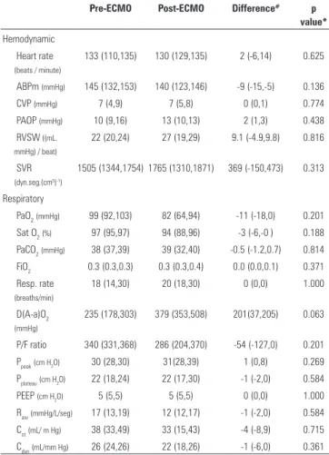
Table 1 and igure 1 show the hemodynamic, respiratory and metabolic consequences of priming and blood-ECMO membrane contact. Despite the absence of statistical signiicance, it is interesting to note the elevation of mean pulmonary artery blood pressure, resulting in higher pulmonary vascular resistance, in addition to lower cardiac output and a reduced left ventricle stroke work index. It is also interesting to note
Table 1 - Hemodynamic, respiratory and metabolic data of animals’ before and thirty minutes after ECMO, beginning without sweeper flow
Pre-ECMO Post-ECMO Difference# p
value*
Hemodynamic
Heart rate (beats / minute)
133 (110,135) 130 (129,135) 2 (-6,14) 0.625
ABPm (mmHg) 145 (132,153) 140 (123,146) -9 (-15,-5) 0.136
CVP (mmHg) 7 (4,9) 7 (5,8) 0 (0,1) 0.774
PAOP (mmHg) 10 (9,16) 13 (10,13) 2 (1,3) 0.438
RVSW ((mL. mmHg) / beat)
22 (20,24) 27 (19,29) 9.1 (-4.9,9.8) 0.816
SVR (dyn.seg.(cm5)-1)
1505 (1344,1754) 1765 (1310,1871) 369 (-150,473) 0.313
Respiratory
PaO2(mmHg) 99 (92,103) 82 (64,94) -11 (-18,0) 0.201
Sat O2(%) 97 (95,97) 94 (88,96) -3 (-6,-0 ) 0.188
PaCO2(mmHg) 38 (37,39) 39 (32,40) -0.5 (-1.2,0.7) 0.814
FiO2 0.3 (0.3,0.3) 0.3 (0.3,0.4) 0.0 (0.0,0.1) 0.371
Resp. rate (breaths/min)
18 (14,30) 20 (18,30) 0 (0,0) 1.000
D(A-a)O2
(mmHg)
235 (178,303) 379 (353,508) 201(37,205) 0.063
P/F ratio 340 (331,368) 286 (204,370) -54 (-127,0) 0.201
Ppeak(cm H2O) 30 (28,30) 31(28,39) 1 (0,8) 0.269
Pplateau(cm H2O) 22 (18,24) 22 (17,30) -1 (-2,0) 0.584
PEEP (cm H2O) 5 (5,5) 5 (5,5) 0 (0,0) 1.000
Raw(mmHg/L/seg) 17 (13,19) 12 (12,17) -1 (-2,0) 0.584
Cst(mL/ m Hg) 38 (33,49) 33 (15,43) -4 (-8,9) 0.715
Cdyn(mL/mm Hg) 26 (24,26) 22 (18,26) -1 (-6,0) 0.361
the trend toward elevation of the alveolo-arterial oxygen gradient. here were trends toward the reduction of the core temperature and chloride elevation.
DISCUSSION
In this study, there were no significant differences in the measured respiratory, hemodynamic and metabolic variables before or after blood contact with an ECMO circuit and fluid priming.
The core temperature decrease and chloride elevation could be explained by the system’s normal saline priming, not necessarily reflecting the contact of blood with the ECMO circuit. We cannot exclude the possibility of chloride elevation due to systemic
inflammation, as described with endotoxemia.(18) One
could expect more metabolic alterations due to the sudden increase in intravascular distribution volume; however, in this model using large animals, those
Table 1 - Continuation
Pre-ECMO Post-ECMO Difference# p
value*
Metabolic Core temperature (ºC)
38.2 (37.8,38.7) 37.5 (37.2,38.1) -0.6 (-0.6,-0.2) 0.058
pH 7.49 (7.47,7.52) 7.48 (7.45,7.54) -0.014 (-0.038,-0.014) 0.588
SBE (mEq/L) 4.1 (3.8,5.8) 3.2 (3.2,5.7) -1.9 (-2.1,-0.6) 0.188
Lactate (mEq/L) 1.8 (1.0,1.9) 1.6 (1.3,2.1) -0.2 (-0.7,0.4) 0.814
Na (mEq/L) 138 (138,140) 139 (138,141) 1 (-1,1) 0.766
K (mEq/L) 3.6 (3.5,3.6) 3.6 (3.6,3.6) 0 (-0.1,0.0) 1.000
Ca (mEq/L) 1.34 (1.32,1.36) 1.34 (1.30,1.39) 0.03 (-0.02,0.03) 0.787
Cl (mEq/L) 102 (101,104) 104 (103,105) 2 (1,2) 0.057
Hemoglobin (g/dL)
11.8 (11.3,13.2) 11.2 (10.7,12.0) -0.9 (-1.1,-0.1) 0.188
Glucose (mg/dL) 121 (111,129) 119 (105,133) -6 (-12,-2) 0.187
ECMO - extracorporeal membrane oxygenation; ABPm - mean arterial blood pressure; CVP - central venous pressure; PAOP - pulmonary artery oxygen in arterial blood; RVSW - right ventricle stroke work; SVR - systemic vascular resistance; PaO2 - partial pressure of carbon dioxide; SatO2 - oxygen arterial saturation; PaCO2 - partial
pressure of carbon dioxide in arterial blood; FiO2 - fraction of inspired oxygen ; Resp. rate - respiratory rate; D(A-a)O2 – alveolar arterial oxygen; P/F ratio - ratio of arterial
oxygen concentration; Ppeak - peak pressure; Pplateau – plateau pressure; PEEP - positive end expiratory pressure; Raw - airway resistance; Cst - respiratory static compliance;
Cdyn - respiratory dynamic compliance; SBE - standard base excess. # Difference = Post-ECMO - Pre-ECMO. * p value of the comparison before and after ECMO.
ECMO circuit resulted in non-significant systemic and metabolic alterations.
RESUMO
Objetivo: Investigar o impacto hemodinâmico, respirató-rio e metabólico do contato do sangue suíno com o volume do primming e com o circuito extracorpóreo da oxigenação por membrana extracorpórea, antes do início da ventilação e da oxi-genação da membrana.
Métodos: Cinco animais foram instrumentados e submeti-dos a oxigenação por membrana extracorpórea. Os dasubmeti-dos foram coletados no basal e 30 minutos depois do início da circulação extracorpórea, ainda sem o luxo de ventilação da membrana.
Resultados: Depois do início da circulação pela membrana, houve elevação não signiicativa da resistência vascular pulmonar de 235 (178,303) para 379 (353,508) dyn.seg.(cm5)-1 (p=0,065),
as-sociada a uma elevação no gradiente alveolo arterial de oxigênio de 235 (178,303) para 379 (353,508) mmHg (p=0,063). Foi observada também uma queda no trabalho sistólico do ventrículo esquerdo de 102 (94,105) para 78 (71,87) (mL.mmHg)/batimento (p=0,064), em paralelo a uma redução do débito cardíaco de 7,2 (6,8-7,6) para 5,9 (5,8-6,3) L/min (p=0,188). O trabalho sistólico do ventrículo direito foi contrabalanceado entre o aumento da resistência vascular pulmonar e a queda do débito cardíaco, mantendo-se estável.
Conclusões: O modelo é seguro e factível. O contato do sangue dos animais com o primming e o circuito extracorpóreo resultou em alterações sistêmicas e metabólicas não signiicativas.
Descritores: Síndrome do desconforto respiratório do adulto; Respiração artiicial; Oxigenação por membrana extracorpórea; Insuiciência de múltiplos órgãos; Suínos
*Participants in the ECMO group:
Marcelo Park, MD
Luciano Cesar Pontes Azevedo, MD, PhD Eduardo Leite Vieira Costa, MD, PhD Pedro Mendes, MD
Alexandre Toledo Maciel, MD Leandro Utino Taniguchi, MD, PhD Fernanda Maria Queiroz Silva, MD André Luiz de Oliveira Martins, MD Edzangela Vasconcelos Santos Barbosa, RN Raquel Oliveira Nardi, RN
Michelle de Nardi Ignácio, RN Cláudio Cerqueira Machtans, RN Wellington Alves Neves, RN Adriana Sayuri Hirota, RT
Marcelo Brito Passos Amato, MD, PhD
Guilherme de Paula Pinto Schettino, MD, PhD Carlos Roberto Ribeiro Carvalho, MD, PhD expected alterations did not occur.
The remaining alterations were likely related directly or indirectly to blood contact with the membrane. The increase in the alveolar-arterial oxygen gradient and the increase in the pulmonary vascular resistance were in line with the pulmonary injury and systemic inflammation in animals supported
by a polypropylene-based ECMO device,(7) added
to the pulmonary white-out of patients supported
by polymethylpentene-based ECMO systems.(9) The
increase in the pulmonary vascular resistance could per se explain the reductions in cardiac output and in left ventricle stroke work. The right ventricle stroke work did not increase, most likely due to the counterbalance between the higher pulmonary arterial pressure and the reduced cardiac output. Although more biocompatible than polypropylene-based membranes, contact of the blood with polymethylpentene-based ECMO circuits still induces changes noticeable at the bedside with potential clinical importance.(9) In other words, the contact of blood with the priming volume and the ECMO circuit could be responsible for left ventricle dysfunction, likely via an inflammatory
pathway,(19) causing pulmonary hypertension and the
elevation of the alveolar-arterial gradient of oxygen. Another point to consider is the temperature decrease, which could be at least partially responsible for the alterations measured.
Weighting our indings, the non-signiicant alterations in respiratory physiology and hemodynamics, the advantage of using protective or ultra-protective mechanical ventilation will likely be greater than the lung injury in this
The strength of this study is its first characterization of immediate systemic alterations due to blood contact with the priming volume and the circuit in the ECMO at the beginning, without the influence of oxygenation. However, the study has several limitations: 1) the small number of animals rendered the power of the study poor and was likely responsible for the non-significant findings; and 2) the absence of measurements of inflammatory markers precluded an adequate conclusion correlating the study findings with a systemic inflammatory process.
CONCLUSIONS
REFERENCES
1. Sidebotham D, McGeorge A, McGuinness S, Edwards M, Willcox T, Beca J. Extracorporeal membrane oxygenation for treating severe cardiac and respiratory disease in adults: Part 1--overview of extracorporeal membrane oxygenation. J Cardiothorac Vasc Anesth. 2009;23(6):886-92.
2. Peek GJ, Mugford M, Tiruvoipati R, Wilson A, Allen E, Thalanany MM, Hibbert CL, Truesdale A, Clemens F, Cooper N, Firmin RK, Elbourne D; CESAR trial collaboration. Efficacy and economic assessment of conventional ventilatory support versus extracorporeal membrane oxygenation for severe adult respiratory failure (CESAR): a multicentre randomised controlled trial. Lancet. 2009;374(9698):1351-63. Erratum in Lancet. 2009;374(9698):1330.
3. Leprince P, Combes A, Bonnet N, Ouattara A, Luyt CE, Theodore P, et al. Circulatory support for fulminant myocarditis: consideration for implantation, weaning and explantation. Eur J Cardiothorac Surg. 2003;24(3):399-403.
4. Noah MA, Peek GJ, Finney SJ, Griffiths MJ, Harrison DA, Grieve R, et al. Referral to an extracorporeal membrane oxygenation center and mortality among patients with severe 2009 influenza A(H1N1). JAMA;2011;306(15):1659-68.
5. Patroniti N, Zangrillo A, Pappalardo F, Peris A, Cianchi G, Braschi A, et al. The Italian ECMO network experience during the 2009 influenza A(H1N1) pandemic: preparation for severe respiratory emergency outbreaks. Intensive Care Med. 2011;37(9):1447-57.
6. Chauhan S, Subin S. Extracorporeal membrane oxygenation, an anesthesiologist’s perspective: physiology and principles. Part 1. Ann Card Anaesth. 2011;14(3):218-29.
7. McILwain RB, Timpa JG, Kurundkar AR, Holt DW, Kelly DR, Hartman YE, et al. Plasma concentrations of inflammatory cytokines rise rapidly during ECMO-related SIRS due to the release of preformed stores in the intestine. Lab Invest. 2010;90(1):128-39.
8. Kurundkar AR, Killingsworth CR, McILwain RB, Timpa JG, Hartman YE, He D, et al. Extracorporeal membrane oxygenation causes loss of intestinal epithelial barrier in the newborn piglet. Pediatr Res. 2010;68(2)128-33.
9. Khoshbin E, Dux AE, Killer H, Sosnowski AW, Firmin RK, Peek GJ. A comparison of radiographic signs of pulmonary inflammation during ECMO between silicon and poly-methyl pentene oxygenators. Perfusion. 2007;22(1):15-21.
10. Zimmermann AK, Weber N, Aebert H, Ziemer G, Wendel HP. Effect of biopassive and bioactive surface-coatings on the hemocompatibility of membrane oxygenators. J Biomed Mater Res B Appl Biomater. 2007;80(2):433-9.
11. Meliones JN, Moler FW, Custer JR, Snyder SJ, Dekeon MK, Donn SM, et al. Hemodynamic instability after the initiation of extracorporeal membrane oxygenation: role of ionized calcium. Crit Care Med. 1991;19(10):1247-51. 12. de Azevedo LC, Park M, Noritomi DT, Maciel AT, Brunialti MK, Salomão
R. Characterization of an animal model of severe sepsis associated with respiratory dysfunction. Clinics (Sao Paulo). 2007;62(4):491-8.
13. da Silva Almeida JR, Machado FS, Schettino GP, Park M, Azevedo LC. Cardiopulmonary effects of matching positive end-expiratory pressure to abdominal pressure in concomitant abdominal hypertension and acute lung injury. J Trauma. 2010;69(2):375-83.
14. Rosário AL, Park M, Brunialti MK, Mendes M, Rapozo M, Fernandes D, et al. SvO2-guided resuscitation for experimental septic shock: effects of fluid infusion and dobutamine on hemodynamics, inflammatory response, and cardiovascular oxidative stress. Shock. 2011;36(6):604-12.
15. Siggaard-Andersen O. The van Slyke equation. Scand J Clin Lab Invest Suppl. 1977;146:15-20.
16. Douglas AR, Jones NL, Reed JW. Calculation of whole blood CO2 content. J Appl Physiol. 1988;65(1):473-7.
17. R Development Core Team (Viena - Austria). R: A language and environment for statistical computing. R Foundation for Statistical Computing; 2009. 18. Kellum JA, Bellomo R, Kramer DJ, Pinsky MR. Etiology of metabolic acidosis
during saline resuscitation in endotoxemia. Shock. 1998;9(5):364-8. 19. Parker MM, Shelhamer JH, Bacharach SL, Green MV, Natanson C,
Frederick TM, et al. Profound but reversible myocardial depression in patients with septic shock. Ann Intern Med. 1984;100(4):483-90. 20. Gattinoni L, Carlesso E, Langer T. Towards ultraprotective mechanical