Varicella associated acute respiratory distress
syndrome in an adult patient: an example for
extracorporeal respiratory support in Brazilian
endemic diseases
INTRODUCTION
Worldwide, several infectious diseases have a high mortality rate. hree of the top ten causes of death listed by the World Health Organization are
infectious diseases.(1) Additionally, new infectious agents are occasionally
detected, some of which result in a high number of fatalities, such as inluenza A (H1N1) in 2009.
Primary varicella, or chickenpox, is usually a benign childhood illness. Despite the fact that fewer than 10% of cases occur in adults, the risk of death for adults with chickenpox is 23 to 29 times higher than in children.(2) Furthermore, pneumonitis is the most serious complication and the most common cause
of death among adults with this illness.(3) We report a case of chickenpox in
a non-immunocompromised adult with severe pneumonitis, central nervous vasculitis and acute renal failure who needed veno-venous extracorporeal membrane oxygenation (ECMO) support.
CASE PRESENTATION
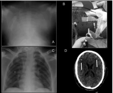
A previously heathy 30 year-old man was admitted to the emergency room with a ive day history of cutaneous vesicles, fever and dyspnea. His daughter was in the convalescence phase of chickenpox. He was cyanotic and in moderate respiratory distress. he chest x-ray and appearance of the skin at the time the patient presented are shown in igures 1A and 1B, respectively.
he clinical diagnosis was chickenpox with pulmonary manifestation. At this time, renal and hemodynamic functions were normal. herapy with acyclovir
Marcela da Silva Mendes1, Ho Yeh-Li1, Thiago Gomes Romano1, Edzangela Vasconcelos Santos1, Adriana Sayuri Hirota1, Bruna Mitiyo Kono1, Marilia Francesconi Felicio1, Marcelo Park1, on behalf of Hospital das Clínicas de São Paulo ECMO group*
1. Intensive Care Unit, Hospital das Clínicas, Faculdade de Medicina, Universidade de
São Paulo - São Paulo (SP), Brazil. A case of a 30 year-old man presenting
with severe systemic chickenpox with refractory hypoxemia, central nervous system vasculitis and anuric renal failure is described. Ambulance transportation and support using veno-venous extracorporeal membrane oxygenation were necessary
Conflicts of interest: None.
Submitted on July 27, 2014 Accepted on October 10, 2014
Corresponding author:
Marcelo Park
Disciplina de Emergência
Rua Enéas Carvalho de Aguiar, 255, 5º andar Zip code: 05403-000 - São Paulo (SP), Brazil E-mail: [email protected]
Responsible editor: Thiago Costa Lisboa
Síndrome da angústia respiratória aguda associada à varicela em
paciente adulto: exemplo de suporte respiratório extracorpóreo em
doenças endêmicas brasileiras
ABSTRACT
Keywords: Extracorporeal membrane oxygenation; Respiratory failure; Respiration, artiicial; Chickenpox; Intensive care units; Case reports
until the patient recovered. Ultimately, the potential use of extracorporeal membrane oxygenation support in low-middle income countries to manage common diseases is discussed.
was initiated, and the patient was admitted to the intensive care unit (ICU) for monitoring and respiratory support. On the irst day of ICU stay, he developed unrelenting respiratory distress; he was intubated, and mechanical ventilation was initiated. His hemodynamic and renal status deteriorated. During the irst week of ICU stay, he required up to 0.3mcg/kg/minute of norepinephrine, and the cumulative luid balance was positive at approximately 40 liters despite peritoneal dialysis. In addition to the renal and hemodynamic dysfunctions, his pulmonary function also deteriorated, and he developed severe hypoxemia on the eighth day after admission (Table 1). his condition was unresponsive to an alveolar recruitment maneuver and
a positive end-expiratory pressure (PEEP) of 22cmH2O
(Table 1). At this time, it was thought that the patient had ventilator associated pneumonia, and he was treated with vancomycin and imipenem. Next, ECMO support
was indicated, and the ECMO group Hospital das Clínicas
de São Paulo was contacted. he patient was cannulated
in situ using a percutaneous technique with 19 and 21
French cannulaes through the left jugular (atrial cannula) and femoral (drainage cannula) veins, respectively. he venous-venous ECMO support was initiated with 5L/minute of blood and gas low. he mechanical
ventilation was adjusted with PEEP=10cmH2O, driving
pressure=10cmH2O, respiratory rate=10 breaths/minute,
and the ventilator FiO2 was progressively lowered to 0.6. he norepinephrine infusion was promptly stopped due to hemodynamic improvement. he patient developed difuse cutaneous and conjunctive petechiaes, highly suggestive of vasculitis; therefore, anticoagulation with heparin was not initiated due to the possibility of central nervous system bleeding, once tomography was not locally available and the Richmond agitation sedation scale (RASS) was minus ive.
After four hours of clinical stabilization, the ECMO support was adjusted to 6L/minute of blood low and 8L/minute of gas low, and the mechanical ventilation settings were kept the same. Transportation by ambulance to downtown São Paulo City was required, which is a 156km journey lasting approximately two hours. For the transport, the ventilator FiO2 was increased to 1.0.
After transportation, the ventilator FiO2 was reduced again to 0.6, and the other ECMO and ventilator variables were similar to those used in the stabilization period. Continuous venous - venous hemoiltration was initiated with a progressively higher ultrailtration rate, up to 350mL/hour. During the irst day, the peripheral oxygen
saturation (SpO2) dropped to 65%, despite ventilator
FiO2 and ECMO blood low elevations to 1.0 and
6500mL/minute, respectively. At this point, a diagnosis of ECMO refractory hypoxemia was made. ECMO refractory
hypoxemia has been deined as a PaO2<50mmHg (taking
precedence) or an arterial saturation persistently lower
than 85%.(4) In our patient, as the ECMO running
time was <24 hours, the diference of color between the drainage and atrium cannulaes was signiicant (despite high ECMO blood low), and the pulmonary injury was severe (white lung on the x-ray - Figure 1A and
respiratory static compliance=1.2mL/cmH2O - Table 1),
the diagnosis of ECMO blood re-circulation and artiicial lung dysfunction was dismissed. ECMO refractory hypoxemia was attributed to the association between severely decreased native lung function and a low ECMO blood low/cardiac output ratio.(4)
herefore, a stepwise alveolar recruitment maneuver
was performed using PEEPs from 25 to 45cmH2O, with
a driving pressure=15cmH2O, an inspiratory time=3
seconds and a respiratory rate=10 breaths/minutes. After
the initial PEEP=25cmH2O, each step lasted 2 minutes,
and the PEEPs used were 30, 25, 35, 25, 40, 25, 45, 25cmH2O sequentially, as previously described.(5) Finally, the ideal PEEP was titrated using the pressure point
2cmH2O above the best dynamic respiratory compliance
(Cdyn), measuring the Cdyn after the alveolar recruitment
maneuver and decreasing the PEEP from 25cmH2O each
2 minutes in 2cmH2O steps, as previously described.(6)
he ideal PEEP measured was 25cmH2O, which was used
with a driving pressure of 5cmH2O, inspiratory time=1
second, and a respiratory rate=10 breaths/minute. he
SpO2 30 minutes after the alveolar recruitment maneuver
and PEEP titration increased to 84%.
During the next twelve hours, the SpO2 progressively
increased to 92%. After this period, the ventilator
FiO2 and PEEP were gradually reduced, keeping the
SpO2≥85%. After four days, the patient was comfortable
and ventilating with a PEEP=15cmH2O, pressure
support=6cmH2O and FiO2=0.3. he ECMO autonomy
test was performed, setting the gas low=0L/minute for 1 hour, as previously described.(7) he chest x-ray at this point is shown in igure 1C. After ECMO decanulation, a central nervous system tomography was performed and showed multiple bleeding areas, which were highly suggestive of vasculitis (Figure 1D).
Table 1 - Respiratory, hemodynamic and metabolic characteristics of the patient
Pre-ECMO After-ECMO Day 1 Day 2 Day 3 Day 4 Day 5
Blood gases
pH 7.51 7.40 7.38 7.37 7.47 7.49 7.42
PaO2(mmHg) 39 54 31 89 58 54 60
PaCO2(mmHg) 39 51 40 44 44 32 35
SBE (mEq/L) +8.6 +6.1 +2.0 +0.1 +7.7 +1.1 -1.3
Mechanical ventilation
Ventilatory mode PCV PCV PCV PCV PCV PCV PSV
Tidal volume (mL) 500 12 15 110 150 160 200
FIO2 1.0 0.6 1.0 1.0 0.6 0.4 0.4
PEEP (cmH2O) 22 10 10 25 23 19 15
Plateau pressure (cmH2O) 45 20 20 30 28 24 20
Respiratory rate (breaths/minute) 30 10 10 10 10 10 10
Static compliance (mL/cmH2O) 22 1.2 1.5 20 30 32 40
Hemodynamic support
Norepinephrine dosage (mcg/kg/minute) 0 0 0.1 0.1 0.05 0 0
Renal support
Cumulative fluid balance (mL) --- 0 -754 -5124 -9824 -14960 -21266
Creatinine (mg/dL) 13.64 --- 11.71 10.98 6.37 2.77 2.73
BUN (mg/dL) 187 --- 135 123 62 65 61
CVVH (dosage - mL/kg) No No 37.5 37.5 50.0 50.0 37.5
ECMO support
Blood flow (mL/minute) --- 6500 6500 4800 4000 4000 4000
Gas flow (L/minute) --- 11 12 10.5 8 4 1.5
The column Pre-ECMO indicates the eighth day after ICU admission. ECMO - extracorporeal membrane oxygenation; PaO2 - partial pressure of oxygen; PaCO2 - partial pressure of carbon dioxide;
SBE - standard base excess; BUN - blood urea nitrogen; PCV - pressure controlled ventilation; PSV - pressure support ventilation; FIO2 - fraction of inspired oxygen; PEEP - positive end-expiratory
pressure; CVVH - continuous venous-venous hemofiltration. The post-dilutional CVVH run was performed using adenosine citrate dextrose 2.2% to enhance the hemofilter protection.
Figure 1 - Thoracic and central nervous system images. (A) The chest X-Ray after ECMO cannulation; (B) The cutaneous appearance of the patient. (C) The chest X-Ray on the day of ICU discharge; (D) The central nervous system tomography, the arrows indicate two of the multiple focal bleedings.
eyes. A tracheostomy was performed, and the mechanical ventilation was withdrawn. After eleven days, the patient was discharged from the ICU, the neurologic and renal function gradually recovered, and after thirty three days he was discharged from the hospital to his home.
DISCUSSION
ECMO respiratory support has been used worldwide to sustain the lives of patients with severe respiratory failure. he mortality associated with infectious diseases has been reduced in Brazil during the last decade. However, these infectious diseases are still the main cause of severe respiratory failure in middle income countries.(8) herefore, we would like to discuss the use of ECMO to support patients with some endemic infectious diseases in low-middle income countries, such as Brazil.
those 75 patients, sixty died primarily due to chickenpox with severe respiratory failure. he majority of deaths
were in young adult men (81%).(9) Among adult and
pediatric patients, viral pneumonia was the cause of severe respiratory failure in 27% of patients who required ECMO. Furthermore, up to 18% of these ECMO supported patients were young (mean age of 33 years old) and were diagnosed with severe chickenpox.(10) Overall survival was
57-71%,(10,11) the median time on ECMO support was
seven days, and renal replacement therapy was necessary in
71% of patients.(11) ECMO support has been considered
an important adjunct therapy in severe chickenpox in high-income countries.(12) he severe renal and respiratory dysfunction both contributed to the hypoxemia of our patient. Moreover, the ECMO support allowed for the safe transportation of the patient in addition to suicient time for the renal replacement therapy to be efective. he clinical approach for severe hypoxemia during ECMO support is discussed elsewhere.(4)
he inluenza A H1N1 epidemics in 2009 had a hugely negative impact in Brazilian tertiary hospitals,(13) resulting
in unprecedented organizational responses.(14) However,
despite the local and government emergency measures, the case-fatality was higher than in other countries, mainly due to severe respiratory failure.(13) In high income countries, the H1N1 pandemics led to extreme advanced resource mobilization in order to ofer ECMO support to a high number of patients concomitantly. In Australia, elective surgeries, especially cardiac surgery, were suspended to open ICU beds and to re-allocate perfusion devices for
ECMO support in the ICU.(15,16) his policy resulted in
a high number of ECMO supported patients and a high
survival rate (78%).(17) In England, three novel ECMO
centers were built during the epidemics, and there was a high survival rate (76%).(18) Other countries also needed to improve their ECMO support facilities during the H1N1 crisis, resulting in encouraging outcomes.(19,20)
Malaria is another low-middle income country disease that potentially results in severe respiratory failure needing
ECMO support. Plasmodium vivax and Plasmodium
falciparum are the etiologies of respiratory failure in patients who required ECMO support. It is interesting to note that the ECMO support that is described in the literature was in patients who emigrated from low-middle income regions to high-income regions, likely relecting
the cultural - economical incapacity of low-middle income countries to control the disease or to ofer adequate support for the victims of severe malaria.(21,22)
he Hantaan virus cardio-respiratory syndrome presentation with severe hypoxemia and refractory cardiovascular failure is associated with 100% mortality.(23) Although veno-arterial ECMO support has been described to support (cardiovascular and respiratory support) those patients with high death probability, with a survival rate of 61%.(24,25)
Leptospirosis is a frequent tropical fever disease that can present with severe hypoxemia and alveolar
hemorrhage.(26) he severe cardiovascular and respiratory
failure can be refractory to optimized ICU therapy;(26)
therefore, in this scenario, respiratory and cardiovascular
ECMO support has been successfully used.(27) We would
like to emphasize that the hemorrhagic lung disease did
not prevent the success of ECMO support.(28)
Tuberculosis is a prevalent disease in low-middle income countries and is a major health concern. However, severe respiratory and cardiovascular failure are not common. hose more severely ill patients have been supported
with veno-arterial and/or veno-venous ECMO.(29,30)
hose descriptions of ECMO supported patients with tuberculosis were also in high income countries.
he incidence of dengue has increased 30 fold worldwide over the last 50 years. In 2014, several Brazilian states noted a signiicant rise in cases, with more than two
hundred deaths.(31) Although pulmonary hemorrhage is
not frequent in severe dengue, the presence of pulmonary edema, pleural efusion and ARDS are associated with a high mortality.(32,33) ECMO can be a feasible alternative support for this condition.
CONCLUSION
Descreveu-se aqui o caso de um homem de 30 anos de idade com quadro de varicela grave, hipoxemia refratária, vasculite do sistema nervoso central e insuiciência renal anúrica. Foi necessário transporte por ambulância com suporte respiratório extracorpóreo veno-venoso, sendo este utilizado até a recuperação
do paciente. Discute-se o potencial uso de oxigenação por membrana extracorpórea em países em desenvolvimento para o controle de doenças comuns nestas áreas.
RESUMO
Descritores: Oxigenação por membrana extracorpórea; Insu-iciência respiratória; Respiração artiicial; Varicela; Unidades de terapia intensiva; Relatos de casos
REFERENCES
1. World Health Organization. The top 10 causes of death: World Health Organization; 2014 [updated 2014-05-20 19:17:4420 Jul 2014]. [cited 2014 Oct 15]. Available from: http://www.who.int/mediacentre/ factsheets/fs310/en/
2. Heininger U, Seward JF. Varicella. Lancet. 2006;368(9544):1365-76. Review.
3. Joseph CA, Noah ND. Epidemiology of chickenpox in England and Wales, 1967-85. Br Med J (Clin Res Ed). 1988;296(6623):673-6.
4. Nunes LB, Mendes PV, Hirota AS, Barbosa EV, Maciel AT, Schettino GP, Costa EL, Azevedo LC, Park M; ECMO Group. Severe hypoxemia during veno-venous extracorporeal membrane oxygenation: exploring the limits of extracorporeal respiratory support. Clinics (São Paulo). 2014;69(3):173-8. 5. Borges JB, Okamoto VN, Matos GF, Caramez MP, Arantes PR, Barros F,
et al. Reversibility of lung collapse and hypoxemia in early acute respiratory distress syndrome. Am J Respir Crit Care Med. 2006;174(3):268-78. 6. Suarez-Sipmann F, Böhm SH, Tusman G, Pesch T, Thamm O, Reissmann
H, et al. Use of dynamic compliance for open lung positive end-expiratory pressure titration in an experimental study. Crit Care Med. 2007;35(1):214-21. 7. Park M, Azevedo LC, Mendes PV, Carvalho CR, Amato MB, Schettino
GP, et al. First-year experience of a Brazilian tertiary medical center in supporting severely ill patients using extracorporeal membrane oxygenation. Clinics (São Paulo). 2012;67(10):1157-63.
8. Barreto ML, Teixeira MG, Bastos FI, Ximenes RA, Barata RB, Rodrigues LC. Successes and failures in the control of infectious diseases in Brazil: social and environmental context, policies, interventions, and research needs. Lancet. 2011;377(9780):1877-89.
9. Rawson H, Crampin A, Noah N. Deaths from chickenpox in England and Wales 1995-7: analysis of routine mortality data. BMJ. 2001;323(7321):1091-3. 10. Lee WA, Kolla S, Schreiner RJ Jr, Hirschl RB, Bartlett RH. Prolonged
extracorporeal life support (ECLS) for varicella pneumonia. Crit Care Med. 1997;25(6):977-82.
11. White RW, Peek GJ, Jenkins DR, Killer HM, Firmin RK. Extracorporeal membrane oxygenation for chickenpox pneumonia: a single institution’s experience. ASAIO J. 2003;49(4):378-82.
12. Roberts N, Peek GJ, Jones N, Firmin RK, Elbourne D. Deaths from Chickenpox. Extracorporeal membrane oxygenation has important role. BMJ. 2002;324(7337):610-1.
13. Schout D, Hajjar LA, Galas FR, Uip DE, Levin AS, Caiaffa Filho HH, et al. Epidemiology of human infection with the novel virus influenza A (H1N1) in the Hospital das Clinicas, São Paulo, Brazil-June-September 2009. Clinics (São Paulo). 2009;64(10):1025-30.
14. Hajjar LA, Schout D, Galas FR, Uip DE, Levin AS, Caiaffa Filho HH, et al. Guidelines on management of human infection with the novel virus influenza A (H1N1)-a report from the Hospital das Clínicas of the University of São Paulo. Clinics (São Paulo). 2009;64(10):1015-24.
15. Forrest P, Ratchford J, Burns B, Herkes R, Jackson A, Plunkett B, et al. Retrieval of critically ill adults using extracorporeal membrane oxygenation: an Australian experience. Intensive Care Med. 2011;37(5):824-30.
16. Nair P, Davies AR, Beca J, Bellomo R, Ellwood D, Forrest P, et al. Extracorporeal membrane oxygenation for severe ARDS in pregnant and postpartum women during the 2009 H1N1 pandemic. Intensive Care Med. 2011;37(4):648-54.
17. Australia and New Zealand Extracorporeal Membrane Oxygenation (ANZ ECMO) Influenza Investigators, Davies A, Jones D, Bailey M, Beca J, Bellomo R, Blackwell N, et al. Extracorporeal Membrane Oxygenation for 2009 Influenza A(H1N1) Acute Respiratory Distress Syndrome. JAMA. 2009;302(17):1888-95.
18. Noah MA, Peek GJ, Finney SJ, Griffiths MJ, Harrison DA, Grieve R, et al. Referral to an extracorporeal membrane oxygenation center and mortality among patients with severe 2009 influenza A(H1N1). JAMA. 2011;306(15):1659-68.
19. Pham T, Combes A, Rozé H, Chevret S, Mercat A, Roch A, Mourvillier B, Ara-Somohano C, Bastien O, Zogheib E, Clavel M, Constan A, Marie Richard JC, Brun-Buisson C, Brochard L; REVA Research Network. Extracorporeal membrane oxygenation for pandemic influenza A(H1N1)-induced acute respiratory distress syndrome: a cohort study and propensity-matched analysis. Am J Respir Crit Care Med. 2013;187(3):276-85.
20. Cianchi G, Bonizzoli M, Pasquini A, Bonacchi M, Zagli G, Ciapetti M, et al. Ventilatory and ECMO treatment of H1N1-induced severe respiratory failure: results of an Italian referral ECMO center. BMC Pulm Med. 2011;11:2.
21. Alves C, Chen JT, Patel N, Abrams D, Figueiredo P, Santos L, et al. Extracorporeal membrane oxygenation for refractory acute respiratory distress syndrome in severe malaria. Malar J. 2013;12:306.
22. Losert H, Schmid K, Wilfing A, Winkler S, Staudinger T, Kletzmayr J, et al. Experiences with severe P. falciparum malaria in the intensive care unit. Intensive Care Med. 2000;26(2):195-201.
23. Crowley MR, Katz RW, Kessler R, Simpson SQ, Levy H, Hallin GW, et al. Successful treatment of adults with severe Hantavirus pulmonary syndrome with extracorporeal membrane oxygenation. Crit Care Med. 1998;26(2):409-14.
24. Dietl CA, Wernly JA, Pett SB, Yassin SF, Sterling JP, Dragan R, et al. Extracorporeal membrane oxygenation support improves survival of patients with severe Hantavirus cardiopulmonary syndrome. J Thorac Cardiovasc Surg. 2008;135(3):579-84.
25. Wernly JA, Dietl CA, Tabe CE, Pett SB, Crandall C, Milligan K, et al. Extracorporeal membrane oxygenation support improves survival of patients with Hantavirus cardiopulmonary syndrome refractory to medical treatment. Eur J Cardiothorac Surg. 2011;40(6):1334-40.
26. Carvalho CR, Bethlem EP. Pulmonary complications of leptospirosis. Clin Chest Med. 2002;23(2):469-78. Review.
27. Kahn JM, Müller HM, Kulier A, Keusch-Preininger A, Tscheliessnigg KH. Veno-arterial extracorporeal membrane oxygenation in acute respiratory distress syndrome caused by leptospire sepsis. Anesth Analg. 2006;102(5):1597-8.
29. Petrillo TM, Heard ML, Fortenberry JD, Stockwell JA, Leonard MK Jr. Respiratory failure caused by tuberculous pneumonia requiring extracorporeal membrane oxygenation. Perfusion. 2001;16(6):525-9. 30. Homan W, Harman E, Braun NM, Felton CP, King TK, Smith JP. Miliary
tuberculosis presenting as acute respiratory failure: treatment by membrane oxygenator and ventricle pump. Chest. 1975;67(3):366-9.
31. Secretaria de Vigilância em Saúde. Ministério da Saúde. Dengue: monitoramento até a Semana Epidemiológica (SE) 27 de 2014. Bol Epidemiol. 2014;45(16):1-6.
32. Lee IK, Liu JW, Yang KD. Fatal dengue hemorrhagic fever in adults: emphasizing the evolutionary pre-fatal clinical and laboratory manifestations. PLoS Negl Trop Dis. 2012;6(2):e1532.
33. Ong A, Sandar M, Chen MI, Sin LY. Fatal dengue hemorrhagic fever in adults during a dengue epidemic in Singapore. Int J Infect Dis. 2007;11(3):263-7.
* The ECMO group comprises: