CLINICAL SCIENCE
First-year experience of a Brazilian tertiary medical
center in supporting severely ill patients using
extracorporeal membrane oxygenation
Marcelo Park,I,IILuciano Cesar Pontes Azevedo,I,IIPedro Vitale Mendes,I,IICarlos Roberto Ribeiro Carvalho,I Marcelo Brito Passos Amato,IGuilherme Paula Pinto Schettino,IIMauro Tucci,IAlexandre Toledo Maciel,I,II Leandro Utino Taniguchi,I,IIEdzangela Vasconcelos Santos Barbosa,IRaquel Oliveira Nardi,IMichelle de Nardi Igna´cio,ICla´udio Cerqueira Machtans,I Wellington Alves Neves,IAdriana Sayuri Hirota,IEduardo Leite Vieira CostaI,II
IHospital das Clı´nicas da Faculdade de Medicina da Universidade de Sa˜o Paulo, Sa˜o Paulo/SP, Brazil. IIHospital Sı´rio-Libaneˆs, Sa˜o Paulo/SP,
Brazil.
OBJECTIVES: The aim of this manuscript is to describe the first year of our experience using extracorporeal membrane oxygenation support.
METHODS:Ten patients with severe refractory hypoxemia, two with associated severe cardiovascular failure, were supported using venous-venous extracorporeal membrane oxygenation (eight patients) or veno-arterial extra-corporeal membrane oxygenation (two patients).
RESULTS:The median age of the patients was 31 yr (range 14–71 yr). Their median simplified acute physiological score three (SAPS3) was 94 (range 84–118), and they had a median expected mortality of 95% (range 87–99%). Community-acquired pneumonia was the most common diagnosis (50%), followed byP.jirovecipneumonia in two patients with AIDS (20%). Six patients were transferred from other ICUs during extracorporeal membrane oxygenation support, three of whom were transferred between ICUs within the hospital (30%), two by ambulance (20%) and one by helicopter (10%). Only one patient (10%) was anticoagulated with heparin throughout extracorporeal membrane oxygenation support. Eighty percent of patients required continuous venous-venous hemofiltration. Three patients (30%) developed persistent hypoxemia, which was corrected using higher positive end-expiratory pressure, higher inspired oxygen fractions, recruitment maneuvers, and nitric oxide. The median time on extracorporeal membrane oxygenation support was five (range 3–32) days. The median length of the hospital stay was 31 (range 3-97) days. Four patients (40%) survived to 60 days, and they were free from renal replacement therapy and oxygen support.
CONCLUSIONS:The use of extracorporeal membrane oxygenation support in severely ill patients is possible in the presence of a structured team. Efforts must be made to recognize the necessity of extracorporeal respiratory support at an early stage and to prompt activation of the extracorporeal membrane oxygenation team.
KEYWORDS: Extracorporeal Membrane Oxygenation; Respiratory Failure; Mechanical Ventilation; Patient Care Team; Intensive Care Unit.
Park M, Azevedo LC, Mendes PV, Carvalho CR, Amato MB, Schettino GP, et al. First-year experience of a Brazilian tertiary medical center in supporting severely ill patients using extracorporeal membrane oxygenation. Clinics. 2012;67(10):1157-1163.
Received for publication onApril 25, 2012;First review completed onJune 11, 2012;Accepted for publication onJune 12, 2012 E-mail: [email protected]
Tel.: 55 11 2661-7221
INTRODUCTION
Since the first successful description of extracorporeal membrane oxygenation (ECMO) in 1971 (1), this modality of
respiratory support has been investigated in several trials. In 1979, the first randomized trial of ECMO in acute respiratory distress syndrome (ARDS) patients resulted in nonsignificant differences in mortality between the control and ECMO groups (2). In 1986, Gattinoni et al. described, with good outcomes, the use of ECMO to remove carbon dioxide from the blood, enabling the use of low-frequency mechanical ventilation, which was reported to have resulted in a lower incidence of ventilation-associated lung injury (3). In 1994, Morris et al. conducted a randomized trial to test the effectiveness of this strategy of carbon dioxide (CO2) Copyrightß2012CLINICS– This is an Open Access article distributed under
the terms of the Creative Commons Attribution Non-Commercial License (http:// creativecommons.org/licenses/by-nc/3.0/) which permits unrestricted non-commercial use, distribution, and reproduction in any medium, provided the original work is properly cited.
removal associated with low-frequency ventilation com-pared with conventional treatment (4). The trial was stopped early due to futility. Mechanical ventilation with suboptimal settings and high airway pressures in the CO2 removal arm may have been associated with the negative final result. In 2009, the CESAR trial demonstrated a reduction in mortality and disability at six months among severe ARDS patients using a combined strategy of ECMO and protective mechanical ventilation compared with conventional protective mechanical ventilation (5).
The renewed interest in the use of ECMO was also fostered by the 2009 epidemic of severe pneumonia due to influenza-A H1N1(6-13). Young patients with refractory hypoxemia were common among those affected by this disease, and respiratory rescue with ECMO was carried out with excellent outcomes in Australia (14), the United Kingdom (15), and Italy (16). These data suggest a survival benefit of using ECMO in addition to conventional support (15). In Brazil, many patients with influenza-A H1N1 required mechanical ventilation (17), and some of them developed severe hypoxemia, in whom the case mortality reached 11.6% (11). The absence of centers in Brazil that specialize in ECMO limited the availability of adequate support for those patients (18).
We implemented an ECMO service in two hospitals in Sa˜o Paulo, Brazil. We followed a stepwise approach, which included theoretical training of ECMO, the use of wet lab practice with ECMO in animals in the research facility, and finally the use of ECMO in patients at the bedside. The aim of this manuscript is to describe the first year of our experience using ECMO support in patients with severe hypoxemic respiratory failure.
METHODS
The ECMO group comprised nurses, respiratory thera-pists, and physicians. The care for ECMO-supported patients was delivered by one physician, one nurse, two nursing assistants, and one respiratory therapist, all of whom were also in charge of three additional critically-ill patients (19).
ECMO support was indicated based on individual judgment of the following objective criteria (20):
Major criteria (both required):
-
Acute or acute-on-chronic pulmonary disease-
Possibility of disease reversionComplementary criteria (at least one required):
-
P/F ratio#50 with FiO2= 1 for at least 1 hour, with or without rescue maneuvers-
P/F ratio#50 with FiO2$0.8 for at least 3 hours, despite rescue maneuvers-
Hypercapnia with pH#7.2 using a respiratory rate of at least 35 breaths per minute, with a tidal volume of 4– 6 mL/kg of ideal body weight and a plateau pressure#30 cmH2O-
Murray’s score (lung injury score).3.0 with the patient presenting a clinical deteriorationECMO support was not administered to patients with severe chronic illness without the possibility of improve-ment of their quality of life.
The canulation of patients was performed percutaneously at the bedside with the Seldinger technique using femoral and jugular canulae (Edwards Lifesciences, Irvine, CA, EUA). In the three patients who were transferred from other hospitals, we initiated the ECMO support before the patient was transported. We primed the ECMO circuit with 600 mL of normal saline solution at room temperature. A centrifugal magnetic pump with a polymethylpentene oxygenation membrane (Rotaflow/Jostra Quadrox - D, Maquet Cardio-pulmonary AG, Hirrlingen, Germany) was used for all patients.
Initially, the blood flow was maintained at 500 mL per minute until the system was filled with blood. Blood flow and sweep (gas) flow were subsequently increased to 2000 mL per minute when using the venous-venous (VV) configuration. The blood flow and sweep flow were then elevated in a 1:1 ratio, with a peripheral oxygen saturation target of at least 90%. When using the veno-arterial (VA) configuration, the blood flow was gradually elevated to 5000 mL per minute, and the sweep flow was set at 5 L per minute. The ECMO support settings were adjusted based on the arterial blood gases (in the VV configuration) or on the arterial blood pressure, vasopressor requirement, and serum lactate levels (in the VA configuration).
No prophylactic antibiotics were administered. Anticoa-gulation with heparin was initiated at the team’s discretion, beginning with 1000 IU per hour without bolus and adjusted to reach an activated partial thromboplastin time ratio of 1.5–2.5.
Mechanical ventilation was adjusted to a positive end-expiratory pressure (PEEP) of 10-15 cmH2O, an inspired fraction of oxygen (FiO2) of 0.3 or the least possible value, a driving pressure of less than 10 cmH2O, and, if necessary, a respiratory rate of 10 breaths per minute (5). The parameters checked daily included arterial blood gases, clots in the system visible through transillumination, pump campanula auscultation, and flowmeter lubrication to maintain a good signal quality. The ECMO blood flow was adjusted to maintain a PaO2greater than 55 mmHg, and the sweep flow was adjusted to keep the pH$7.3 (through the PaCO2 modulation). Typical sedatives and analgesics were used if necessary to reach a Richmond agitation sedation scale (RASS) score of zero with no pain. Body temperature was kept between 36 and 37 degrees Celsius with an external apparatus adapted to the ECMO system.
A test to evaluate whether a patient could be weaned from ECMO support was carried out daily. The mechanical ventilation settings remained constant in patients with pressure support ventilation; in patients under controlled ventilation, the tidal volume was raised to 6 mL/ideal body weight or was maintained at a level sufficient to keep the plateau pressure ,25 cmH2O and the driving pressure
,15 cmH2O. In both situations, the FiO2was set to 0.6. In the VV configuration, the test consisted of stopping the sweep flow. In the VA configuration, the test consisted of reducing both the blood flow and sweep flow to 1,000 mL per minute, with an ECMO FiO2of 0.21. The weaning test was interrupted if the patient presented a mean arterial blood pressure ,65 mmHg, peripheral oxygen saturation
weaning test) if he or she was hemodynamically stable without elevations in inotropics or vasopressors, without respiratory distress, with a PaO2$55 mmHg and a pH$7.30. The decanulation was performed at the bedside in the venous-venous configuration after the reversal of anticoagulation. In the veno-arterial configuration, the arterial canulae withdrawal was performed in the surgical room.
Data are presented as medians (minimum–maximum), which were obtained using the R – free source statistical package (Vienna, Austria, 2009) (21).
RESULTS
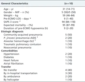
During a one-year period, ten patients with severe respiratory and/or cardiac dysfunction were supported with ECMO. The general characteristics of patients are shown in Table 1. The patients were young and had extremely severe disease, as demonstrated by their high expected mortality rate (Table 1). The most common etiology of the acute cardiopulmonary dysfunction was community-acquired pneumonia, and six of the ten patients were transferred from outside ICUs.
The patients’ support and clinical statuses prior to ECMO are shown in Table 2. They had low partial pressure of oxygen to inspired fraction of oxygen (P/F) ratios, as well as elevated PEEP and plateau pressures. At least one rescue therapy strategy for refractory hypoxemia had been performed on each patient prior to the initiation of ECMO. We used five sets of 20 French canulae (five long venous and five short arterial canulae) and five sets of 22 French canulae to administer ECMO.
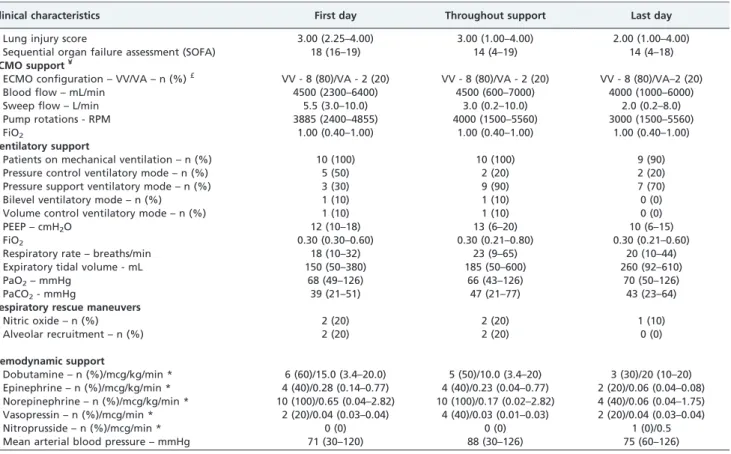
Data regarding the monitoring and physiological vari-ables associated with ECMO support are shown in Tvari-ables 3
and 4. ECMO support enabled a significant reduction in tidal volume and FiO2during its use.
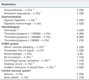
Table 5 presents the patient complications identified during ECMO support. Pneumothorax, thrombocytopenia, and hypoxic hepatitis were common complications of the procedure, and there were no significant vascular complica-tions in our patients.
Regarding the weaning tests, our patients were supported by ECMO for a total of 92 days, during which time 89 autonomy tests were performed; nine tests were performed in patients with the VA configuration, and 80 tests were performed in patients with the VV configuration. Ten tests were compatible with the removal of ECMO support; however, in two tests (both in the venous-venous configura-tion), at the discretion of the team, the patient was maintained on ECMO support for one additional day. Seven tests failed due to hypotension in patients using the veno-arterial Table 1 -Characteristics of patients supported by ECMO.
General Characteristics (n = 10)
Age – yr 31 (14–71)
Gender – M/F – n (%) 5 (50)/5 (50)
Weight – kg 59 (46–84)
Pre-ECMO LOS – days * 9 (1–40)
SAPS 3 score# 94 (84–118)
Expected mortality – (%) 95 (87–99) Duration of pre-ECMO hypoxemia (h) 9 (2–20)
Etiologic diagnosis
Community-acquired pneumonia 5 (50) P.jirovecipneumonia+AIDS1
2 (20) Alveolar hemorrhage+SLE"
1 (10) Traumatic pulmonary contusion 1 (10)
Nosocomial pneumonia 1 (10)
Comorbidities
Hypertension 2 (20)
Diabetes 1 (10)
Heart failure 1 (10)
Atrial fibrillation 1 (10)
Transfer
No transfer 4 (40)
By in-hospital transportation 3 (30)
By ambulance 2 (20)
By helicopter 1 (10)
Quantitative data are shown as medians (minimum, maximum). *ECMO denotes extracorporeal membrane oxygenation LOS denotes
length of stay.
#SAPS denotes simplified acute physiological score. 1
AIDS denotes acquired immunodeficiency syndrome. "SLE denotes systemic lupus erythematosus.
Table 2 -Pre-ECMO clinical status and support1.
Arterial blood gases (n = 10)
PaO2– mmHg 50 (36-56)
PaCO2– mmHg 57 (31–142)
P/F ratio 50 (36-56)
Mechanical ventilation
PEEP – cmH2O 15 (10–23)
FiO2 1 (1–1)
Respiratory rate – breaths/minute 28 (18–90)
Tidal volume – mL 275 (130–400)
Plateau pressure – cmH2O 31 (25–46)
Pre-ECMO respiratory rescue maneuvers
Alveolar recruitment – n (%) 7 (70) Neuromuscular blockade – n (%) 6 (60)
Corticosteroids – n (%) 4 (40)
High frequency ventilation – n (%) * 3 (30) Tracheal gas insufflation – n (%) 1 (10)
Hemodynamics
Norepinephrine – n (%)/dosage (mcg/kg/minute)
10 (100)/0.64 (0.28– 5.12)
Epinephrine – n (%)/dosage (mcg/kg/minute)
2 (20)/0.44 (0.19–0.70)
Dobutamine – n (%)/dosage (mcg/kg/minute)
3 (30)/15 (4-20)
Vasopressin – n (%)/dosage (mcg/minute)
1 (10)/0.03
Mean arterial blood pressure – mmHg 63 (30–94) Heart rate – beats/minute 145 (109-180)
Metabolic
Temperature -˚C 37.4 (36.6–41.2)
Lactate – mEq/L 2.8 (1.6–4.1)
pH 7.17 (6.9–7.41)
SBE – mEq/L#
-2.3 (-21.9–5.6)
Sedation, analgesia and neuromuscular blockers
SAS 1.5 (1.0–3.0)
Midazolam - n (%)/dosage (mg/kg/hour) 4 (40)/0.16 (0.11–0.42) Propofol - n (%)/dosage (mg/kg/hour) 4 (40)/0.73 (0.21–1.69) Fentanyl - n (%)/dosage (mcg/kg/hour) 7 (70)/2.25 (0.85–6.28) Thionembutal - n (%)/dosage
(mg/kg/hour)
1 (10)/3
Atracurium - n (%)/dosage (mg) 5 (50)/50 (50–50) Cisatracurium - n (%)/dosage (mg) 1 (10)/20
All quantitative data are presented as medians (minimum, maximum). 1
ECMO denotes extracorporeal membrane oxygenation.
*High-frequency ventilation denotes high-frequency positive pressure
ventilation (HFPPV).
configuration, and 73 tests failed due to a peripheral saturation,85% associated with respiratory distress.
Seven patients were ventilated with pressure support during the positive weaning test, with a respiratory rate of 25 (20-42) breaths per minute, tidal volume of 310 (290-500) mL, and PEEP of 8 (5-15) cmH2O. Analysis of the arterial blood gases after one hour into the weaning test revealed a pH of 7.392 (7.320-7.474), PaO2of 81 (56-197) mmHg, PaCO2 of 41 (37-55) mmHg and SBE of 0.7 (-4–5.2) mEq/L.
Table 6 shows the clinical outcomes of the ten patients. The median time of ECMO support was five days. The majority of the patients were successfully weaned from ECMO, and four patients survived to 60 days after hospital discharge.
DISCUSSION
In these severely injured patients with an expected survival of less than 13%, this study demonstrated a survival of 40% when using ECMO for respiratory and/or cardiovascular support. Another interesting aspect of this study was the enrollment of patients with acquired immunodeficiency syndrome (AIDS), who are not candi-dates for ECMO support in classical ECMO referral centers due to their high morbidity and mortality rates (22). Of note, the two AIDS patients included in our study died; one death was due to meningitis and the other was due to severe liver failure, and both occurred after the removal of ECMO
support. One of these AIDS patients required 32 days of ECMO support and died 11 days after ECMO weaning. However, AIDS is a common condition among the patients in the Brazilian public health care services (23), and the use of ECMO support for those patients cannot be denied a prioriuntil it is evaluated in a prospective trial.
The high SOFA and simplified acute physiological score (SAPS) 3 scores at the beginning of ECMO support in our patients suggest late enrollment and late initiation of ECMO support. This issue could be corrected by the early recognition of severe ARDS and the early activation of the ECMO team. To this end, promoting the awareness of the methodology and the existence of a referral center within the local medical community is likely an important step.
The VV configuration has lower oxygen-transfer efficacy compared with the VA configuration due to recirculation (24). Therefore, one can expect to have patients with persistent hypoxemia during the administration of VV ECMO support, as occurred in three of our patients. The presence of pneumothorax was diagnosed at the time of ECMO support weaning (data not published). We postulate that these pneumothoraxes were the result of barotrauma caused by the increase in transpulmonary pressure neces-sary to maintain alveolar ventilation.
Central nervous system injury during ECMO support is very common (25). Among the patients in our study, it is interesting to note that two experienced a seizure; one occurred during the system pause for lubrification of the Table 3 -Respiratory and hemodynamic support and monitoring during extracorporeal membrane oxygenation.
Clinical characteristics First day Throughout support Last day
Lung injury score 3.00 (2.25–4.00) 3.00 (1.00–4.00) 2.00 (1.00–4.00)
Sequential organ failure assessment (SOFA) 18 (16–19) 14 (4–19) 14 (4–18)
ECMO support¥
ECMO configuration – VV/VA – n (%)£ VV - 8 (80)/VA - 2 (20) VV - 8 (80)/VA - 2 (20) VV - 8 (80)/VA–2 (20)
Blood flow – mL/min 4500 (2300–6400) 4500 (600–7000) 4000 (1000–6000)
Sweep flow – L/min 5.5 (3.0–10.0) 3.0 (0.2–10.0) 2.0 (0.2–8.0)
Pump rotations - RPM 3885 (2400–4855) 4000 (1500–5560) 3000 (1500–5560)
FiO2 1.00 (0.40–1.00) 1.00 (0.40–1.00) 1.00 (0.40–1.00)
Ventilatory support
Patients on mechanical ventilation – n (%) 10 (100) 10 (100) 9 (90)
Pressure control ventilatory mode – n (%) 5 (50) 2 (20) 2 (20)
Pressure support ventilatory mode – n (%) 3 (30) 9 (90) 7 (70)
Bilevel ventilatory mode – n (%) 1 (10) 1 (10) 0 (0)
Volume control ventilatory mode – n (%) 1 (10) 1 (10) 0 (0)
PEEP – cmH2O 12 (10–18) 13 (6–20) 10 (6–15)
FiO2 0.30 (0.30–0.60) 0.30 (0.21–0.80) 0.30 (0.21–0.60)
Respiratory rate – breaths/min 18 (10–32) 23 (9–65) 20 (10–44)
Expiratory tidal volume - mL 150 (50–380) 185 (50–600) 260 (92–610)
PaO2– mmHg 68 (49–126) 66 (43–126) 70 (50–126)
PaCO2- mmHg 39 (21–51) 47 (21–77) 43 (23–64)
Respiratory rescue maneuvers
Nitric oxide – n (%) 2 (20) 2 (20) 1 (10)
Alveolar recruitment – n (%) 2 (20) 2 (20) 0 (0)
Hemodynamic support
Dobutamine – n (%)/mcg/kg/min * 6 (60)/15.0 (3.4–20.0) 5 (50)/10.0 (3.4–20) 3 (30)/20 (10–20) Epinephrine – n (%)/mcg/kg/min * 4 (40)/0.28 (0.14–0.77) 4 (40)/0.23 (0.04–0.77) 2 (20)/0.06 (0.04–0.08) Norepinephrine – n (%)/mcg/kg/min * 10 (100)/0.65 (0.04–2.82) 10 (100)/0.17 (0.02–2.82) 4 (40)/0.06 (0.04–1.75) Vasopressin – n (%)/mcg/min * 2 (20)/0.04 (0.03–0.04) 4 (40)/0.03 (0.01–0.03) 2 (20)/0.04 (0.03–0.04)
Nitroprusside – n (%)/mcg/min * 0 (0) 0 (0) 1 (0)/0.5
Mean arterial blood pressure – mmHg 71 (30–120) 88 (30–126) 75 (60–126)
*This is the maximum dosage administered during the analyzed period when on drug use.
#This is the median dosage during the analyzed period when on drug use.
blood flowmeter, and the other occurred due to meningitis. Three patients underwent brain death; one case was due to a trauma-related carotid dissection, one was due to meningi-tis and one was due to severe and prolonged hypoxemia (,12 h) prior to ECMO administration. The former
patient-also developed severe hypoxia-related rhabdomyolysis and hypoxic hepatitis, and he was the only patient who was determined to be brain dead while receiving ECMO support, which is a challenging diagnosis (26).
Sixty percent of our patients were transported from an ICU to our referral ICUs, and all of them had ECMO support initiated in loco. The transportation of ECMO-supported patients with good results and survival has been described previously (27,28). Three out of six of our patients survived. It thus appears that the transportation of patients during ECMO support is feasible in our service.
Another important point to discuss is the ECMO team. All members were trained to perform the canulation at the bedside using the Seldinger technique, and the team was also the staff responsible for the care of the patients in the ICU. There was no particular staff member designated exclusively for the care of the ECMO-supported patients,
which is a staff configuration that can potentially reduce the costs of ECMO support.
Our patients were allowed to awaken after the ECMO installation and transport. Their comfort was maintained despite their severe lung injuries. Some patients presented respiratory compliances as low as 6 mL/cmH2O; however, when the membrane sweep flow was sufficiently high, those patients were usually comfortable while alert and oriented.
Only one patient was anticoagulated during the entire ECMO support. The coexistence of bleeding or severe coagulopathy in the remaining patients resulted in the contraindication of anticoagulation (22). The new ECMO systems have a heparin-bound surface, which allows the ECMO support to proceed with minimal or no antic-oagulation (29). In addition to the surface technology, the use of a high blood flow through the system is likely associated with the preservation of the ECMO circuits.
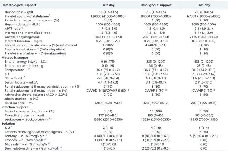
Renal replacement therapy is frequently applied through the ECMO circuit (30). Eighty percent of our patients required continuous venous-venous hemofiltration, which was applied through an individual catheter, a choice we made to minimize manipulations of the ECMO system. Table 4 -Hematological, metabolic, infection and sedation support and monitoring of patients during extracorporeal membrane oxygenation.
Hematological support First day Throughout support Last day
Hemoglobin – g/dL 7.6 (4.7–11.5) 7.5 (4.7–11.5) 7.0 (6.0–8.5)
Platelet count – platelets/mm3 120000 (41000–400000) 84000 (7000–40000) 67000 (19000–254000)
Patients on heparin therapy – n (%) 5 (50) 6 (60) 3 (30)
Heparin dosage – (IU/h) 1000 (500–1000) 1000 (500–1200) 1000 (500–1000)
APTT ratio"
1.7 (0.8–3.0) 1.3 (0.8–3.3) 2.1 (1.9–2.1)
International normalized ratio 1.5 (1.3–4.0) 1.3 (1.1–4.4) 1.4 (1.1–3.0)
Lactate dehydrogenase 1882 (1111–16173) 2281 (491–31413) 2175 (1322–31143)
Indirect bilirubin – mg/dL 0.23 (0.01–2.27) 0.29 (0.01–3.10) 0.38 (0.10–1.38)
Packed red cell transfusion – n (%)/units/patient 1 (10)/2 4 (40)/4 (3–11) 1 (10)/2
Plasma transfusion – n (%)/units/patient 0 (0)/0 3 (30) 1 (10)
Platelet transfusion – n (%)/units/patient 0 (0)/0 6 (60) 1 (10)
Metabolic support
Enteral energy intake – kCal 0 (0–475) 825 (0–1200) 638 (0–1200)
Enteral protein intake - g 0 (0–19) 36 (0–48) 26 (0–48)
Temperature - ˚C 36.4 (35.0–41.2) 36.4 (33.1–41.2) 36.2 (34.2–37.9)
pH 7.38 (7.11–7.51) 7.39 (7.11–7.51) 7.37 (7.29–7.47)
SBE – mEq/L1
-5.0 (-18.9–8.4) 4.4 (-18.9–17) 1.6 (-13.3–11.1)
Serum lactate – mEq/L 4.4 (1.1–17.3) 3.1 (0.8–19.7) 2 (1.2–17.0)
Renal replacement therapy administration – n (%) 7 (70) 8 (80) 7 (70)
Renal replacement therapy mode – n (%) CVVHD 1(10)/CVVHF 6 (60)H CVVHF 8 (80)H CVVHF 7 (70)H
Adenosine citrate dextrose (ACD-A 2.2%) administration – n (%)
2 (20) 5 (50) 5 (50)
Fluid balance – mL 1203 (-1028–7564) 428 (-4997–8612) 200 (-1355–3037)
Infection support
Patients using antibiotics – n (%) 9 (90) 10 (100) 9 (90)
C-reactive protein – mg/dL 177 (43–465) 165 (8–465) 167 (40–336)
Leukocytes – leuckocytes/mm3 13620 (2510–60350) 13620 (2510–60350) 11995 (7000–41980)
Sedation
SASJ
2 (1–5) 4 (1–6) 3 (1–4)
Patients receiving sedatives/analgesics – n (%) 9 (90) 9 (90) 5 (50)
Fentanyl – n (%)/mcg/kg/h# 8 (80)/1.1 (0.4–6.3) 8 (80)/1.0 (0.3–6.3) 5 (50)/0.8 (0.3–2.3)
Propofol – n (%)/mg/kg/h#
3 (30)/0.8 (0.5–2.1) 3 (30)/0.9 (0.2–2.1) 0 (0)
Midazolam – n (%)/mg/kg/h# 1 (10)/0.08 1 (10)/0.10 0 (0)
Dexmedetomidine – n (%)/mcg/kg/h#
1 (10)/0.5 2 (20)/0.2 (0.2–0.5) 0 (0)
*This is the maximum dosage used during the analyzed period.
£ VV and VA denote the venous-venous and veno-arterial ECMO configuration, respectively.
#This is the median dosage during the analyzed period.
HCVVHF denotes continuous venous-venous hemofiltration, and CVVHD denotes continuous venous-venous hemodialysis.
¥ ECMO denotes extracorporeal membrane oxygenation. "APTT denotes activated partial thromboplastin time. 1SBE denotes standard base excess.
When oxygenation was improving and there was low respiratory compliance, we sometimes decided to apply a strategy of permissive hypercapnia to wean patients from ECMO support early. In this situation, if the kidney is normal, the elimination of chloride with metabolic compen-sation is normally promptly conducted (31); however, during continuous hemofiltration therapy, we occasionally found it useful to reduce the chloride concentration in the replacement fluid.
The use of ECMO support in severely ill patients in Brazil is possible in the presence of a structured team. Efforts must be made to recognize the necessity of extracorporeal respiratory support at an early stage and to prompt activation of the ECMO team.
ACKNOWLEDGMENTS
The authors received a donation of ECMO membranes from MAQUETH Cardiovascular in Brazil.
AUTHOR CONTRIBUTIONS
Park M, Azevedo LC, Carvalho CR, Schettino GP and Costa EL were responsible for the data collection, patients support and manuscript drafting. Mendes PV, Amato MB, Tucci M, Maciel AT, Taniguchi LU, Barbosa EV, Nardi RO, Ignacio MN, Machtans CC, Neves WA and Hirota AS were responsible for data collection and patients support.
REFERENCES
1. Hill JD, O’Brien TG, Murray JJ, Dontigny L, Bramson ML, Osborn JJ, et al. Prolonged extracorporeal oxygenation for acute post-traumatic respira-tory failure (shock-lung syndrome). Use of the Bramson membrane lung. N Engl J Med. 1972;286(12):629-34, http://dx.doi.org/10.1056/ NEJM197203232861204.
2. Zapol WM, Snider MT, Hill JD, Fallat RJ, Bartlett RH, Edmunds LH, et al. Extracorporeal membrane oxygenation in severe acute respiratory failure. A randomized prospective study. JAMA. 1979;242(20):2193-6, http://dx.doi.org/10.1001/jama.1979.03300200023016.
3. Gattinoni L, Pesenti A, Mascheroni D, Marcolin R, Fumagalli R, Rossi F, et al. Low-frequency positive-pressure ventilation with extracorporeal CO2 removal in severe acute respiratory failure. JAMA. 1986;256(7):881-6, http://dx.doi.org/10.1001/jama.1986.03380070087025.
4. Morris AH, Wallace CJ, Menlove RL, Clemmer TP, Orme JF, Jr., Weaver LK, et al. Randomized clinical trial of pressure-controlled inverse ratio ventilation and extracorporeal CO2 removal for adult respiratory distress syndrome. Am J Respir Crit Care Med. 1994;149(2 Pt 1):295-305. 5. Peek GJ, Mugford M, Tiruvoipati R, Wilson A, Allen E, Thalanany MM, et al. Efficacy and economic assessment of conventional ventilatory support versus extracorporeal membrane oxygenation for severe adult respiratory failure (CESAR): a multicentre randomised controlled trial. Lancet. 2009;374(9698):1351-63, http://dx.doi.org/10.1016/S0140-6736(09)61069-2.
6. Cao B, Li XW, Mao Y, Wang J, Lu HZ, Chen YS, et al. Clinical Features of the Initial Cases of 2009 Pandemic Influenza A (H1N1) Virus Infection in China. N Engl J Med. 2009;361(26):2507-17, http://dx.doi.org/10.1056/ NEJMoa0906612.
7. Dominguez-Cherit G, Lapinsky SE, Macias AE, Pinto R, Espinosa-Perez L, de la TA, et al. Critically Ill patients with 2009 influenza A(H1N1) in Mexico. JAMA. 2009;302(17):1880-7, http://dx.doi.org/10.1001/ jama.2009.1536.
8. Bishop JF, Murnane MP, Owen R. Australia’s Winter with the 2009 Pandemic Influenza A (H1N1) Virus. N Engl J Med. 2009;31; 361(27):2591-4, http://dx.doi.org/10.1056/NEJMp0910445.
9. Hackett S, Hill L, Patel J, Ratnaraja N, Ifeyinwa A, Farooqi M, et al. Clinical characteristics of paediatric H1N1 admissions in Birmingham, UK. Lancet. 2009;374(9690):605, http://dx.doi.org/10.1016/S0140-6736(09)61511-7.
10. Kumar A, Zarychanski R, Pinto R, Cook DJ, Marshall J, Lacroix J, et al. Critically ill patients with 2009 influenza A(H1N1) infection in Canada. JAMA. 2009;302(17):1872-9, http://dx.doi.org/10.1001/jama.2009.1496. 11. Oliveira W, Carmo E, Penna G, Kuchenbecker R, Santos H, Araujo W,
et al. Pandemic H1N1 influenza in Brazil: analysis of the first 34,506 notified cases of influenza-like illness with severe acute respiratory infection (SARI). Euro Surveill. 2009;14(42).pii:19362.
12. Memish ZA, McNabb SJ, Mahoney F, Alrabiah F, Marano N, Ahmed QA, et al. Establishment of public health security in Saudi Arabia for the 2009
Table 5 -Complications during ECMO support *.
Respiratory
Pneumothorax – n (%)# 3 (30)
Persistent hypoxemia – n (%)1
3 (30)
Gastrointestinal
Hypoxic hepatitis – n (%)"
5 (50)
Digestive hemorrhage – n (%) 2 (20)
Hematological
HemolysisH 3 (30)
Thrombocytopenia (,150000) – n (%) 9 (90) Thrombocytopenia (,100000) – n (%) 8 (80) Thrombocytopenia (,50000) – n (%) 7 (70)
ECMO system
Minor canulae bleeding – n (%)ß
2 (20) Flowmeter loss of signal – n (%) 2 (20)
Blood leakage – n (%) 0 (0)
Re-circulation – n (%)¥ 1 (10)
Centrifugal pump cavitation – n (%)£ 1 (10)
Shaking circuit – n (%)£ 1 (10)
Sudden reduction in blood flow – n (%)£ 3 (20)
Central nervous system
Seizure – n (%) 2 (20)
Brain death – n (%) 1 (10)
*ECMO denotes extracorporeal membrane oxygenation.
#The pneumothoraxes occurred at the end of ECMO support in all patients.
1Persistent hypoxemia was defined as a PaO
2#50 mmHg despite an ECMO blood flow.5500 L/minute, a PEEP$10 cmH2O and a FiO2$0.6. "Hypoxic hepatitis was diagnosed when alanine transaminase and aspartate aminotransferase were acutely elevated by at least five-fold soon after the initiation of ECMO support.
HHemolysis was considered in patients who had brown urine, brown
effluent fluid from renal replacement therapy and/or a haptoglobin level,36 mg/dL (low limit of normality in our laboratory). ß
Sufficient bleeding to require a cannula insertion dressing change more than twice a day.
¥ Re-circulation was considered when persistent hypoxemia occurred with ECMO system drainage and an oxygen blood saturation of,70%. £ Centrifugal pump cavitation, a shaking circuit and a sudden decrease in blood flow constitute the ‘‘suck-up’’ phenomena, which are secondary to the pre-pump lower pressure associated with drainage cannula misplacement or hypovolemia.
Table 6 -Outcomes of patients treated with ECMO support.
General outcomes
ICU LOS – days * 18 (3–50)
Hospital LOS – days 31(3–97)
Time on ECMO support – days# 5 (3–32)
Weaning from ECMO support – n (%)1
8 (80)
ICU discharge – n (%) 4 (40)
Hospital discharge – n (%)"
4 (40)
Survival to 60 days – n (%) 4 (40)
Causes of death
Brain death – n (%)ß
3 (30) Multiple organ failure – n (%) 2 (20)
Liver failure – n (%) 1 (10)
*ICU denotes the intensive care unit, and LOS denotes the length of stay.
#ECMO denotes extracorporeal membrane oxygenation. 1
n (%) denotes the number and percentage of patients weaned from ECMO support.
"All patients were discharged free from dialysis and oxygen support. ß
Hajj in response to pandemic influenza A H1N1. Lancet. 2009;374(9703):1786-91, http://dx.doi.org/10.1016/S0140-6736(09)61927-9.
13. Webb SA, Pettila V, Seppelt I, Bellomo R, Bailey M, Cooper DJ, et al. Critical care services and 2009 H1N1 influenza in Australia and New Zealand. N Engl J Med. 2009;361(20):1925-34.
14. Extracorporeal Membrane Oxygenation for 2009 Influenza A(H1N1) Acute Respiratory Distress Syndrome. JAMA. 2009;302(17):1888-95. 15. Noah MA, Peek GJ, Finney SJ, Griffiths MJ, Harrison DA, Grieve R, et al.
Referral to an Extracorporeal Membrane Oxygenation Center and Mortality Among Patients With Severe 2009 Influenza A(H1N1). JAMA. 2011;306(15):1659-68, http://dx.doi.org/10.1001/jama.2011.1471. 16. Patroniti N, Zangrillo A, Pappalardo F, Peris A, Cianchi G, Braschi A, et al. The Italian ECMO network experience during the 2009 influenza A(H1N1) pandemic: preparation for severe respiratory emergency outbreaks. Intensive Care Med. 2011;37(9):1447-57, http://dx.doi.org/ 10.1007/s00134-011-2301-6.
17. Schout D, Hajjar LA, Galas FR, Uip DE, Levin AS, Caiaffa Filho HH, et al. Epidemiology of human infection with the novel virus influenza A (H1N1) in the Hospital das Clinicas, Sao Paulo, Brazil–June-September 2009. Clinics. 2009;64(10):1025-30, http://dx.doi.org/10.1590/S1807-59322009001000014.
18. Extracorporeal life support organization (ELSO). Extracorporeal life support bed status map: checked on 03/04/2012. http://www.elso. med.umich.edu.Maps.html.2012.
19. Park M, Costa EL, Azevedo LC, Afonso Junior JE, Samano MN, Carvalho CR. Extracorporeal membrane oxygenation as a bridge to pulmonary transplantation in Brazil: are we ready to embark upon this new age? Clinics. 2011;66(9):1659-61, http://dx.doi.org/10.1590/S1807-59322011000900027.
20. Azevedo LC, Park M, Costa EL, Santos EV, Hirota A, Taniguchi LU, et al. Extracorporeal membrane oxygenation in severe hypoxemia: time for reappraisal? J Bras Pneumol. 2012;38(1):7-12.
21. R Development Core Team (Viena - Austria). R: A language and environment for statistical computing. R Foundation for Statistical Computing 2009.
22. Combes A, Bacchetta M, Brodie D, Muller T, Pellegrino V. Extracorporeal membrane oxygenation for respiratory failure in adults. Curr Opin Crit Care. 2012;18(1):99-104, http://dx.doi.org/10.1097/MCC.0b013e32834ef412. 23. Grangeiro A, Escuder MM, Castilho EA. Magnitude and trend of the
AIDS epidemic in Brazilian cities, from 2002 to 2006. Rev Saude Publica. 2010;44(3):430-40, http://dx.doi.org/10.1590/S0034-89102010005000013. 24. Knight GR, Dudell GG, Evans ML, Grimm PS. A comparison of
venovenous and venoarterial extracorporeal membrane oxygenation in the treatment of neonatal respiratory failure. Crit Care Med. 1996;24(10):1678-83, http://dx.doi.org/10.1097/00003246-199610000-00013.
25. Mateen FJ, Muralidharan R, Shinohara RT, Parisi JE, Schears GJ, Wijdicks EF. Neurological injury in adults treated with extracorporeal membrane oxygenation. Arch Neurol. 2011;68(12):1543-9, http://dx.doi.org/ 10.1001/archneurol.2011.209.
26. Muralidharan R, Mateen FJ, Shinohara RT, Schears GJ, Wijdicks EF. The challenges with brain death determination in adult patients on extracorporeal membrane oxygenation. Neurocrit Care. 2011;14(3):423-6, http://dx.doi.org/10.1007/s12028-011-9516-9.
27. Forrest P, Cheong JY, Vallely MP, Torzillo PJ, Hendel PN, Wilson MK, et al. International retrieval of adults on extracorporeal membrane oxygenation support. Anaesth Intensive Care. 2011;39(6):1082-5. 28. Forrest P, Ratchford J, Burns B, Herkes R, Jackson A, Plunkett B, et al.
Retrieval of critically ill adults using extracorporeal membrane oxygena-tion: an Australian experience. Intensive Care Med. 2011;37(5):824-30, http://dx.doi.org/10.1007/s00134-011-2158-8.
29. Zimmermann AK, Weber N, Aebert H, Ziemer G, Wendel HP. Effect of biopassive and bioactive surface-coatings on the hemocompatibility of membrane oxygenators. J Biomed Mater Res B Appl Biomater. 2007;80(2):433-9.
30. Sidebotham D. Troubleshooting adult ECMO. J Extra Corpor Technol. 2011;43(1):27-32.