Use of a dual lumen cannula for venovenous extra
corporeal membrane oxygenation in a patient with
acute respiratory distress syndrome and a previously
inserted inferior vena cava ilter: a case report
INTRODUCTION
Extracorporeal membrane oxygenation (ECMO) has been increasingly used to support patients with refractory hypoxemia. To provide adequate blood low for oxygenation, the cannulation site and cannula size must be carefully selected. In acute respiratory distress syndrome (ARDS) patients, the venovenous mode is preferred, in which the femoro-jugular and right internal jugular (with double lumen cannulas) cannulation strategy is the
most commonly used.(1) Anticoagulation is usually required during the
ECMO run to prevent thrombotic complications in the extracorporeal circuit and patient.
Trauma patients usually have bleeding and/or thrombotic complications
and require multiple surgical procedures,(2) and anticoagulation in this
context is not always safe or even possible. We present a case of a blunt thoracic trauma patient complicated with severe ARDS placed on venovenous-ECMO with a dual lumen (DL) cannula via right internal jugular due to the presence of an inferior vena cava ilter.
Fernando Palizas Jr.1, Christian Casabella García1, Mariano Norese1
1. Clínica Bazterrica - Buenos Aires, Argentina.
Extracorporeal membrane oxygenation is used in refractory hypoxemia in many clinical settings. horacic trauma patients usually develop acute respiratory distress syndrome. Due to high risk of bleeding, thrombotic complications present in this context are particularly diicult to manage and usually require insertion of an inferior vena cava ilter to prevent embolism from the distal veins to the pulmonary circulation. Here, we present a case of a
Conflicts of interest: None. Submitted on September 21, 2015 Accepted on December 9, 2015
Corresponding author:
Fernando Palizas Jr.
Juncal 3002 (1425), Ciudad Autónoma de Buenos Aires,
Argentina
E-mail: [email protected]
Responsible editor: Luciano César Pontes de Azevedo
Uso de cânula venovenosa com duplo-lúmen para oxigenação por
membrana extracorpórea em paciente com síndrome de angústia
respiratória aguda com prévia inserção de iltro na veia cava
inferior: relato de caso
ABSTRACT
Keywords: Extracorporeal membrane oxygenation; Respiratory distress syndrome, adult; Inferior vena cava ilter; horacic injury; Vena cava ilters; Venous thrombosis; Case reports
thoracic trauma patient with severe acute respiratory distress syndrome requiring venovenous extracorporeal membrane oxygenation via a right internal jugular double lumen cannula due to a previously inserted inferior vena cava ilter caused by distal bilateral calf muscle vein deep vein thrombosis.
CASE REPORT
A 68-year-old man with a prior history of smoking (30 pack/year) without chronic respiratory symptoms was admitted to our intensive care unit due to blunt thoracic trauma after a car accident. A computed tomography scan revealed a grade I left-side anterior pneumothorax, left lower lung contusion and 4 left rib fractures (rib number 1 to 4). hree ribs were fractured in two places. A complete examination revealed left scapulae fracture and mild traumatic brain injury without neurological symptoms and a normal computed tomography scan. His injury severity score was 18.
During the irst day, the patient developed signs of respiratory insuiciency requiring supplemental oxygen via a Venturi mask, and eventually, non-invasive ventilation was started due to hypoxemia and chest wall paradoxical motion. Due to the progression of pulmonary iniltrates and worsening hypoxemia, he was intubated and placed on mechanical ventilation with a protective ventilatory strategy, and a left-side chest tube was inserted to drain the pneumothorax. Distal bilateral calf muscle vein thrombosis was diagnosed by ultrasound, and anticoagulation with enoxaparin was started. Antibiotics were started because the patient was febrile, and a Methicillin-sensitive Staphylococcus Aureus was recovered from a tracheal aspirate. On the following day, the patient developed a left-side hemothorax, which was drained using a chest tube, and the anticoagulation was stopped, and a retrievable inferior vena cava ilter was implanted.
Two days later, the patient condition deteriorated;
the patient developed ARDS (mean PaO2/FiO2
160mmHG) and hemodynamic instability requiring vasopressors. Ventilator-associated pneumonia was
diagnosed, and Klebsiella Pneumoniae was recovered
from a bronchoalveolar lavage.
On the 7th day, venovenous-ECMO was indicated
because of severe ARDS with a PaO2/FiO2 ratio of
90 despite the use of lung protective ventilation and neuromuscular blockers. Prone positioning previous to ECMO was not considered due to left hemi-thorax instability and inhaled nitric oxide was not available in our institution.
A DL 27 French Avalon®
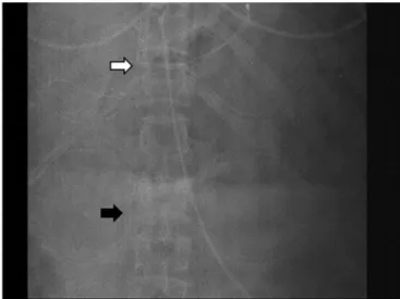
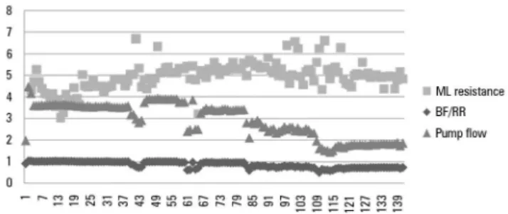
(Maquet Cardiopulmonary AG, Rastatt, Germany) right internal jugular cannula was placed percutaneously under luoroscopic and transesophageal echocardiography control with a previous right internal jugular ultrasound showing the absence of thrombi. Because there were some concerns about inferior vena cava ilter migration due to the suction generated by the ECMO pump, daily abdominal and chest x-rays were performed, and the distance between the inferior vena cava ilter and the tip of the DL cannula remained unchanged during the entire ECMO run (Figure 1). No problems were detected related to the presence of the inferior vena cava ilter and ECMO system. he ECMO low, resistance of the oxygenator and blood low to the rotation of the pump ratio, and system pressures remained within acceptable ranges (Figure 2). Recirculation was not measured; however, inlet venous saturation remained below 72%, suggesting little or no recirculation.
Figure 1 - Double lumen cannula and inferior vena cava filter. Inferior vena cava filter and tip of the double lumen cannula was confirmed with daily x-rays due to potential filter migration. The white arrow indicates the tip of the double lumen cannula, and the black arrow indicates the inferior vena cava filter.
Respiratory rest settings were selected (Table 1).
Sweep gas was adjusted to achieve an arterial CO2 of
40mmHg. Initial ECMO blood low was set at 4L/min and then adjusted to achieve arterial oxygen saturation between 88 and 95%.
acenocoumarol. he inferior vena cava ilter was left in place due to evidence of thrombi trapped in it (Figure 3). After a short period on the general ward, he was discharged home without organ dysfunctions.
Table 1 - Respiratory and organ function parameters before and after extracorporeal membrane oxygenation
Pre ECMO On ECMO
Respiratory rate (bpm) 22 10
Tidal volume (mL/Kg) 6.2 5
Pplat (cmH2O) 29 25
PEEP (cmH2O) 12 10
Driving pressure (cmH2O) 17 15
FiO2(%) 90 30
SaO2(%) 90 96
Cestrs (mL/cmH2O) 25.8 26
Respiratory minute volume (L/m) 9.7 3.9
SOFA 9 1
Noradrenaline (mcg/Kg/min) 0.2 0
ECMO - extracorporeal membrane oxygenation; Pplat - inspiratory plateau pressure; PEEP - positive end-expiratory pressure; FiO2 - fraction of inspired oxygen; SaO2 - arterial oxygen saturation; Cestrs - static compliance of the respiratory system; SOFA - Sequential Organ Failure Assessment.
Figure 2 - Extracorporeal membrane oxygenation circuit monitoring the entire run. There were no problems during the run with resistance to blood flow or with the pump load conditions, which was evaluated as blood flow to the rotation ratio. ML resistance - membrane lung resistance to blood flow; BF/RR - blood flow to rotation ratio.
Figure 3 - Inferior vena cava filter with trapped thrombi. The inferior vena cava filter was left in place after the extracorporeal membrane oxygenation run due to the trapped thrombi in the filter despite adequate anticoagulation treatment during extracorporeal membrane oxygenation.
used during the irst 24 hours. Heparin was subsequently added in increasing doses (mean 22 units per kilogram per hour), and full anticoagulation (mean activated partial thromboplastin time: 66”) was achieved by
ECMO on the 35th hour.
Oxygenation improved, and after a full day of weaning from ECMO, the patient was percutaneously decannulated. Few ibrin deposits were observed on the venous side of the oxygenator without signiicantly afecting membrane function.
Due to diicult weaning, the patient was tracheostomized on day 14. He required video-assisted
thoracoscopy for left-side empyema due to Klebsiella
Pneumoniae and was weaned from the ventilator on
day 18 and decannulated on day 21. Partial right internal jugular vein thrombosis was diagnosed on follow-up, and anticoagulation was switched to oral
DISCUSSION
In thoracic trauma patients, ECMO has the ability to artiicially maintain cardiopulmonary function while the damaged organ recovers.(3) It has been demonstrated
to be a feasible and safe method in this particular
population.(4) hrombotic complications are one of
the reasons for the increase in morbidity and mortality
after major trauma.(5) However, in particular cases,
anticoagulation is contraindicated due to bleeding in the traumatized areas. Our patient developed distal bilateral calf muscle vein thrombosis, and anticoagulation was indicated due to the high risk imposed by his condition.(6)
while leaving the inferior vena cava ilter in place. he strategy to leave the tip of the cannula below the renal veins has the potential to limit ECMO low by inferior vena cava wall collapse due to suction,(7) and this option
was discarded. inferior vena cava ilter extraction was considered but immediate full anticoagulation was deemed not to be safe due to previous hemothorax. Due to the risk of thrombus progression, if the femoral vein were to be cannulated, we decided to use a 27
French Avalon®
. As was previously described, there was no interaction between the inferior vena cava ilter and ECMO system, and the distance between the two intravascular devices remained unchanged. In our patient, anticoagulation had been associated with thoracic bleeding, and heparin was withheld during the beginning of the run, as has been previously reported.(4)
Gothner et al.(8) described the use of the DL cannula
in trauma patients, but none have been described to have a previously inserted inferior vena cava ilter. Luk et al.(9)
described the insertion of an inferior vena cava ilter prior to ECMO decannulation as a prophylactic approach to lower the risk of embolism. Femoro-jugular cannulation is traditionally preferred in ARDS patients due to higher
lows permitted by the cannulas.(7) However, Pappalardo
et al.(10) recently described no diference in blood low or
oxygenation with the use of the DL cannula compared with femoro-jugular cannulation. To the best of our knowledge, this is the irst description of the need to use a DL cannula for venovenous-ECMO due to a previously inserted inferior vena cava ilter.
CONCLUSION
We described a case of thoracic trauma with severe acute respiratory distress syndrome patient who underwent venovenous extracorporeal membrane oxygenation via a double lumen right internal jugular with a previously inserted inferior vena cava. here were no complications related to inferior vena cava ilter migration due to the extracorporeal membrane oxygenation system generated suction in the inferior vena cava. he use of the double lumen right internal jugular cannula could be a safe and feasible option in patients who cannot be cannulated via the femoral veins due to a previously inserted intravascular device.
Author´s contribution
F Palizas Jr., CC García and M Norese conceived the study, F Palizas Jr. and CC García drafted the manuscript, and all authors read and approved the inal manuscript.
A oxigenação por membrana extracorpórea é utilizada em casos de hipoxemia refratária em diversas condições clínicas. Pacientes vítimas de traumatismo torácico geralmente desenvolvem síndrome da angústia respiratória aguda. Em razão do elevado risco de sangramentos, as complicações trombóticas que se apresentam neste contexto são particularmente difíceis de tratar e, geralmente, demandam a inserção de um iltro na veia cava inferior, com a inalidade de prevenir a migração de êmbolos oriundos das veias distais para a circulação pulmonar. Neste
artigo, apresentamos o caso de um paciente com traumatismo torácico, que apresentou grave síndrome de angústia respiratória aguda, com necessidade de utilizar oxigenação por membrana extracorpórea aplicada por meio da introdução de uma cânula com duplo-lúmen na veia jugular interna direita. Este procedimento foi realizado tendo em vista a prévia inserção de um iltro na veia cava inferior, por conta da ocorrência de trombose venosa profunda em ambas as panturrilhas.
RESUMO
Descritores: Oxigenação por membrana extracorpórea; Síndrome do desconforto respiratório do adulto; Traumatismos torácicos; Filtros de veia cava; Trombose venosa; Relatos de casos
REFERENCES
1. Peek G, Harvey C, Faulkner G. Adult respiratory ECMO. In: Annich GM, Lynch WR, MacLaren G, Wilson JM, Bartlett RH, editors. ECMO extracorporeal cardiopulmonary support in critical care. 4th ed. Ann Arbor, Michigan, USA: Extracorporeal Life Support Organization; 2012. p. 309-21.
2. Hess JR, Brohi K, Dutton RP, Hauser CJ, Holcomb JB, Kluger Y, et al. The coagulopathy of trauma: a review of mechanisms. J Trauma. 2008;65(4):748-54.
3. Bassi E, Azevedo LC, Costa EL, Maciel AT, Vasconcelos E, Ferreira CB, et al. Hemodynamic and respiratory support using venoarterial extracorporeal membrane oxygenation (ECMO) in a polytrauma patient. Rev Bras Ter Intensiva. 2011;23(3):374-9.
4. Arlt M, Philipp A, Voelkel S, Rupprecht L, Mueller T, Hilker M, et al. Extracorporeal membrane oxygenation in severe trauma patients with bleeding shock. Resuscitation. 2010;81(7):804-9.
6. Kearon C, Akl EA, Comerota AJ, Prandoni P, Bounameaux H, Goldhaber SZ, Nelson ME, Wells PS, Gould MK, Dentali F, Crowther M, Kahn SR; American College of Chest Physicians. Antithrombotic therapy for VTE disease: Antithrombotic Therapy and Prevention of Thrombosis, 9th ed: American College of Chest Physicians Evidence-Based Clinical Practice Guidelines. Chest. 2012;141(2 Suppl):e419S-94S.
7. Pranikoff T, Hines M. Vascular access for extracorporeal support. In: Annich GM, Lynch WR, MacLaren G, Wilson JM, Bartlett RH, editors. ECMO extracorporeal cardiopulmonary support in critical care. 4th ed. Ann Arbor, Michigan, USA: Extracorporeal Life Support Organization; 2012. p. 133-47.
8. Gothner M, Buchwald D, Strauch JT, Schildhauer TA, Swol J. The use of double lumen cannula for veno-venous ECMO in trauma patients with ARDS. Scand J Trauma Resusc Emerg Med. 2015;23:30.
9. Luk YS, Fung KH, Leung KW. Insertion of inferior vena cava filter prior to removal of prolonged indwelling extracorporeal membrane oxygenation catheters. Hong Kong J Radiol. 2014;17(4):293-6.