(1) Universidade Federal de Sergipe, Aracaju, Sergipe, Brasil.
(2) Departamento de Medicina, serviço de Reumatologia do Hospital Universitário da Universidade Federal de Sergipe (UFS), São Cristóvão, Sergipe, Brasil. (3) Departamento de Fonoaudiologia,
Universidade Federal de Sergipe (UFS), Lagarto, Sergipe, Brasil.
(4) Departamento de Fonoaudiologia, Universidade Federal de Sergipe (UFS), São Cristóvão, Sergipe, Brasil. Aid supply to the survey: Pibic / CNPq Program - bag COPES/UFS (2012-13).
Conlict of interest: non-existent
Speech Therapy in sclerosis systemic: a case report
Intervenção fonoaudiológica na esclerose sistêmica: relato de casos
Leylane Fonseca Almeida(1) Milena Cabral de Lima(1) José Caetano Macieira(2) Carla Patrícia Hernandez Alves Ribeiro César(3) Sílvia Elaine Zuim de Moraes Baldrighi(4)
Received on: October 29, 2015 Accepted on: November 26, 2015
Mailing address: Leylane Fonseca Almeida Rua Carlos Pereira de Melo 421 – Residencial Sul - apto 204 – Bloco Atalaia Farolândia
Aracaju – SE – Brasil CEP: 49030-150
E-mail: [email protected]
doi: 10.1590/1982-0216201618117715
ABSTRACT
Systemic sclerosis is a rare autoimmune rheumatic connective tissue disease, progressive of unknown etiology and variable evolution. Reaches vital organs and perioral tissues, causing limitation of mouth opening, stiffness of the articulators, face mask with appearance, dificulty in chewing and swallowing. Aiming to compare the pre and post intervention miofuncional speech orofacial in subjects with systemic sclerosis was performed qualiquantitativo exploratory clinical study in Rheumatology department of the University Hospital and Clinic, School of Speech Pathology, Federal University of Sergipe, from July / 2012 to December / 2013. The sample consisted of ive individuals, of both genders, aged between 24 and 60 years and conirmed medical diagnosis of that condition. The subjects underwent evaluation by MBGR protocol and speech therapy (myofunctional and myoterapica), totaling 36 sessions. At the end, the initial and inal results were compared from clinical indicators worked. After analyzing the results, improvements in all aspects worked were observed. The previous assessment of speech therapy revealed restriction of mouth opening in all subjects and the inal results showed gains between 5.3 and 14.2mm opening (ave
-rage: 9.26). Regarding tone, mobility of the articulators and orofacial numbness observed improvements and evidence of adequacy of oral functions. Can be concluded that although some patients still show changes in speech rehabilitation promoted signiicant improvements in clinical and quality of life of these
individuals.
Keywords: Speech, Language and Hearing Sciences; Scleroderma, Systemic; Speech Therapy
RESUMO
A Esclerose Sistêmica é uma doença reumática autoimune do tecido conjuntivo, progressiva, pouco fre -quente, de etiologia desconhecida e com evolução variável. Atinge órgãos nobres e tecidos periorais,
causando limitação na abertura da boca, hipertonia dos órgãos fonoarticulatórios, face com “aparência de máscara”, diiculdades na mastigação e deglutição. Com o objetivo decomparar os resultados pré e
pós-intervenção fonoaudiológica miofuncional orofacial em sujeitos com esclerose sistêmica, realizou
--se estudo clínico exploratório qualiquantitativo no setor de Reumatologia do Hospital Universitário e na Clínica escola de Fonoaudiologia da Universidade Federal de Sergipe, no período de julho/2012 a dezem
-bro/2013. A amostra foi composta por cinco indivíduos, de ambos os gêneros, com faixa etária entre 24 e 60 anos e diagnóstico médico conirmado da referida afecção. Os indivíduos passaram por avaliação por meio do protocolo MBGR e 36 sessões de terapia fonoaudiológica (miofuncional e mioterápica). Ao término, os resultados iniciais e inais foram comparados a partir dos indicadores clínicos trabalhados. Diante da análise dos resultados obtidos, foram observadas melhoras em todos os aspectos trabalhados. A avaliação anterior à fonoterapia revelou restrição na abertura da boca em todos os indivíduos e os resultados inais apresentaram ganhos entre 5.3 e 14.2 mm de abertura (média: 9,26 mm). Com relação à tonicidade, mobilidade dos órgãos fonoarticulatórios e dormência orofacial, observaram-se melhoras e evidências de adequação das funções orais. Pode-se concluir que, apesar de alguns pacientes ainda apresentarem alterações, a reabilitação fonoaudiológica promoveu melhoras signiicativas no quadro clí -nico e qualidade de vida dos indivíduos do grupo de estudo.
Descritores: Fonoaudiologia; Escleroderma Sistêmico; Fonoterapia
Case reports
INTRODUCTION
The Systemic Sclerosis (SS) is part of a heteroge-neous group of rheumatic diseases. It is relatively rare and its etiology factor is unknown, producing systemic alterations involving the connective tissue throughout the body. Its evolution is slow, progressive and incapacitating. However, it may occur rapidly, due to the impairment of internal organs1.
The SS affects multiple organs, including skin, cardiovascular system, lungs, gastrointestinal tract and kidneys2. It is clinically characterized by vascular impairment and ibrotic changes in the skin and internal
organs3.
It can be classiied into two subgroups according
to the cutaneous impairment: limited and diffuse.
Limited SS presents limited cutaneous ibrosis in
hands, forearms, legs, neck and face. Patients usually present Raynaud’s phenomenon (RyP) for years and
may have telangiectasia, skin calciication and a late
incidence of pulmonary hypertension. The diffuse SS
is characterized by proximal skin ibrosis of knees or
elbows, excluding the face and neck. Patients may present Raynaud’s phenomenon (RyP) within a year of developing the disease and are more likely to present pulmonary, kidney or heart involvement2.
In Brazil, rheumatic diseases present themselves as the third leading cause of inability for working, supplanted only by psychiatric and cardiovascular diseases4.
Regarding the matters involving the orofacial motricity, there are gaps in the literature. In researches conducted by rheumatologists and dentists, were cited a few manifestations of Speech, Language and Hearing Sciences studies, including microstomia, xerostomia, loss of gengival mucosa5, laryngeal impairment, oropharyngeal dysphagia 6,7, dificulty in chewing, tongue rigidity, changes in the production of bilabial sounds due to the limitation of mandibular movements and vocal changes8-10.
It is estimated with this study, a contribution to the
elucidation of gaps in the scientiic literature (in the area
of Speech, Language and Hearing Sciences), seeking better understanding of the pathophysiology of this
disease, in the interdisciplinarity of related ields. As well as it is believed that it presents signiicant clinical
impact in conducting these cases. Therefore, the aim of this study was to compare the orofacial myofunctional Speech, Language and Hearing Sciences pre and post-intervention results in subjects with systemic sclerosis.
CASE REPORT
Exploratory, longitudinal, prospective, descriptive, with a series of clinic interventional cases, qualitative-quantitative, non-randomized and uncontrolled study. According to the established by the National Council of Health Resolution No. 466/12, of 12 December 2012, the present study was submitted to the Ethics Committee in Research of the University Hospital of the Federal University of Sergipe (UFS), and approved under the CEP-0132.0.107.000-10 registration.
This study is the result of a partnership between the Rheumatology Clinic of the University Hospital and the School Clinic of the Speech, Language and Hearing Sciences course, both located at UFS, from July 2012 to December 2013.
The sample was composed by convenience, and the inclusion criteria adopted were that all subjects should
have a conirmed medical diagnosis of SS, present a
speech therapy disorder, be an adult or an elderly and have cognitive abilities that allowed their participation at all stages of the study. The exclusion criteria were the refusal and abandonment of participating in the survey.
It was initially requested a list of patients with
conirmed diagnosis of SS assisted by a rheumatol
-ogist doctor of the University Hospital aforementioned. Then, the subjects were contacted by telephone and informed about the research procedures – those who have shown interest in participating conducted an initial interview.
Five individuals were selected, four females and one male, aged between 24 and 60 years (average age of 44.6 years).
The initial interview followed a previously elaborated script to collect data about personal records, speech therapy complaints, a previous history of the disease (age of the diagnosis and initial symptoms, medicines used etc.) and other inferences of the SS on the living conditions of the participants. The instrument for data collection was applied orally by the examiners and the data obtained were recorded on proper sheet. At this meeting, it was also asked the signature of the letter of information, the free and informed consent and the authorization form for the image use. In addition to the information collected, there was a consultation to the medical records of individuals in order to get more information about each case.
Following, the orofacial myofunctional evaluation was initiated. During the procedure, the subject was
asked to remain seated with the feet on the loor, in
patient. Data collection and clinical examination had the total of one hour. The MBGR11 protocol was adopted for pre and post Speech, Language and Hearing Sciences intervention.
After the completion of the evaluation, it was decided on the need or not of Speech, Language
and Hearing Sciences therapy. This need was notiied
individually to the participants that, during the feedback session, decided for their participation in the thera-peutic process.
The Speech, Language and Hearing Sciences intervention was performed once a week, in individual sessions that lasted for about 50 minutes. Initially, all
the participants were fed back on the indings, the
work objectives and the therapeutic approaches, with two approaches being selected: a) the myofunctional
– engaged in muscular modiication through the resto
-ration of orofacial functions and b) the myotherapy
– that seeks the modiication of the muscle behavior
through the execution of exercises12.
We worked to prepare the shoulder, cervical,
facial and speciic musculature (temporal, masseter
and sternocleidomastoid) regions. And isotonic and isometric exercises were performed13,14, settled according to the needs of each patient.
Oral functions of chewing and swallowing were crafted with solid and liquid food, relying on the guideline of perception of how the patient performs them and in the demonstration of the ideal achievement standards, reviewing the position, speed, rhythm, sequence and coordination between functions12. To expand this perception, visual aids (camera, video camera and mirror) were used in the sessions.
During the pre and post-therapeutic intervention moments, photographic records were also made with the patient positioned standing, with spine and head erect and one meter distant of the camera (Cyber-shot DSC-WX7 Sony), except for the analysis of wrinkles, in which the participants were positioned sitting, requesting that they should look ahead, without smiling. They observed the presence or absence of marks and wrinkles (primary or secondary) in the forehead, glabella, eyes, cheeks, lips, and mentalis15.
For the analysis of reduced orofacial myofunc-tional disorders and complaints of patients in the sample, the MBGR11 Protocol was partially used, which the instrument features scores that enable the measurement (even if subjective in the “muscle tonus” of the evaluation) of the patients’ performance in the evaluated aspects. Therefore, the items revaluated daily
were: subjective facial analysis, frontal norm (items jaw and usual position of lips), mandibular movement (maximum active interincisal distance), breathing (item
nasal airlow), partial mobility (lips, tongue, cheeks and
jaw), muscle tonus and oral functions such as breathing
(mode and nasal airlow), chewing and swallowing. The
procedure post-intervention was considered with an effective clinical outcome when the scores obtained in this stage were “zero”.
For the measurement of the maximum active interin-cisal distance (MAID) in pre- and post-intervention (start and end of the session), we used a digital caliper, Western 6” with precision of 0.01 mm, verifying the maximum active interincisal distance in frontal vision and the result was transcribed in millimeters in a sheet of the protocol11.
In addition to the above, the participants were also informed about the performance of each exercise at home, using mirror, every day (twice a day), as
prescribed and recorded the dificulties encountered. In
every initiation of a session, there was a consultation about the home exercises, solving possible doubts.
Were stipulated as part of the research protocol, the procedures below, running in all sessions:
• Consultation, by the therapist, of the home
exercises, solving possible doubts;
• Measurement of nasal airlow (with and without
cleaning)11 and MAID11 with the use of a caliper (in the beginning and the end of therapy);
• Application of global relaxation maneuvers and
loosening of cervical muscles, concomitant with breathing (breathing capacity);
• Stretching maneuvers of orofacial muscles;
• Orofacial exercises conduction with application of
parts of the protocol11 before and after the exercises;
• Rehabilitation of oral functions, with application of
part of the protocol11 before and after the imple-mentation of therapeutic activities planned for this purpose.
The exercises were selected and organized as described in the literature15,16. Orofacial massages were composed by sliding maneuvers to promote relax-ation and to reduce and undo the tensions. Kneading maneuvers were carried out to improve the power of
contractility, elasticity, lexibility and to stimulate the
and swallowing, foods of different consistencies were offered.
It is noteworthy that for the selection of the speech therapy protocol of research in orofacial motricity, it was conducted a pilot project with a patient with SS18. From the therapeutic success in that project, the thera-peutic steps of this study were established, including the improvement of facial movements and mobility of lips, tongue, cheeks and jaw; the decrease of the muscle tonus of lips, tongue and cheeks; decrease of the sensation of dormancy; the reestablishment of the nasal breathing mode; the adequacy of the standards of chewing and swallowing and the smoothing of wrinkles and expression marks.
The Speech, Language and Hearing Sciences inter-vention lasted eighteen months (36 sessions) and, to compare the effectiveness of the work, revaluation was performed using the same protocol used during the initial evaluation. Every two months and at the end of the study, were given individual feedbacks to the participants.
The therapeutic sessions, including evaluation and therapy, were performed by two students of the Speech, Language and Hearing Sciences course at the Federal University of Sergipe, São Cristóvão campus. Before the beginning of the study, the students were trained for the application of the protocols used by the guiding professor. This, a specialist in orofacial motricity and
with experience in the ield, monitored the implemen -tation of all stages of the procedure proposed, in order to facilitate the implementation of the proposal.
The obtained data were entered in Excel
spread-sheet software (Microsoft Ofice® package) for
descriptive data analysis and the statistical analysis was performed at the Department of Statistics and Actuarial Science of the original institution. We used the Statistical Package for Social Sciences program – IBM SPSS version 16.0 for Windows (SPSS Inc., 1989-2006, Chicago, Illinois, USA).
RESULTS
With the exception of the subject 1 (the only male participant in the survey), most subjects presented the beginning of the disease between 30 and 55 years (average: 34.6 years old). The participant was male, 24, and reported the beginning of the disease when he was 22.
The interval between the diagnosis of SS and the
irst symptoms lasted a year and a half. Xerostomia
was a frequent complaint in the sample group (80% – n = 4), as well as Raynaud’s phenomenon – cited
by the majority of the sample (in three out of ive
patients – subjects 2, 3 and 5) as an early symptom. Other symptoms reported were: weakness in the limbs (subject 1), swelling (subjects 4 and 5) and joint pain (subject 4).
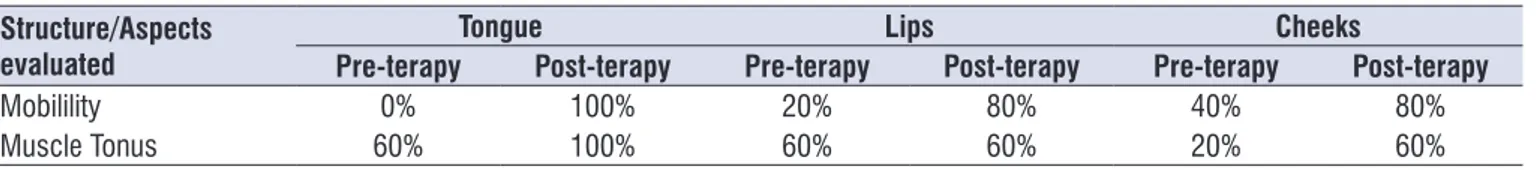
The results of the comparison of the general mobility of lips, tongue and cheeks during the evaluation and after eight months of Speech, Language and Hearing Sciences intervention and the data related to the muscle tonus of structures related to lips, tongue and cheeks are in Table 1.
Table 1. Comparison of the pre and post Speech, Language and Hearing Sciences intervention regarding the mobility and muscle tonus of tongue, lips and cheeks of the ive patients with Systemic Sclerosis.
Structure/Aspects evaluated
Tongue Lips Cheeks
Pre-terapy Post-terapy Pre-terapy Post-terapy Pre-terapy Post-terapy
Mobilility 0% 100% 20% 80% 40% 80%
Muscle Tonus 60% 100% 60% 60% 20% 60%
Regarding the chewing pattern, 100% of the subjects presented alterations in the evaluation. In the post-therapy moment, the pattern remained unilateral only in subject 3.
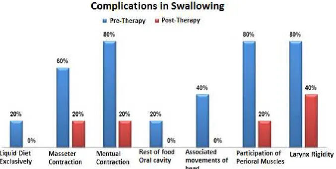
Regarding swallowing, Figure 1 shows the signs and symptoms observed during the evaluation of swallowing in the pre and post therapy periods.
Figure 1. Demonstration of the causative factors of complications in swallowing
Regarding the maximum mouth opening, it can be observed the evolution in all patients of the study, although only two (subjects 4 and 5) obtained values considered within the normal range (Table 2).
In Figure 2, it is possible to notice the presence of lines and wrinkles (primary or secondary) of the studied subjects in the pre- and post- Speech, Language and Hearing Sciences intervention.
Through the pre-therapy evaluation, changes were observed in the rigidity of the facial expression muscles,
absence of expression lines, face with the aspect of a ‘mask’ and glare. Furthermore, it was observed
anteriorization of the head compared to the cervical axis, rigidity of the sternocleidomastoid and trapezius
muscles in 100% of the sample.
Table 2. Measures, in millimeters, of the mouth opening in the pre- and post-Speech, Language and Hearing Sciences intervention moments
Number of the Subject Pre-intervention (in mm)
Post-intervention
(in mm) Difference
1 37,0 42,3 5,3
2 30,0 36,5 6,5
3 26,9 37,2 10,3
4 37,0 47,0 10,0
5 40,0 54,2 14,2
Figure 2. Presentation of the subjects involved in the study, comparing the presence of lines and wrinkles (primary and secondary) in pre- and post-therapy moments
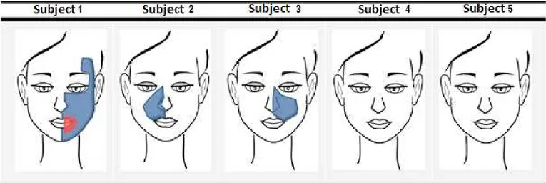
Figure 3. Face regions affected by dormancy, reported by subjects in the pre (in blue) and post (in red) Speech, Language and Hearing Sciences intervention moments
After the Speech, Language and Hearing Sciences
intervention, in 100% of the sample, the rigidity
previ-ously found in the muscles of facial expression
was minimized, the face with a ‘mask’ appearance
was attenuated by smoothing the nasolabial folds
and reduction of the glow existing in the skin. The
anteriorization and rigidity of cervical muscles were also attenuated in 100% of the cases.
hygiene, performance of facial movements; choking and feeling of dormancy and tension (especially in masseter, temporomandibular joint, mentual and orbicularis oris regions).
For the clinical care, some special cares have been established, because of the affection, as the concern about the climate of the physical space. The sessions were always conducted in a room that was purposely not climatized18, because the refrigeration (either by air conditioner or fan) can worsen the RyP condiiton, which causes clinical signs such as vasospasms of extrem-ities, associated with typical color changes, and also can occur a limitation of joint movements, restricting, thus, the continuity of the therapeutic session19.
The sample was composed by ive individuals, 80%
were female (n = 4) and 20% (n = 1) were male. The overall average age was 44.6 years (24-60 years), the average age of the disease start was 34.6 years old. The SS starts between 30 and 50 years old7, this is also found in most of the studied subjects. The average of years of the disease progression was ten years and the time to diagnose it, 1.6 years.
Regarding gender, the female dominance
corrobo-rated the indings of the literature review that cited
a higher prevalence of SS in women, reaching the proportion of three women affected for every man8. In addition to the higher prevalence in women, the SS is also cited as rare in men under 30 years and with a worse prognosis20, something was also observed in this sample.
The indings relating to the initial symptoms of the disease disagree with most of the literature indings.
In 20% of the sample there was, as initial symptom, weakness in the limbs, another 20% presented swelling and spots over the body. And the symptoms of only body spots appear in 40% and swelling and pain in joints in other 20% of the sample. Therefore, most individuals reported noticing early signs of spots over
the body, swelling and pain in joints. The inding differs
from the bibliographic data, because, according to the literature 7,19, the presence of Raynaud’s Phenomenon (RyP) is a more frequent symptom in about 90% of the cases. A bias to be highlighted is about the small sample size of this study, not being possible to gener-alize the results.
Regarding the speech organs (OFAs), it can be observed impairment in mobility, muscle tonus and normal resting posture.
The muscle tonus, mobility and posture of the OFAs
become signiicant during the function, that is, they
DISCUSSION
The interaction between Speech, Language and Hearing Sciences and Rheumatology is relevant to the expansion of knowledge between the areas and a better assistance in Health to individuals who have rheumatic diseases. The SS is highlighted, because, although
relatively rare, it produces signiicant progressive
systemic changes, with slow or fast course, depending on the impairment of the connective corporeal tissue1, especially by reaching multiple2, external and internal3, which requires a specialized and interdisciplinary team.
Another aspect to be highlighted is the need for constant assistance and protective measures in Public Policies, since the SS can disable the subject in his work4, favoring social exclusion.
The SS may be classiied as diffuse or limited2, with
the limited being more interesting to Speech, Language and Hearing Sciences, since it includes changes in the face and neck. Despite the above, the literature is still scarce in reporting the orofacial myofunctional alterations in subjects with SS and Speech, Language and Hearing Sciences intervention possibilities in this condition.
One of the hypotheses that justify incipient researches in the area is related to the relative rarity of
the SS. This hypothesis was conirmed in this study, in which it is possible to see the dificulty in recruiting
cases for the composition of the sample. In the liter-ature1, it is shown an annual incidence of four to 19 individuals per one million inhabitants.
Another hypothesis concerns the dificulty of estab
-lishing partnerships among professionals, although
the researches are justiied in the area by the need to
expand the interdisciplinary treatment. Some publica-tions have shown structural and functional involvement of the stomatognathic system 5-10, justifying the impor-tance of including Speech, Language and Hearing Sciences in Rheumatology clinics.
Although this study has been conducted with a small sample group, during the therapeutic process, it was observed the effective participation of the participants, that rarely missed the sessions. The results discussed here are related to the initial evaluation and revalu-ation after a year and a half of Speech, Language and Hearing Sciences therapy (average of 36 sessions). The original intention was to determine if the Speech, Language and Hearing Sciences intervention with these subjects could improve the clinical presentation.
The biggest complaints were the dificulty in respiratory
period). Regarding the tongue, it was observed in the pre-therapy evaluation 60% of the sample without
changes. At the inal revaluation, 100% of the sample
was considered normal regarding the tongue tension. The muscle tonus of the cheeks was normal in 20% of the studied sample in the evaluation, and, after the intervention, 60% of subjects presented themselves normal.
In the usual posture of lips at rest, 100% of the sample began to show lip seal after performing the Speech, Language and Hearing Sciences intervention. It is hypothesized that the gain occurred due to the reduction in the oral and facial rigidity acquired due
to the lengthening of oromyofacial ibers. It should
be noted, however, that the subject 2 would present,
because of malocclusion Angle Class II, dificulties
in lip seal. It can be observed, in Figure 2, that such subject performs the sealing, but with hypertonia of the mentual, not being, therefore, required such a posture to this subject.
Researchers10 reported changes in the aspects related to mobility, muscle tonus of the OFAs and in the rest position of patients with SS. These changes probably occur due to the decrease of the functions of the muscles of the face, indicating increased rigidity, which may help to reduce the amplitude of mandibular movements and, hence, cause damages to the oral functions.
The explanation of the abovementioned aspects
may be justiied also due to the chronic deposition of
collagen, as well as in the restriction of mouth opening, often cited in literatura1,9 in subjects with SS, also displayed in Table 2. In such cases, the skin becomes thickened and gradually loses elasticity, limiting the facial movements. On the face, a thickening skin leads to orofacial events, including rigidity and skin atrophy, loss of expression (reduction of the lips and tapering of the nose) and alteration in mouth opening. These characteristics lead to mask facies1,7. The improvement in the abovementioned aspects had been checked by researchers18 in a clinic case after the institution of Speech, Language and Hearing Sciences therapy initiation.
As shown, the chewing function was altered in 100% of subjects – before therapy, it was
charac-terized by ineficient grinding, atypical muscle contrac
-tions, decreased speed and incidence of masticatory preference. The achievement of the ideal mastication (bilateral) post-therapy was present in 80%. The
dificulty in chewing in subjects with SS was cited
must not be seen alone only12. Despite the studied individuals can perform the OFAs mobility activities, it was observed muscle rigidity of the cheeks and mentual. The functions of these structures are important not only at rest, but also during chewing, since some individuals had unsystematic closure of lips, abnormal chewing and swallowing functions (Figure 1), indicating malfunction of orofacial muscles and consequent loss in the functionality of cheeks and mentual.
Regarding mobility, the entire sample presented
dificulties in tongue mobility (Table 1), which is
associated with the presence of rigidity. However, after the intervention, all subjects achieved the normal pattern. Regarding the lips, only 20% of the sample presented score ‘zero’ during the pre-intervention and, after the treatment, 80% of the sample managed to get score ‘zero’. The mobility of the cheeks was observed ‘normal’, initially, in 40% of subjects, and after the Speech, Language and Hearing Sciences intervention, this value rose to 80%.
Despite the improvement in most of the sample (80% – as shown in Table 1), one of the subjects (S5) has not yet obtained complete improvement of the mobility of lips and cheeks (S5 tries to perform the movement and it is approximately normal).
The praxic orofacial movements are the most important ones to the execution of oral functions, especially chewing, swallowing, speeking and phonation, and are essential to both the accuracy of the motion as to their temporal sequence. In the protocol used, there is the possibility of annotating for both isolated and praxic movements of different grada-tions (zero for normality, score one for approximate movement, score two for the attempt to perform a movement and score three for nonexistent mobility), which facilitates monitoring the progress of patients in attendance. However, it would be interesting adding parameters such as time-hesitation, whether there was or not a need for demonstration and help from the therapist, whether through proprioceptive, tactile or kinesthetic clues – which was not used in this research, which is a factor that can be considered as a limitation of the present study.
muscle contraction in 60%. The contraction of the mentual, the perioral involvement and the larynx rigidity were noted in 80% of the sample. The head associated movement (anterior-posterior) occurred in 40%. After the Speech, Language and Hearing Sciences therapy, the subject S3, that initially only ate in a liquid diet, after the 17th session, started to eat pasty consistency foods. The contraction of the masseter, mentual and the perioral participation still exist in 20% of the sample – even so, it was observed some improvements in the clinical condition of the participants. The food residue in the oral cavity and the associated head movement was no longer noticed in 100% of subjects. Regarding the rigidity of the larynx, 40% of the sample still presents the alteration, but with an improvement in the condition. During the anamnesis, some participants reported the presence of swallowing disorder symptoms, such as coughing after swallowing (60% of cases) and choking (80%). It is noteworthy that these symptoms, suggestive of swallowing disorders, were not observed during the evaluation or during the Speech, Language and Hearing Sciences intervention process. When these data were taken up during the revaluation, there were no more complaints of these symptoms by the sample group.
Another factor that impairs chewing and swallowing is the presence of xerostomia. According to the liter-ature, swallowing22 is a subjective sensation of dry mouth. When there is a quantitative decrease in the
salivary low, among the most important signs is the non-accumulation of saliva in the loor of the mouth:
the lips become dry and with a changed texture (white,
foamy, ibrous or sticky saliva), and can occur a persis
-tence of caries in the tooth cervix, dental erosion,
chronic pain or burning sensation, dificulty in speaking,
chewing and swallowing. In conducting the anmnesis, 80% of the individuals presented such complaint. During the revaluation, this data was not reported by the studied subjects.
It is hypothesized that increasing the tongue mobility, as well as providing guidance on the need for more oral hydration were aspects that have led to the improvement obtained regarding xerostomia.
Regarding respiratory issues, authors12,23 argue that nasal breathing is a process of vital importance for the individual, and it is essential for the growth and balanced development of the orofacial muscles. So, when there is a persistence of the altered respiratory condition (oral or oronasal), it is put in danger the balance of other oral functions.
in literature8-10. The altered chewing function can be related to temporomandibular joint disorder and limita-tions of mandibular movements. In addition, the rigidity leads to a change in the movements of the structures involved in this act as a result of collagen deposition in perioral tissues1.
Thus, multifactorial aspects can justify the losses in the masticatory process of individuals with SS, as increased muscle tonus of tongue, lips and cheeks; the rigidity with loss of elasticity of the skin, temporoman-dibular disorders, the poor condition of dental elements and the periodontal and xerostomia and limited mobility
lips, tongue, cheeks and jaw. It is justiied, greatly, the
need for Speech, Language and Hearing Sciences intervention as soon as possible, since those functions are performed daily and do not affect only the nutri-tional aspects, but also interfere in the emonutri-tional and social aspects involving the subjects.
Another function that will also suffer the same impacts is swallowing, which depends on complex neuromuscular action (sensitivity, taste, proprio-ception, mobility, muscle tonus and tension). In SS, swallowing was reported in the literature8-10,19 as altered. The disorder in swallowing can lead to conditions of pulmonary complications which, if not recognized and treated in time, will lead to frequent hospitalizations and worsening of the health situation, which can, severely, compromise the lives and health condition of these individuals.
Researchers21 reported a clinical case of a female patient, 50 years old, who presented mixed disease of connective tissue (SS, systemic lupus erythematosus
and polymyositis) with dificulty to swallow, frequent choking, limited mouth opening and relux symptoms.
Weekly Speech, Language and Hearing Sciences therapy has been established, for a year, demon-strating effectiveness in the speech therapy process.
Through videoluoroscopy, it was evidenced the difi -culties in ejection of the bolus, with stasis in valleculae and piriform recess, demonstrating decreased pharyngeal motility. By esophageal manometry, it was found a motor disorder characterized by hypotonia and absence of contractile activity.
In Table 2, it can be seen the results obtained in pre and post- Speech, Language and Hearing Sciences intervention regarding the maximum mouth opening. It can be seen that the initial values, of the entire sample group, were lower than 40 millimeters (mm). In the literature, the reference values for the normality of the opening are discussed, because there are authors13,14,25 that cited the normality of values above 40mm, while others26 claim normality above 45mm in adults.
It was decided, in this study, for the normal index above 45 mm according to the one cited by Bianchini26. During the therapeutic process, it was observed that,
in the therapeutic pathway, there was a signiicant
improvement in the opening values of the entire sample. Measurements were performed in all sessions, so that, the individual could observe how the stretching
maneuvers and exercises resulted in signiicant gain
of mouth opening, valuing the therapeutic process. Thus, measurements served to encourage in the adherence to the therapeutic process and in continuing the exercises at home. However, although still below the normal standard of mouth opening26, the subjects S1, S2, and S3 presented signiicant gains. S1 and S2
showed gains ranging from 5 to 6mm, S3 increased 10.3mm and S4 and S5 achieved the normality pattern, with a gain varying from 10 to 14.2mm.
As mentioned, the SS is a rare connective tissue disease having as prominent manifestation the
thick-ening or skin ibrosis27. Regarding the facial movements, oral muscles (static or moving) were stiff, especially the orbicularis oris. This condition is most evident in the upper lip region, as reported by researchers in the area9. The reduction of the facial muscles is reported in the literature as a fairly frequent alteration in these individuals1,8,28-30.
The presence of lines and wrinkles (primary or secondary) was analyzed in this study through subjective clinical examination carried out by visual observation and photographic records, as the images contained in Figure 2. In the pre-therapy evaluation, changes were observed, regarding the rigidity of the muscles of the facial expression, absence of expression lines, face the aspect of a mask and glare. Also were observed anteriorization of the head regarding the cervical axis and rigidity of the sternocleidomastoid and trapezius muscles in 100% of the sample.
After the Speech, Language and Hearing Sciences intervention, there was a decrease of the face muscle rigidity in 100% of the sample, making it possible to perform facial movements. Moreover, the characteristic In the SS, more than 70% of patients present
pulmonary impairment, manifested from pulmonary
ibrosis to pulmonary vascular disease. The condition
can evolve to pulmonary hypertension, cited in the literature as one of the leading causes of death in this condition24.
There is a change report8 in the breathing mode in the SS. This is justiied by pulmonary problems and the dificulty presented for systematically lip closure due to
the rigidity in the orofacial muscles and skin thickening.
Initially, during the anamnesis, the subjects were asked about complaints regarding the breath awareness. All the subjects (100%) reported symptoms such as fatigue and shortness of breath when performing physical exertion activities. In the literature, this could be explained by the shortness of breath
caused by pulmonary ibrosis – the individual begins
to present a stiff lung1. After checking that no organic obstruction factor prevented the nasal breathing mode, working with raising awareness and exercises involving the nasal breathing mode were performed, with the possibility of improvement in the sixth session. In the post-intervention evaluation, it was found that neither the complaint nor the altered breathing mode were present in the sample. It is noteworthy that the subject 2, because of occlusal changes and although breathing through the nose, still remained in a posture with open lips.
Regarding the limitation of mouth opening, some subjects start to present, during the course of the
disease, the microstomia. This is a common inding in
cases of SS and it is possibly related to the thickening of the skin, a characteristic of the disease1.
Measures that provide an increase in the mouth opening amplitude promote improvements of oral functions and hygiene measures, justifying the action as early as possible in patients with rheumatic diseases such as the SS. It is avoided, thus, the installation of microstomy13,14.
In this study, this inding was conirmed in the clinical
examination of the stomatognathic system: during
reports in the anamnesis, dificulties were reported
in performing oral hygiene maneuvers (setbacks in handling the toothbrush caused by the scleroderma claw and mouth opening for hygienisation) aspects also cited by literature1. This inding was conirmed by another study9, that commented on the implications of the limitation in opening the mouth for speech and for
of the face with the aspect of a mask was attenuated due to the softening of the nasolabial furrow, and also occurred a decrease of the existing glow in the skin. The anteriorization and rigidity of the cervical muscles were also attenuated in 100% of cases.
For the improvement of such aspects, it has become indispensable that the study group presented body and orofacial proprioception, so that, there would be the
regulation of the habitual posture and modiication of
tension conditions (when possible), as stated by the literature15.
Moreover, relaxation movements in the orofacial region favor, according to the literature15, the sensory receptors and increase the low of blood and lymph
circulation by increasing the dilation of blood vessels and, therefore, can remove toxins even after the massages performed in the orofacial region. Thus, it allows more agile, coordinated and without much effort muscle movements – sensations not perceived from the beginning of the disease progression for all subjects of
the sample. This was also signiicant for the adhesion
of the research subjects.
An important observation in the pre and post Speech, Language and Hearing Sciences intervention facial analysis was the reduction of lines and wrinkles in 80% of the sample (subjects 2 to 5), as can be seen in Figure 2. According to the literature15, wrinkles may be caused by repetitive, normal and excessive muscle contrac-tions. In the case of SS, the rigidity of the orofacial muscles, combined with the vascular involvement and
ibrotic skin changes3 are suficient to premature aging
in these subjects. Thus, another beneit of the perfor -mance of speech therapists in the SS regards the facial aesthetics, working on the subject’s self-esteem and, consequently, on compliance with the treatment.
Another symptom, cited and possible to be observed in Figure 3, was orofacial dormancy. Accordingly,
two hypotheses can be listed. The irst, about the
concomitant presence of trigeminal neuropathy (V cranial nerve), reported as possible by the literature6. The second due to the lack of blood supply, due to the
speciic vascular impairment of the disease3. During the
massage application for increasing the blood low in
the facial area, it occurs an increase in the mobility and improves the blood circulation. Consequently, there is a decrease in the sensation of dormancy, present in 60% of the study participants. After Speech, Language and Hearing Sciences, the dormancy persisted only in 20% of the sample (S1), but only in the lower intraoral third
and with a smaller intensity than the aforementioned therapeutic intervention.
It can be said that the established therapeutic goals, from the proposed clinical outcome, have been successfully achieved, since it was observed in the subjects:
• the improvement of facial movements and mobility
of lips, tongue, cheeks and jaw;
• decreased muscle tonus of lips, tongue and cheeks;
• decreased sensation of dormancy;
• the restoration of the nasal breathing mode;
• the adequacy of chewing and swallowing patterns;
and
• the smoothing of marks and wrinkles in most of the
study subjects.
The reports obtained also cited as gains in the therapeutic process the possibility to better express their emotions; decreasing in the sensation of “dry mouth”; the possibility of feeding more properly; the sensation of breathing more easily; and the perception of “release” of the orofacial muscles. Are evident,
therefore, the impacts of bio-psychosocial beneits
and quality of life caused by the proposed procedure – especially considering that the subjects are aware that the SS is a progressive and chronic disease.
Larger groups and longitudinal follow-ups study are necessary to observe whether or not there is mainte-nance of the affected Speech, Language and Hearing Sciences goals.
CONCLUSION
Comparing the initial evaluation results and the results obtained in pre and post- Speech, Language and Hearing Sciences intervention moments, it was
observed a signiicant improvement in the orofacial
myofunctional aspects, especially in the ones concerning the maximum mouth opening; the mobility and muscle tonus of tongue, cheeks and jaw; the implementation of facial movements; the functions of chewing, breathing and swallowing; the reduction of orofacial dormancy and xerostomia complaints; the usual posture of lips.
REFERENCES
2. Godoi ETAM, Barbosa AT, Godoi JTAM, Ramos MAM, Marques SRB, Duarte ALBP. Envolvimento macrovascular e esclerose sistêmica. J Vasc Bras. 2009;8(1):65-76.
3. Correa MJU, Perazzio SF, Andrade LEC, Kayser C.
Laser doopler imaging para quantiicação do luxo sanguíneo de polpa digital em condições basais e
após estímulo frio em pacientes com ES. Rev Bras reumat. 2010;50(2):128-34.
4. Miguel RCC, Resende GG, Garcia PP, Gresta LT. O tecido conjuntivo e o sistema musculoesquelético. In: Carvalho MAP, Lana CCD, Bértolo MB. (Org.) Reumatologia: diagnóstico e tratamento. 3 ed. Rio de Janeiro: Guanabara Koogan; 2008. p. 3-11.
5. Wada T, Ram S. Limited mouth opening secondary to diffuse systemic sclerosis. case Rep. Dent. 2013;2013:937487.
6. Baron M, Hudson M, Tatibouet S, Steele R, Lo E, Gravel S et al. The Canadian systemic sclerosis oral health study: orofacial manifestations and oral health-related quality of life in systemic sclerosis compared with the general population. Rheumatology (Oxford). 2014;53(8):1386-94.
7. Pereira DB, Amaral JLA, Szajubok JCM, Lima SMA,
Chahade WH. Manifestações otorrinolaringológicas nas doenças reumáticas autoimunes. Rev Bras
Reumatol. 2006;46(2):118-25.
8. Magro PCFM. Esclerodermia: revisão da literatura
e caracterização da população observada na
consulta de Reumatologia do Centro Hospitalar
Cova da Beira, E.P.E. [dissertação]. Covilhã
(Portugal). Universidade da Beira Interior; 2009. 9. Baldrighi SEZM, Brito AF, Teixeira JP. Lima APLV.
Esclerodermia Sistêmica: relato de caso. Revista extensão & Sociedade. 2011 [acesso em 30 set
2015]; 2(3). Disponível em http://www.periodicos.
ufrn.br/ojs/index.php/extensaoesociedade
10. Baldrighi SEZM, Nascimento LT. Repercussões
orofaciais na Esclerodermia Sistêmica: relato de caso. In: 19º Congresso Brasileiro e 8º Internacional de Fonoaudiologia, 2011, São Paulo. Disponível em: <www.sbfa.org.br/portal/suplementossbfa>. Acesso em: 25 set. 2013.
11. Baldrighi SEZM, Lopes LD, Lima MC, Almeida LF, Macieira JC. Achados Fonoaudiológicos na Esclerose Sistêmica. In: 21º Congresso Brasileiro de Fonoaudiologia, 2013, Porto de Galinhas. Disponível em: <http://sbfa.org.br/fono2013/pdf/ anais_poster.pdf. Acesso em: 25 set. 2013.
12. Genaro KF, Berretin-Félix G, Rehder MIBC,
Marchesan IQ. Avaliação miofuncional orofacial:
protocolo MBGR. Rev CEFAC. 2009;11(2):237-55.
13. Marchesan IQ. Deglutição – diagnóstico e
possibilidades terapêuticas. In: Marchesan IQ. Fundamentos em Fonoaudiologia – aspectos clínicos da motricidade oral. 2ª. ed. Rio de Janeiro: Guanabara Koogan; 2005. p. 51-8.
14. Vincent C, Agard C, Barbarot S, N’Guyen JM, Planchon B, Durant C et al. Orofacial manifestations of systemic sclerosis: A study of 30 consecutive patients. Rev Stomatol Chir Maxillofac.. 2010;111(3):128-34.
15. Yuen HK, Marlow NM, Reed SG, Mahoney S, Summerlin LM, Leite R et al. Effect of orofacial exercises on oral aperture in adults with systemic sclerosis. Disabil Rehabil. 2012;34(1):84-9.
16. Tasca SMT. Programa de aprimoramento muscular em fonoaudiologia estética facial. Barueri: Pró-Fono; 2002.
17. Toledo PN. Programa de tratamento miofuncional
e estomatognático para estética facial.
fonoaudiologia e estética. São Paulo: Lovise; 2006. 18. Bianchini EMG. Bases da terapia de motricidade
orofacial. In: Marchesan IQ, Silva HJ, Berretin-Felix G. Terapia fonoaudiológica em motricidade orofacial. São José dos Campos: Pulso; 2012. p. 31-42.
19. Baldrighi SEZM, Almeida LF, Lima MC, César
CPHAR, Macieira JC. Impacto da intervenção
fonoaudiológica na esclerose sistêmica. Distúrb Comum. 2014;26(3):596-605.
20. Kayser C, Correa MJU, Andrade LEC. Fenômeno de Raynaud. Rev Bras Reumatol. 2009;49(1):48-63.
21. Marcucci M, Abdala N. Análise do masseter,
por espectroscopia de próton, em pacientes com esclerose sistêmica. Radiol Bras. 2009;42(3):145-50.
22. Mendes LMA, Mendes CG, Rehder MIBC. Atuação
fonoaudiológica em um caso de doença mista do
tecido conjuntivo. Rev CEFAC. 2006;8(1):79-83. 23. Michels-VanAmelsfort JM, Brand HS, Van Laar JM.
Oral manifestation of systemic sclerosis. Review Dutch. 2013;120(9):446-50.
24. Silva MAA, Marchesan IQ, Ferreira LP, Schimitd R, Ramires RR. Postura, tônus e mobilidade de
lábios e língua de crianças respiradoras orais. Rev
CEFAC. 2012;14(5):853-60.
através da Técnica de Oscilações Forçadas [Dissertação]. Rio de Janeiro (RJ): Universidade do
Rio de Janeiro; 2010.
26. Paquette DL, Falanga V. Cutaneous concerns of scleroderma patients. J Dermatol. 2003;30:438-43.
27. Bianchini EMG. Mastigação e ATM: avaliação
e terapia. In: Marchesan IQ. Fundamentos em Fonoaudiologia – aspectos clínicos da motricidade oral. 2ªed. Rio de Janeiro: Guanabara Koogan. 2005; p.45-57.
28. Kayser C, Andrade LEC. Esclerose sistêmica. In: Sato E. Guias de medicina ambulatorial e hospitalar-reumatologia. São Paulo: Manole; 2010. p. 113-23.
29. Pedroza AMA, Mota MHA, Carvalho AGC, Oliveira EA, Cardia MCG, Lucena NMG et al. Atuação
da isioterapia em pacientes com esclerodermia
sistêmica: relato de casos. Rev Bras Cien Saúde. 2012;16(2):115-24.