Arterial stiffness and 25-hydroxyvitamin D levels in chronic kidney
disease patients
HAKAN AKDAM1*, ALPER ALP2
1Division of Nephrology, Department of Internal Medicine, Faculty of Medicine, Adnan Menderes University, Aydın, Turkey 2İzmir Tepecik Training and Research Hospital, Nephrology Clinic, İzmir, Turkey
S
UMMARYStudy conducted at Faculty of Medicine, Adnan Menderes University, Aydın, Turkey
Article received: 1/15/2017
Accepted for publication: 3/12/2017
*Correspondence:
Division of Nephrology, Department of Internal Medicine Faculty of Medicine, Adnan
Menderes University Aydın, Turkey Postal code: 09100 [email protected]
http://dx.doi.org/10.1590/1806-9282.63.10.910
Objective: Arterial stiffness refers to arterial wall rigidity, particularly develop-ing in central vessels. Arterial stiffness increases in early stage chronic kidney disease (CKD), and it is a strong predictor of cardiovascular and all cause mortality. Vitamin D has beneficial effects on blood pressure, vascular endo-thelial function and arterial stiffness. 25-hydroxyvitamin D (25(OH)D) defi-ciency is quite common worldwide and in the CKD population. We aimed to evaluate the prevalence of 25(OH)D deficiency and its relation with arterial stiffness in CKD.
Method: Our study included 101 patients (51 male, 50 female), with stages 3B–5 CKD not on dialysis. A single-cuff arteriograph device (Mobil-O-Graph) was used to evaluate arterial stiffness parameters of pulse wave velocity (PWV) and augmentation index (Alx@75). The patients were divided into two groups: group I vitamin D non-deficient [25(OH)D > 15 ng/mL] and group II vitamin D deficient [25(OH)D ≤ 15 ng/mL].
Results: Overall, the mean 25(OH)D level was 14.1±7.9 ng/mL and 70 patients (69.4%) were vitamin D deficient. The mean Alx@75 value was significantly higher in group II (28.6±10.8% vs. 23.3±13.5%, p=0.038). PWV was higher in group II, but the difference was not significant. Group II exhibited significantly lower serum albumin (p<0.001), hemoglobin (p=0.005), calcium (p=0.041) and estimated glomerular filtration rate (eGFR) (p=0.041), but significantly higher 24-hour proteinuria (p=0.011) and more females (p=0.006). Vitamin D was negatively correlated with Alx@75 augmentation pressure, parathyroid hormone, proteinuria and body mass index, and positively correlated with albumin, hemoglobin, eGFR, calcium and transferrin. 25(OH)D was independently associated with Alx@75 (beta=-0.469, p=0.001) and albumin (beta=0.447, p=0.002).
Conclusion: In CKD patients 25(OH)D deficiency was common, particularly in
females. Level of 25(OH)D was independently associated with Alx@75.
Keywords: chronic kidney disease, vitamin D, vascular stiffness, pulse wave analysis.
I
NTRODUCTIONArterial stiffness is a term used to define the viscoelastic properties of the vessel wall and refers to arterial wall rigidity.1 Ageing of the arterial system, progressive
struc-tural changes, disintegration and degeneration of elastin, collagen deposition, arterial wall thickening, endothelial damage and progressive arterial dilatation are all associ-ated with arterial stiffness.2,3 Deterioration of coronary
perfusion and left ventricular hypertrophy secondary to arterial stiffness increase morbidity and mortality.1,3,4
Cardiovascular events and cardiovascular mortality in-crease in the early stage of chronic kidney disease (CKD). Arterial stiffness is an independent predictor of cardio-vascular disease in CKD.5-7 Increase in arterial stiffness
emerges before atherosclerosis and has been evaluated as an early marker of systemic atherosclerosis. Arterial stiff-ness is independently associated with cardiovascular events and all-cause mortality.4,6
and measuring 25-hydroxyvitamin D (25(OH)D) is the best method for determining vitamin D deficiency due to its longer half-life.8-10 Values of 25(OH)D above 30 ng/mL
are considered normal.8,10,11 Vitamin D plays a crucial role
in bone and calcium metabolism. In addition, it has ben-eficial effects on the immune system, blood pressure regulation, vascular endothelial function, and body growth
and development.9,10,12 Previous studies have
demon-strated the association between 25(OH)D deficiency and increased arterial stiffness in chronic diseases such as lupus, diabetes, hyperparathyroidism, and kidney disease.13-15
Pulse wave velocity (PWV) and augmentation index normalized with 75/min heart rate (Alx@75) are consid-ered to be gold standard index for arterial stiffness.1,3,6 Al
Mheid et al.16 demonstrated that 25(OH)D levels
nega-tively correlated with PWV and Alx in healthy subjects, and reported that vitamin D deficiency increased arterial stiffness effecting by vascular dysfunction.
In our study, we aimed to evaluate the prevalence of 25(OH)D deficiency and the relationship between 25(OH) D and PWV, and Alx@75 in patients with stage 3B-5 CKD.
M
ETHODOne hundred and one (101) patients over 18 years of age with stage 3B-5 CKD not on dialysis, diagnosed by using the specified in the KDIGO 2012 Clinical Practice Guide-line for the Evaluation and Management of Chronic Kidney Disease17 were recruited. Patients were excluded from the
study if they were known to have coronary artery disease, heart failure, valvular heart disease, metal valves, stents, metal prosthesis, peripheral artery disease, malignancy, active autoimmune disorders, recent acute coronary syn-drome (within the last three months), or if already on vi-tamin D supplementation. The study was conducted dur-ing winter (December through March) throughout the years 2012-2015. Our study was conducted in accordance with the Helsinki declaration, and was approved by the local ethics committee of clinical research (decision num-ber 2016/870, dated 12.05.2016). The patients were in-formed about the nature of the study, and inin-formed consent was obtained from all participants before entering the study. All patients underwent a physical examination that included height and weight measurements, age, sex, and etiology of CKD as recorded in the medical chart of each patient. Body mass index was calculated by dividing body weight (kg) by the square of the body height (m2).
Study group
Vitamin D insufficiency was defined as serum 25(OH)D levels of between 15-30 ng/mL, and deficiency as lower than
15 ng/mL, according to the NKF/KDOQI guidelines.11
Pa-tients were divided in two groups according to their vita-min D levels: group I vitavita-min D non-deficient, had 25(OH) D > 15 ng/mL; group II vitamin D deficient, had 25(OH)D ≤ 15 ng/mL.
Biochemical and 25(OH)D measurements
Twelve-hour fasting blood samples were obtained in order to assess serum creatinine, urea, glucose, albumin, uric acid, calcium, phosphorus, parathyroid hormone, 25(OH)D and hemoglobin levels at the time of arterial stiffness measure-ment. 25(OH)D measurement was performed by radio-immune assay (RIA) method. All hormonal and biochem-ical tests were measured by Architect c8000 Clinbiochem-ical Chemistry Analyzer device (©2015 Abbott Laboratories. Abbott Park, Illinois, USA). Estimated glomerular filtra-tion rate (eGFR) calculated by the short Modificafiltra-tion of Diet in Renal Disease (MDRD) formula.
Arterial stiffness measurement
Arterial stiffness measurements were performed using the oscillometric method. After 15 minutes of rest, measure-ments were performed with a single-cuff arteriograph de-vice (Mobil-O-Graph PWA, a model pulse wave analysis device, I.E.M. GmbH, Stolberg, Germany). An appropriate blood pressure cuff was placed on the brachial artery trace of the upper arm. The arteriograph device performed three consecutive measurements automatically at 30-sec intervals. The arteriograph measures the blood pressure in the upper arm; then, the device inflates the pressure cuff to 35 mmHg above the systolic blood pressure. This enables detection of fluctuations in brachial artery pres-sure. A tonometric sensor amplifies the fluctuations and transmits them to the arteriograph. The device software program decomposes early and late systolic and diastol-ic waves. Pulse wave velocity, pulse pressure, central sys-tolic – diassys-tolic pressure calculated by the central pressure changes, early (direct, P1), late (backward, P2) systolic and diastolic waves. Augmentation pressure is the difference between the first and second systolic peak, and Alx is cal-culated as the proportion of augmentation pressure to pulse pressure. Augmentation index was normalized to a heart rate of 75 bpm (AIx@75) for comparison for differ-ent heart rates.3,18
Statistical analysis
distribu-tion. Qualitative variables were expressed as number and percentage. The quantitative normally distributed variables were expressed as mean±standard deviation. Abnormally distributed variables were expressed as median and 25th-75th
percentiles. Chi-square test was used for comparison of qualitative variables. For intergroup comparisons of quan-titative variables, Student’s t-test and Mann-Whitney U test were used for data with normal and abnormal distri-butions, respectively. A Kruskal-Wallis test was used to make comparisons between CKD stages. Normally dis-tributed variable associations were analyzed by Pearson’s correlation coefficient and variables with abnormal dis-tributions were analyzed by Spearman’s correlation coef-ficient. Multiple regression analysis was used to determine the factors affecting 25(OH)D and Alx@75 variables. A value of p<0.05 was considered significant.
R
ESULTSOne hundred and one (101) CKD patients who were not on dialysis were included in the study. The mean age was 56.7±11.3 years, 49.5% were female. The mean 25(OH) D level was 14.1±7.9 ng/mL. Out of the 101 analyzed patients, 70 (69.4%) had deficient levels of 25(OH)D, 25
(24.7%) had insufficient levels of 25(OH)D and only six (5.9%) had normal 25(OH)D levels (above 30 ng/mL). Levels of 25(OH)D in CKD stages 3B, 4 and 5 expressed as median and 25th-75th percentile range were: stage 3B:
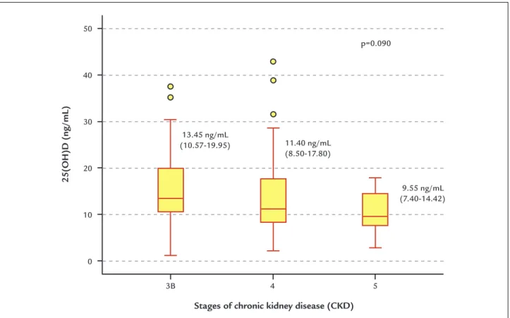
13.45 ng/mL (10.57-19.95); stage 4: 11.40 ng/mL (8.50-17.80); and stage 5: 9.55 ng/mL (7.40-14.42). Decreased levels were observed in all CKD stages, with a tendency to decrease as the CKD stage increased (p=0.090). Dis-tribution of 25(OH)D according to CKD stages is shown in Figure 1.
The CKD patients were grouped as follows based on their 25(OH)D levels: 31 patients (30.6%) in group I [25(OH)D > 15 ng/mL] and 70 patients (69.4%) in group II [25(OH)D ≤ 15 ng/mL]. The causes of kidney disease in group I and group II were diabetes mellitus at 19.4-25.7%, hypertension at 41.9-31.4% and glomerulonephri-tis at 16.1-11.4%, respectively. In group I, 50% of the dia-betic patients were using insulin and 50% were using oral antidiabetic drugs, and in group II 66.7% of the diabetic patients were using insulin and 33.3% were using oral antidiabetic drugs. In all, 64.5% of group I and 52.9% of group II were using a single antihypertensive. Use of an-tihypertensive drugs is shown in Table 1.
FIGURE 1 25-hydroxyvitamin D levels in chronic kidney disease stages 3B, 4 and 5.
[25(OH)D; median (25-75 percentile)±min, max value]
25(OH)D (ng/mL)
Stages of chronic kidney disease (CKD) 13.45 ng/mL
(10.57-19.95) 11.40 ng/mL (8.50-17.80)
9.55 ng/mL (7.40-14.42) p=0.090
3B 4 5
50
40
30
20
10
TABLE 1 Comparison of clinical and biochemical characteristics of groups.
Parameter Group I
[25(OH)D > 15 ng/mL]
Group II
[25(OH)D ≤ 15 ng/mL]
p
Female / Male (n) 9 / 22 41 / 29 0.006
Age (year) 55.8±12.8 57.1±10.6 0.591
BMI (kg/m2) 26.4±3.5 28.0±5.7 0.152
Glucose (mg/dL) 113.1±48.1 103.4±30.4 0.225
Creatinine (mg/dL) 2.64±0.89 3.08±1.76 0.195
eGFR (ml/min/1.73 m2) 27.5±8.7 23.4±9.4 0.041
Albumin (g/dL) 3.9±0.2 3.5±0.6 <0.001
Uric acid (mg/dL) 8.1±2.1 7.6±2.1 0.375
Calcium (mg/Dl) 9.2±0.6 8.8±0.9 0.041
Phosphorus (mg/dL) 3.65±0.84 4.15±1.34 0.060
ALP (U/L) 101.8±52.4 87.2±34.1 0.254
PTH (pg/mL) 175.1 (81.3-287.6) 200.7 (129.9-315.7) 0.117
Hemoglobin (g/dL) 11.9±1.3 10.9±1.8 0.005
Ferritin (μg/dL) 65.2 (34.2-129.2) 103.1 (40.9-187.7) 0.183
Transferrin (mg/dL) 208.0±46.7 178.4±48.5 0.079
Proteinuria (g/day) 396.8 (213.0-910.0) 895.1 (332.3-1835.9) 0.011
PWV (m/sec) 8.09±1.74 8.25±1.47 0.650
Alx@75 (%) 23.3±13.5 28.6±10.8 0.038
AugP (mmHg) 7.2±6.6 8.7±5.3 0.250
CSBP (mmHg) 115.8±20.3 117.5±18.5 0.682
CDBP (mmHg) 85.0±15.4 85.9±13.4 0.768
Antihypertensive usage (n, %)
ACEI or ARB 19 (61.3) 33 (47.1) 0.189
CCB 10 (32.3) 34 (48.6) 0.127
Beta blocker 7 (22.6) 10 (14.3) 0.304
Thiazide diuretic 4 (12.9) 11 (15.7) 1.000
Alpha blocker 2 (6.5) 4 (5.7) 1.000
BMI: body mass index; eGFR: estimated glomerular iltration rate; ALP: alkaline phosphatase; PTH: parathyroid hormone; PWV: pulse wave velocity; Alx@75: augmentation index normalized with 75/min heart rate; AugP: augmentation pressure; CDBP: central diastolic blood pressure; CSBP: central systolic blood pressure; ACEI: angiotensin converting enzyme inhibitor; ARB: angiotensin receptor blocker; CCB: calcium channel blocker.
Variable values were expressed as number (n, %), mean±SD, or median (25-75 percentile).
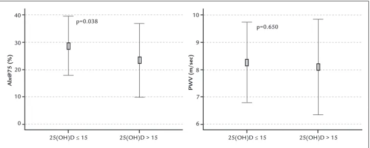
Mean Alx@75 value was significantly higher in group II (28.6±10.8% vs. 23.3±13.5%, p=0.038). The PWV values in group I and group II were 8.0±1.7 and 8.2±1.4 m/s, re-spectively, but the difference was not significant (Figure 2). Serum albumin (p<0.001), hemoglobin (p=0.005), calcium (p=0.041), and eGFR (p=0.041) were significantly lower in group II. Mean age, BMI, parathyroid hormone and uric acid were similar in both groups. Twenty four-hour proteinuria was significantly higher in group II. Group II had a significantly higher ratio of female patients; 82% of all the females in the study were in group II (p=0.006). Demographic features of the groups are shown in Table 1. There was a significant gender difference in mean 25(OH) D levels: 12.2±7.8 ng/mL among female patients and 16.0±7.7 ng/mL for male patients (p=0.016).
D
ISCUSSIONVitamin D deficiency is a worldwide problem. Studies con-ducted with healthy subjects found 25(OH)D deficiency at rates of 42% in America,19 40.4% in Europe20 and 60% in
Eastern countries.21 A meta-analysis showed that 88% of
healthy individuals have 25(OH)D lower than 30 ng/mL.22
The prevalence of vitamin D deficiency in CKD patients not on dialysis has been reported between 21.5-58.3%,15,23-25
and mean 25(OH)D was determined as 18±8 ng/mL.15 In
the present study, the prevalence of vitamin D deficiency was 69.4%; only 5.9% of patients had 25(OH)D levels high-er than 30 ng/mL, and mean 25(OH)D was 14.1±7.9 ng/ mL. Vitamin D deficiency is common in CKD, which has been attributed to many factors such as reduced sun expo-sure, decreased vitamin D-binding protein, proteinuria, reduced dietary intake, advanced age, malabsorption, and down-regulation of megalin levels.8,15,26,27
In Middle Eastern and South Asian developing coun-tries, levels of 25(OH)D < 10 ng/mL were reported in 50% of the population and higher risk was noted among fe-males.28 Of females aged 24-77 years living in Rabat, 91%
had lower than 30 ng/mL 25(OH)D, and female sex was considered a predictor of 25(OH)D deficiency.29 Reports
of 25(OH)D deficiency in females tend to be particularly common in the Asia/Pacific and Middle East/Africa re-gions, possibly due to women wearing a more covered clothing style compared to females in other parts of the world.22 Vitamin D deficiency in females was significantly
higher in our study, in accordance with previous studies.23,28
This may be attributable to the covered clothing style worn by Turkish females due to cultural factors.
It has been shown that PWV and Alx are negatively associated with 25(OH)D in healthy subjects, postmeno-pausal women, and patients with diabetes mellitus, periph-eral arterial disease, rheumatologic diseases and kidney disease.13-16,30,31 Brachial-ankle PWV was found to be
sig-nificantly lower in CKD patients with 25(OH)D deficiency.15
It has been reported that flow-mediated dilatation as a marker of endothelial function was lower in vitamin D deficiency in patients with CKD, and improved with re-placement of 25(OH)D. In addition, vitamin D supple-mentation significantly reduced biomarkers of endothe-lial dysfunction such as intracellular adhesion molecule (ICAM), vascular adhesion molecule (VCAM), endothelial leukocyte adhesion molecule (E-Selectin) and von Wille-brand factor (vWF). It was concluded that vitamin D sup-plementation recovers vascular endothelial function.32
Cholecalciferol supplementation of 40,000 IU every week for 8 weeks had no significant impact on both PWV and Alx@75 in CKD patients as previously shown.33 In
another study, Chitalia et al.32 administered two doses of
300,000 IU cholecalciferol at baseline and 8-weeks to CKD stage 3-4 patients, PWV decreased from 7.9±1.9 to 7.7±2.2 m/s and Alx decreased from 22±16% to 18±20% at week 16. However, they concluded that PWV and Alx tended to improve but did not reach statistical significance.
In our study, Alx@75, a marker of arterial stiffness, was higher among patients with 25(OH)D deficiency. An inverse relation between 25(OH)D level and Alx@75 was determined, and serum 25(OH)D was independently associated with Alx@75 in patients with CKD. These results may indicate that vitamin D deficiency contributes to the development
Alx@75 (%)
PWV (m/sec)
p=0.650
25(OH)D ≤ 15 25(OH)D > 15 25(OH)D ≤ 15 25(OH)D > 15 40
30
20
10
0
10
9
8
7
6
FIGURE 2 Augmentation index and pulse wave velocity values of the groups.
of arterial stiffness in CKD. Vitamin D has a beneficial impact on the renin-angiotensin system, endothelium mediated vasodilatation, insulin resistance, inhibition of vascular smooth muscle proliferation, macrophage activa-tion and cytokine producactiva-tion.10,12,34 Vascular smooth
muscle cell proliferation, the renin-angiotensin-aldosterone system and macrophage invasion of blood vessel walls are activated by vitamin D deficiency. In addition, parathyroid hormone secretion and inflammatory cytokine gene expres-sion are increased. All these pathological processes result in decreased vascular compliance, increased vascular cal-cification and inflammation, which may contribute to the development of arterial stiffnesss.3,15,16,27
Vitamin D deficiency is associated with cardiovascular disease, all-cause mortality, morbidity, poor clinical out-comes and rapid decline of eGFR in patients with CKD.25,34,35
Our study has some limitations. First, 25(OH)D tended to decrease as PWV and CKD stage increased. How-ever, we were unable to definitively confirm the association of 25(OH)D deficiency with PWV and CKD stage due to a relatively small cohort. Secondly, this was a cross-sec-tional case-control study; therefore, we could not evaluate the effect of vitamin D treatment on arterial stiffness, CKD progression or mortality.
C
ONCLUSIONOur study demonstrates that there is a high prevalence of 25(OH)D deficiency and insufficiency in CKD patients, and 25(OH)D values were lower in female patients in particular. The Alx@75 marker of arterial stiffness was higher with 25(OH)D deficiency and negatively corre-lated with 25(OH)D. Our results revealed that serum 25(OH)D was independently associated with Alx@75. Consequently, 25(OH)D deficiency may be a contributing factor in the development of arterial stiffness in CKD.
C
ONFLICT OF INTERESTThe authors declare no conflict of interest.
R
EFERENCES1. Mackenzie IS, Wilkinson IB, Cockcroft JR. Assessment of arterial stiffness in clinical practice. QJM. 2002; 95(2):67-74.
2. Wang X, Keith JC Jr, Struthers AD, Feuerstein GZ. Assessment of arterial stiffness, a translational medicine biomarker system for evaluation of vascular risk. Cardiovasc Ther. 2008; 26(3):214-23.
3. Shirwany NA, Zou M. Arterial stiffness: a brief review. Acta Pharmacol Sin. 2010; 31(10):1267-76.
4. Mattace-Raso FU, van der Cammen TJ, Hofman A, van Popele NM, Bos ML, Schalekamp MA, et al. Arterial stiffness and risk of coronary heart disease and stroke: the Rotterdam Study. Circulation. 2006; 113(5):657-63. 5. Shioi A, Nishizawa Y. Vascular calcification in chronic kidney disease:
pathogenesis and clinical implications. J Ren Nutr. 2009; 19(1):78-81.
6. Sakuragi S, Abhayaratna WP. Arterial stiffness: methods of measurement, physiologic determinants and prediction of cardiovascular outcomes. Int J Cardiol. 2010; 138(2):112-8.
7. Garg AX, Clark WF, Haynes RB, House AA. Moderate renal insufficiency and the risk of cardiovascular mortality: results from the NHANES I. Kidney Int. 2002; 61(4):1486-94.
8. Kennel KA, Drake MT, Hurley DL. Vitamin D deficiency in adults: when to test and how to treat. Mayo Clin Proc. 2010; 85(8):752-7.
9. Lee JH, O’Keefe JH, Bell D, Hensrud DD, Holick MF. Vitamin D deficiency an important, common, and easily treatable cardiovascular risk factor? J Am Coll Cardiol. 2008; 52(24):1949-56.
10. Adams JS, Hewison M. Update in vitamin D. J Clin Endocrinol Metab. 2010; 95(2):471-8.
11. National Kidney Foundation. K/DOQI clinical practice guidelines for bone metabolism and disease in chronic kidney disease. Am J Kidney Dis. 2003; 42(4 Suppl 3):S1-201.
12. Li YC, Kong J, Wei M, Chen ZF, Liu SQ, Cao LP. 1,25-Dihydroxyvitamin D(3) is a negative endocrine regulator of the renin-angiotensin system. J Clin Invest. 2002; 110(2):229-38.
13. Lee JI, Oh SJ, Ha WC, Kwon HS, Sohn TS, Son HS, et al. Serum 25-hydroxyvitamin D concentration and arterial stiffness among type 2 diabetes. Diabetes Res Clin Pract. 2012; 95(1):42-7.
14. Pirro M, Manfredelli MR, Helou RS, Scarponi AM, Schillaci G, Bagaglia F, et al. Association of parathyroid hormone and 25-OH-vitamin D levels with arterial stiffness in postmenopausal women with vitamin D insufficiency. J Atheroscler Thromb. 2012; 19(10):924-31.
15. Luo Q, Wang LL, Gao YH. Association between serum 25-hydroxyvitamin D and arterial stiffness in non-dialysis-dependent CKD. Eur J Clin Nutr. 2016; 70(2):274-6.
16. Al Mheid I, Patel R, Murrow J, Morris A, Rahman A, Fike L, et al. Vitamin D status is associated with arterial stiffness and vascular dysfunction in healthy humans. J Am Coll Cardiol. 2011; 58(2):186-92.
17. Kidney Disease: Improving Global Outcomes (KDIGO) CKD Work Group. KDIGO 2012 clinical practice guideline for the evaluation and management of chronic kidney disease. Kidney Int Suppl. 2013; 3(1):1-150.
18. Baulmann J, Schillings U, Rickert S, Uen S, Düsing R, Illyes M, et al. A new oscillometric method for assessment of arterial stiffness: comparison with tonometric and piezo-electronic methods. J Hypertens. 2008; 26(3):523-8.
19. Forrest KY, Stuhldreher WL. Prevalence and correlates of vitamin D deficiency in US adults. Nutr Res. 2011; 31(1):48-54.
20. Cashman KD, Dowling KG, Škrabáková Z, Gonzalez-Gross M, Valtueña J, De Henauw S, et al. Vitamin D deficiency in Europe: pandemic? Am J Clin Nutr. 2016; 103(4):1033-44.
21. Haq A, Svobodová J, Imran S, Stanford C, Razzaque MS. Vitamin D deficiency: a single centre analysis of patients from 136 countries. J Steroid Biochem Mol Biol. 2016; 164:209-13.
22. Hilger J, Friedel A, Herr R, Rausch T, Roos F, Wahl DA, et al. A systematic review of vitamin D status in populations worldwide. Br J Nutr. 2014; 111(1):23-45. 23. Diniz HF, Romão MF, Elias RM, Romão Júnior JE. Vitamin D deficiency
and insufficiency in patients with chronic kidney disease. J Bras Nefrol. 2012; 34(1):58-63.
24. Rodríguez Villarreal I, Ortega O, Gallar P, Sánchez M, Callejas R, Gracia C, et al. Clinical and biochemical characteristics of predialysis patients in terms of 25 hydroxy vitamin D levels. Nefrologia. 2011; 31(2):185-91.
25. Ravani P, Malberti F, Tripepi G, Pecchini P, Cutrupi S, Pizzini P, et al. Vitamin D levels and patient outcome in chronic kidney disease. Kidney Int. 2009; 75(1):88-95.
26. Holick MF. Vitamin D deficiency. N Engl J Med. 2007; 357(3):266-81. 27. Nakashima A, Yokoyama K, Yokoo T, Urashima M. Role of vitamin D in diabetes
mellitus and chronic kidney disease. World J Diabetes. 2016; 7(5):89-100. 28. Arabi A, El Rassi R, El-Hajj Fuleihan G. Hypovitaminosis D in developing
countries-prevalence, risk factors and outcomes. Nat Rev Endocrinol. 2010; 6(10):550-61.
29. Allali F, El Aichaoui S, Khazani H, Benyahia B, Saoud B, El Kabbaj S, et al. High prevalence of hypovitaminosis D in Morocco: relationship to lifestyle, physical performance, bone markers, and bone mineral density. Semin Arthritis Rheum. 2009; 38(6):444-51.
31. Rezai MR, Wallace AM, Sattar N, Finn JD, Wu FC, Cruickshank JK. Ethnic differences in aortic pulse wave velocity occur in the descending aorta and may be related to vitamin D. Hypertension. 2011; 58(2):247-53. 32. Chitalia N, Ismail T, Tooth L, Boa F, Hampson G, Goldsmith D, et al. Impact
of vitamin D supplementation on arterial vasomotion, stiffness and endothelial biomarkers in chronic kidney disease patients. PLoS One. 2014; 9(3):e91363. 33. Marckmann P, Agerskov H, Thineshkumar S, Bladbjerg EM, Sidelmann JJ, Jespersen J, et al. Randomized controlled trial of cholecalciferol
supplementation in chronic kidney disease patients with hypovitaminosis D. Nephrol Dial Transplant. 2012; 27(9):3523-31.
34. Pilz S, Verheyen N, Grübler MR, Tomaschitz A, März W. Vitamin D and cardiovascular disease prevention. Nat Rev Cardiol. 2016; 13(7): 404-17.
![TABLE 1 Comparison of clinical and biochemical characteristics of groups. Parameter Group I [25(OH)D > 15 ng/mL] Group II [25(OH)D ≤ 15 ng/mL] p Female / Male (n) 9 / 22 41 / 29 0.006 Age (year) 55.8±12.8 57.1±10.6 0.591 BMI (kg/m 2 ) 26.4±3.5 28](https://thumb-eu.123doks.com/thumbv2/123dok_br/19057179.485066/4.892.86.811.132.727/table-comparison-clinical-biochemical-characteristics-groups-parameter-female.webp)