Reexpansion pulmonary edema in children
Edema pulmonar de reexpansão em crianças
Edema pulmonar de reexpansión en niños
Antonio Lucas L. Rodrigues1, Carlos Eduardo Lopes2, Mariana Tresoldi das N. Romaneli3, Andrea de Melo A. Fraga4,
Ricardo Mendes Pereira5, Antonia Teresinha Tresoldi6
Instituição: Faculdade de Ciências Médicas da Universidade Estadual de Campinas (Unicamp), Campinas, SP, Brasil
1Médico-residente em Pediatria do Departamento de Pediatria da Faculdade de Ciências Médicas da Unicamp, Campinas, SP, Brasil
2Chefe da UTI Pediátrica do Hospital de Clínicas da Faculdade de Ciências Médicas da Unicamp, Campinas, SP, Brasil
3Mestre em Ciências pela Unicamp; Médica-Assistente do Departamento de Pediatria da Faculdade de Ciências Médicas da Unicamp, Campinas, SP, Brasil
4Doutora em Ciências pela Unicamp; Médica-Assistente do Departamento de Pediatria da Faculdade de Ciências Médicas da Unicamp, Campinas, SP, Brasil
5Doutor em Ciências pela Unicamp; Professor-Assistente do Departamento de Pediatria da Faculdade de Ciências Médicas da Unicamp, Campinas, SP, Brasil
6Livre-docente em Pediatria pela Unicamp; Professora-Associada do Departamento de Pediatria da Faculdade de Ciências Médicas da Unicamp, Campinas, SP, Brasil
ABSTRACT
Objective: To present a case of a patient with clinical and radiological features of reexpansion pulmonary edema, a rare and potentially fatal disease.
Case description: An 11-year-old boy presenting fe-ver, clinical signs and radiological features of large pleural effusion initially treated as a parapneumonic process. Due to clinical deterioration he underwent tube thoracostomy, with evacuation of 3,000 mL of fluid; he shortly presen-ted acute respiratory insufficiency and needed mechanical ventilation. He had an atypical evolution (extubated twice with no satisfactory response). Computerized tomography findings matched those of reexpansion edema. He recovered satisfactorily after intensive care, and pleural tuberculosis was diagnosed afterwards.
Comments: Despite its rareness in the pediatric popula-tion (only five case reports gathered), the knowledge of this pathology and its prevention is very important, due to high mortality rates. It is recommended, among other measures, slow evacuation of the pleural effusion, not removing more than 1,500 mL of fluid at once.
Key-words: pulmonary edema; pleural effusion; tuberculosis, pleural; adolescent.
RESUMO
Objetivo: Relatar caso de paciente com quadro clínico e radiológico de edema pulmonar de reexpansão, patologia rara e potencialmente fatal.
Descrição do caso: Menino de 11 anos com febre e quadro clínico-radiológico de derrame pleural volumoso, inicialmente tratado como parapneumônico. Após descom-pensação clínica, realizou-se drenagem torácica, com saída de 3.000mL de líquido. Evoluiu rapidamente com insuficiência respiratória aguda, necessitando de ventilação mecânica. Teve duas extubações malsucedidas e, devido à evolução atípica, realizou-se tomografia computadorizada, cujos achados fo-ram compatíveis com edema de reexpansão. Após suporte intensivo, evoluiu satisfatoriamente e, posteriormente, foi diagnosticado com tuberculose pleural.
Comentários: É importante o conhecimento da patologia, ainda que seja rara na população pediátrica (encontrados apenas cinco casos descritos), para prevenção, visto que a taxa de mortalidade é muito alta. Recomenda-se, entre outras medidas, que o esvaziamento de um derrame pleural seja lento e que o volume total retirado não ultrapasse 1.500mL.
Palavras-chave: edema pulmonar; derrame pleural; tuberculose pleural; adolescente.
Endereço para correspondência: Antonio Lucas L. Rodrigues
Rua Roxo Moreira, 1.724, apto 11 – Cidade Universitária CEP 13083-592 – Campinas/SP
E-mail: [email protected]
Conflito de interesse: nada a declarar
RESUMEN
Objetivo: Relatar caso de paciente con cuadro clínico y radiológico de edema pulmonar de reexpansión, patología rara y potencialmente fatal.
Descripción del caso: Niño de 11 años con fiebre y cuadro clínico-radiológico de derrame pleural voluminoso, inicialmente tratado como parapneumónico. Después de la compensación clínica, se realizó drenaje torácico, con salida de 3000mL de líquido. Evolucionó rápidamente con insufi-ciencia respiratoria aguda, necesitando ventilación mecánica. Tuvo dos extubaciones mal sucedidas y, debido a la evolu-ción atípica, se realizó tomografía computadorizada, cuyos hallazgos fueron compatibles con edema de reexpansión. Después de soporte intensivo, evolucionó satisfactoriamente y, posteriormente, se diagnosticó tuberculosis pleural.
Comentarios: Es importante el conocimiento de la pato-logía, aunque sea rara en la población pediátrica (encontrados solamente cinco casos descriptos), para prevención, una vez que la tasa de mortalidad es muy alta. Se recomienda, entre otras medidas, que el vaciamiento de un derrame pleural sea lento y que el volumen total retirado no sobrepase 1500mL.
Palabras clave: edema pulmonar; derrame pleural; tuberculosis pleural; adolescente.
Introduction
Reexpansion pulmonary edema (RPE) is a rare and poten-tially fatal clinical entity. It usually occurs when a chronically collapsed lung is rapidly reexpanded after the evacuation of large amounts of luid or air from the pleural space, often using high negative intrapleural pressures(1-5).
This study reported the case of a child with large pleural effusion who developed severe RPE after tube thoracostomy, besides performing a review of the literature on the topic.
Case report
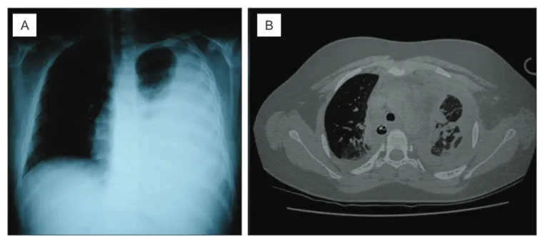
An 11-year-old Caucasian male patient with a history of fever, adynamia, myalgia, and chest pain for three days. Previous intermittent asthma. Physical examination: good general health status, febrile, respiratory rate of 23 breaths per minute, with no respiratory distress; pulmo-nary workup with vesicular breath sounds on the right side and abolished breath sounds on the left side, with dullness on percussion. No other relevant clinical ind-ings. Chest radiograph on admission reveled homogeneous opacity on the left side, with obliteration of costophrenic and cardiophrenic sinuses on the left side (Figure 1). An ultrasound scan was performed, which revealed luid up
Figure 1 - (A) Chest radiograph on admission homogenous opacity on the left side up to the apex, opaciication of costophrenic and
cardiophrenic sinuses, with the presence of mild deviation of the contralateral mediastinum. (B) Chest computerized tomography after tube thoracostomy: axial cut, windowing for lung parenchyma evidencing bilateral lung involvement, with areas of
condensa-tion (alveolar illing), ground-glass areas and bilateral pleural involvement, with the left lung showing the most important indings
to the upper third of the pleural cavity, with no septations or pleural thickening.
The patient underwent thoracocentesis, removing 550mL of citrine luid; blood count showed – hemoglobin: 14.2mg/dL; leukocytes: 7,310/mm3 (73% segmented, 18%
lymphocytes and 9% monocytes); platelets: 445,000/mm3.
Treatment for parapneumonic pleural effusion was started with crystalline penicillin (200,000U/kg/day). Laboratory analysis of the pleural luid revealed: 4,400 erythrocytes/mm3;
730 leukocytes/mm3 (81% lymphocytes and 19%
neutro-phils); glucose: 79mg/dL; proteins: 5.1g/dL.
He remained in good general health status, eupneic, and with daily fever peaks for ive days. Blood cultures, the rapid test for dengue and investigation for cold agglutinins were negative. Due to clinical worsening (chest pain and dyspnea), with no new radiological changes, pleural drainage under general anesthesia was indicated.
During anesthetic induction, he developed bradycardia and hypoxia, which were promptly reversed. Thoracocentesis was performed, with the evacuation of 3000mL of citrine luid during two hours, maintained by water-seal drainage. Fluid laboratory analysis: citrine appearance; 490 eryth-rocytes/mm3; 233 leukocytes/mm3 (97% lymphocytes);
glucose: 94mg/dL; proteins: 4.2g/dL; pH: 7.48; lactate de-hydrogenase (LDH): 623U/L (reference: exudate >300U/L).
After the procedure, when he was already extubated, the patient developed respiratory distress and decreased oxygen saturation, needing invasive ventilation in the pediatric intensive care unit. He remained on mechanical ventilation from the sixth to the 20th day of hospitalization. Within
this 14-day post-procedural period, there were two extuba-tions: one accidental and other planned; in both episodes, he needed to be intubated again, resuming mechanical ven-tilation due to respiratory insuficiency. During the periods when the patient was stable, it was decided to use intermit-tent mandatory ventilation, with peak inspiratory pressure between 20–25cmH2O, positive-end expiratory
pres-sure between 5–8cmH2O, respiratory rate between
12–15 breaths/minute and inspired oxygen concentration between 40–50% (the Horovitz index was calculated to be between 180–190). There was considerable dificulty in the evolution of ventilatory parameters until tracheal cannula was deinitely withdrawn.
Chest computerized tomography was carried out (Figure 1), which evidenced: massive alveolar illing of the left lung and partial illing of the right lung, with bilateral areas of ground-glass opacity; residual pleural effusion on the
left side and laminar effusion on the right side, with bilateral pleural reaction and presence of hilar adenomegaly. Thus, the diagnostic hypothesis was reexpansion pulmonary edema.
Complementary analysis of the pleural luid showed negative results for investigations for neoplastic cells, fungi and bacteria; however, the investigation for adenosine de-aminase (ADA) activity in the pleural luid was positive (137.70U/L; reference: up to 40U/L). A tuberculin skin test was performed, with a result of 19mm. Treatment was started with rifampicin, isoniazid, pirazinamid and etham-butol, associated with prednisone (1mg/kg/day), with the diagnosis of pleural tuberculosis. Serology for HIV and cul-tures for bacteria and mycobacteria were negative. From the second day of treatment on, the patient had no febrile peaks anymore and could be weaned from mechanical ventilation; additionally, the pleural drain could be removed, at the 14th
of intensive care (20th day of hospitalization). The patient
was discharged for outpatient follow-up on the 31rd day of
hospitalization, with clinical and radiological improvement.
Discussion
The pathophysiology of RPE is multifactorial, in-cluding changes in pulmonary capillary permeability and increase in hydrostatic pressure. Both of them cause luid and protein overlow into the pulmonary intersti-tial space and alveoli, leading to pulmonary edema(1-3,5).
Permeability changes occurs due to local hypoxemia, caused by lung collapse, injuring the capillary wall and decreasing surfactant production, with subsequent release of inlammatory mediators – interleukin-8 (IL-8), mono-cyte chemotactic protein-1 (MCP-1), leukotriene B4, nitric oxide, polymorphonuclear and free radicals– which, in turn, amplify the injury and change vascular perme-ability (IL-8 and MCP-1 act also in the contralateral lung, partly justifying the cases of bilateral RPE)(2,3,5). Besides
this aggression, the capillary also undergoes mechanical injury by collapse compression, associated with sudden reexpansion-induced distension. In turn, the increase in hydrostatic pressure occurs due to the abrupt increase in blood low on reinlation(2,3). In the case reported herein,
lung aggression was probably caused by the large cavitary effusion, associated with the increase in intraluminal pres-sure resulting from the reexpansion of the organ due to the evacuation of a great amount of pleural luid.
twentieth century(1,2). It is estimated that its incidence is up
to 1% after drainage of intrapleural air or luid(6). However,
most reports involve adult patients and there are no reviews of published pediatric cases(7). The case reports of children
with RPE are summarized in Table 1(7-11).
Risk factors associated with RPE include: chronicity of lung collapse (usually greater than 72 hours); great amount of pleural air or luid (>1500mL)(1-3,5); high speed of
reexpan-sion and use of high negative pressures to do so(2,5);
hyperten-sion, hypoxemia or other previous lung disease; pre-existing heart disease(2); and male gender(3). In the case described
herein, the patient presented with chronic lung collapse (at least seven days of admission before drainage plus some period prior to hospitalization, considering a possible insidious onset of pleural tuberculosis); great intrapleural volume (3000mL); lung disease (intermittent asthma); and male gender.
Clinical indings are variable, ranging from asymptom-atic patients or presenting only non-speciic symptoms
(fever, nausea, vomiting, tachycardia, hypotension) to patients with severe respiratory insuficiency (dys-pnea, chest pain, cough, foamy sputum, cyanosis)(2,3,5).
A simple chest radiograph may reveal interstitial opaci-ties, consolidations with air bronchograms, and issural inlammation(2). Chest computerized tomography may
show ground-glass opacity, residual pleural effusion, at-electasis, interlobar septum thickening, consolidations, air bronchograms, and pulmonary nodules. Nearly half of patients show diffuse involvement of the previously collapsed lung(4) and bilateral pulmonary involvement is
rarely observed(1,4,5,7,10). The clinical picture of the patient
described herein included respiratory insuficiency and need for mechanical ventilation with high parameters, similarly to some of the reported cases of children with RPE. However, computerized tomography indings as evident as those found by Gleeson et al(4) in adults had
not been described yet in pediatric patients.
Study Age/gender Underlying
pathology
Cause of lung collapse
Reexpansion technique
Time of the onset of symptoms
Duration of the picture
Paksu et al7 9 years/male Bacterial
pneumonia
Left-sided parapneumonic
empyema with history of 7 days
Thoracocentesis and subsequent pleural drainage of 500mL 1 hour Death on the 5th day
(multiple organ failure)
Jardine8 8 years/male
Nephrotic syndrome; infections and recurrent pleural empyema Left-sided large pleural effusion for more than 2 weeks
Tube thoracostomy of
1300mL in 30 minutes
30 minutes 6 hours
Chiang et al9 Data not
available Preterm infant with patent ductus arteriosus Surgical lung retraction for correction of heart
disease
Intraoperative
lung reinlation 1 hour 48 hours
Ozlu et al10 5 years/male Non-Hodgkin
lymphoma Right-sided large neoplastic pleural effusion Tube thoracostomy of 2000mL in a few
minutes 2 minutes 4 hours (invasive ventilatory support)
Tung et al11 16 years/
male Previously healthy Right-sided spontaneous pneumohemothorax
of 12 hours of duration
Tube thoracostomy + subsequent thoracoscopy; total drainage of
3500mL
During
procedure 5 days
Rodrigues et al
(present case) 11 years/ male Pleural tuberculosis Left-sided large tuberculous pleural effusion for at least
7 days
Tube thoracostomy of 3000mL in 2
hours
1 hour 14 days
The onset of RPE symptoms usually occurs in the irst 24 hours after pulmonary reexpansion, most frequently (64-89%) in the irst two hours(3,5). The duration of symptoms
varies from 48 hours to seven days(2,8,10), and mortality from
19 to 21%(2,3,5). In the case reported herein, the symptoms
started in the irst hour after the procedure and the duration of the clinical signs was 14 days, with the need for mechani-cal ventilation, probably due to complications during disease evolution (including unsuccessful extubations and extensive bilateral lung injury).
The treatment of this edema is based on clinical support according to the need of each patient(2,3,5). Some authors
suggest that the use of corticosteroids as stabilizers of the pulmonary vascular membrane (3,11). More invasive measures
are also described, such as occlusion of the pulmonary artery of the affected side(2) and use of extracorporeal membrane
oxygenation(11). In the case reported herein, it was necessary
to use invasive ventilatory support for 14 days, as well as the use of dobutamine (for four days; maximum titration of 0.45mcg/kg/day), (furosemide for 14 days, for stimulation of diuresis and control of the water balance) e corticosteroid (although its exact role in the case could not be deined).
The diagnosis of pleural tuberculosis in the case de-scribed herein was based on clinical data (pleural effusion in febrile non-toxemic child/adolescent)(12,13), suggestive
radiological inding (hilar adenomegaly)(13) and positive
skin tuberculin test(12) (reaching 45 points in the scoring
system proposed by the Brazilian Ministry of Health for children and adolescents, making this diagnosis highly likely)(14). In addition, the analysis of the pleural luid was
characteristic: predominance of lymphocytes; high LDH levels; high ADA levels associated with lymphocyte-to-neutrophils ratio >75%(12,15). Although the culture for
mycobacteria in the luid was negative, it is known that its positivity is low (45-55% of the cases)(13,15). If thoracoscopy
with pleural biopsy was performed, positivity could be higher, as described in the literature(12).
Predicting RPE occurrence is of fundamental impor-tance in patients at risk for developing this condition and was even the subject of recommendations of the British Thoracic Society(6). The evacuation of pleural effusion
should occur slowly, not removing an excessive amount (up to 1500mL), and there should be available means to obtain a deinitive airway and start mechanical ventilation if necessary(2,3,5,6). It is also recommended that drainage is
performed preferably with pleural pressure monitoring, not exceeding -20mmHg(2,6). In the case reported herein,
the procedure was carried out in an operating room, with general anesthesia, by an experienced team, with equip-ment for emergencies available. However, it should be pointed out that the procedure could have had a different outcome if the amount of pleural luid drained was lower or if the measure of cavity pressure was available on that occasion, considering that the patient had more than one risk factor for the development of RPE.
Therefore, despite its low frequency, RPE should be remembered as a potential complication in the evolution of a pediatric patient with large pleural effusion, particu-larly if there were other risk factors for the development of this complication.
References
1. Mahfood S, Hix WR, Aaron BL, Blaes P, Watson DC. Reexpansion pulmonary edema. Ann Thorac Surg 1988;45:340-5.
2. Genofre EH, Vargas FS, Teixeira LR, Vaz MA, Marchi E. Reexpansion pulmonary edema. J Pneumol 2003;29:101-6.
3. Sohara Y. Reexpansion pulmonary edema. Ann Thorac Cardiovasc Surg 2008;14:205-9.
4. Gleeson T, Thiessen R, Müller N. Reexpansion pulmonary edema: computed
tomography indings in 22 patients. J Thorac Imaging 2011;26:36-41.
5. Kesieme EB, Dongo A, Ezemba N, Irekpita E, Jebbin N, Kesieme C. Tube thoracostomy: complications and its management. Pulm Med [serial on the Internet]. 2012;2012 [cited 2012 Feb 20]. Available from: http://www.hindawi. com/journals/pm/2012/256878/cta/
6. Echevarria C, Twomey D, Dunning J, Chanda B. Does re-expansion pulmonary oedema exist? Interact Cardiovasc Thorac Surg 2008;7:485-9.
7. Paksu MS, Paksu S, Akgün M, Kalayci AG, Baysal K. Bilateral reexpansion pulmonary edema associated with pleural empyema: a case report. Eur J Pediatr 2011;170:1205-7.
8. Jardine DS. Reexpansion pulmonary edema. Am J Dis Child 1991;145:1092-4.
9. Chiang MC, Lin WS, Lien R, Chou YH. Reexpansion pulmonary edema following patent ductus arteriosus ligation in a preterm infant. J Perinat Med 2004;32:365-7.
10. Ozlu O, Kiliç A, Cengizlier R. Bilateral re-expansion pulmonary edema in a child: a reminder. Acta Anaesthesiol Scand 2000;44:884-5.
11. Tung YW, Lin F, Yang MS, Wu CW, Cheung KS. Bilateral developing reexpansion pulmonary edema treated with extracorporeal membrane oxygenation. Ann Thorac Surg 2010;89:1268-71.
12. Fischer GB, Andrade CF, Lima JB. Pleural tuberculosis in children. Paediatr Respir Rev 2011;12:27-30.
13. Cruz AT, Ong LT, Starke JR. Childhood pleural tuberculosis: a review of 45 cases. Pediatr Infect Dis J 2009;28:981-4.
14. Conde MB, Melo FA, Marques AM, Cardoso NC, Pinheiro VG, Dalcin PT
et al. III Diretrizes para tuberculose da Sociedade Brasileira de Pneumologia e Tisiologia. J Bras Pneumol 2009;35:1018-48.
15. Krenke R, Korczyński P. Use of pleural luid levels of adenosine deaminase