www.reumatologia.com.br
REVISTA BRASILEIRA DE
REUMATOLOGIA
* Corresponding author.
E-mail: [email protected] (T.L. Skare).
0482-5004/$ - see front matter. © 2013 Elsevier Editora Ltda. All rights reserved.
Original article
Radiographic changes of the cervical spine in rheumatoid
arthritis
Juan Marcelo Fernandez Alcala
a, Diogo Douat
b, Diogo Lago Pinheiro
b, Douglas Jun Kamei
a,
Fábio Raimundo M dos Santos
a, Marilia B Silva
a, Thelma L Skare
a,*
a Faculdade Evangélica de Medicina do Paraná, Curitiba, PR, Brazil
b Unit of Diagnostic Imaging, Hospital Universitário Evangélico de Curitiba, Curitiba, PR, Brazil
a r t i c l e i n f o
Article history:
Received May 13 2012 Accepted March 14 2013
Keywords:
Rheumatoid arthritis Cervical spine Atlanto-axial luxation Basilar invagination Subaxial instability
a b s t r a c t
Introduction: The involvement of the cervical spine is a common feature of rheumatoid
arthritis (RA).
Objective: To study the prevalence of radiographic changes of the cervical spine in patients with RA and their association with clinical and serological proi les of the disease.
Methods: We analysed lateral (neutral position, hyperextension, hyperl exion) and
tran-soral views of cervical spine radiographs from 80 individuals with RA to investigate the presence of atlanto-axial subluxation (AAS), basilar invagination (BI), and subaxial insta-bility (SAI). Demographic, clinical (nodules, interstitial pneumonitis, secondary Sjögren’s syndrome, medications etc.), and serologic (rheumatoid factor - RF, cyclic citrullinated peptide antibody – anti-CCP, and antinuclear factor - ANF) data were obtained from the clinical records.
Results: Cervical spine misalignments were identii ed in 26/80 (32.5%) participants; AAS
occurred in 12/80 (15%) participants, BI in 6/80 (7.5%), and SAI in 13/80 (32.5%). Odontoid erosions were identii ed in 16/80 (20.0%) participants. Cervical spine misalignment exhib-ited associations with age at onset and disease duration (P = 0.03 and 0.02, respectively). No associations were identii ed between the cervical spine changes and the participants’ ethnicity or gender, presence of nodules, interstitial pneumonitis, secondary Sjögren’s syndrome, RF, ANF, or anti-CCP. The participants with cervical spine misalignment exhib-ited higher frequencies of odontoid erosion (P = 0.03).
Conclusions: Cervical spine misalignment was a common radiographic i nding and
Alterações radiográi cas da coluna cervical em artrite reumatoide
Palavras-chave:
Artrite reumatoide Coluna cervical Luxação atlanto-axial Invaginação basilar Instabilidade subaxial
r e s u m o
Introdução: O envolvimento da coluna cervical é comum na artrite reumatoide (AR).
Objetivo: Estudar a prevalência das alterações radiológicas de coluna cervical em pacientes
com AR e sua associação com peri l clinico e sorológico da doença.
Métodos: Analisaram-se as radiograi as de coluna cervical em peri l neutro hiperextensão,
hiperl exão e transoral de 80 pacientes com AR para presença de subluxação atlanto-axial (LAA), invaginação basilar (IB) e instabilidade subaxial (ISA). Dados de peri l demográi co, clínico (nódulos, pneumonite intersticial, síndrome Sjögren secundária, uso de medica-mentos etc.) e sorológico (FR, anti-CCP e FAN) foram obtidos por revisão de prontuários.
Resultados: Havia alguma alteração de eixo de coluna cervical em 26/80 (32,5%); em 12/80
(15%) havia LAA; em 6/80(7,5%) existia IB; em 13/80 (16,2%) existia ISA. Erosões em odon-toide foram vistas 16/80 (20,0%). As alterações do eixo cervical estavam associadas com idade de início da doença e duração da mesma (P = 0,03 e 0,02, respectivamente). Não se encontrou associação das alterações em coluna cervical com raça, gênero, nódulos, pneu-monite intersticial, Sjögren secundário, FR, FAN ou anti-CCP. Pacientes com alterações do eixo cervical apresentavam mais erosões de odontoide (P = 0,03).
Conclusões: Alterações radiológicas em eixo de coluna cervical são comuns e aparecem mais
frequentemente em indivíduos com diagnóstico mais precoce de AR e maior tempo de doença. © 2013 Elsevier Editora Ltda. Todos os direitos reservados.
Introduction
One of the characteristics of rheumatoid arthritis (RA) is the
involvement of the cervical spine.1 The main alterations occur
in the spine’s most mobile region: its upper portion.1 Typical
alterations include anterior atlanto-axial subluxation, atlan-to-axial impaction or basilar invagination (also known as
ver-tical atlanto-axial subluxation), and subaxial disease.1,2 All of
these alterations are due to chronic local inl ammation.1,2
Anterior atlanto-axial luxation occurs when the ligaments that stabilise this area are damaged. Under such circumstanc-es, whenever the neck is moved, the head weight pulls the
at-las away from the axis.2 When inl ammation affects the
atlan-to-axial joints by destroying the cartilage and bone structure, the skull presses the atlas down towards the axis, causing
basilar invagination.2 Subaxial disease is less frequently
ob-served and usually occurs in association with the remainder
of the deformities.1 This pathology is due to inl ammation of
the facet joints below the second cervical vertebra.3
One meta-analysis reported that cervical spine alterations are frequent in patients with RA, occurring in 5.5 to 73% of the cases (average of 32%). These alterations are associated with
neurologic signs and/or symptoms in 17% of cases.1
Atlanto-axial subluxation occurs early,1,2 within the i rst
two years of disease onset.2 Diagnosis is established when a
distance greater than 3 mm is measured between the ante-rior arch of the atlas and the odontoid process of the axis. When the diameter between the posterior arch of the atlas and the odontoid process of the axis is equal to or less than 14 mm, the risk of myelopathy is high. The i rst neurologic sign associated with atlanto-axial subluxation is headache in the occipital area due to compression of the greater occipi-tal nerve (Arnold’s neuralgia), which is followed by sensory
and motor dei cit affecting the arms and legs.4 Further
com-plaints include neck stiffness, earache due to compression of the greater auricular nerve, vertigo, gait abnormalities, loss of balance, and tinnitus due to alterations of the vertebral artery
l ow.5 When the neck is bent forwards, Lhermitte’s sign could
be triggered, which is an electric-like shock sensation running
down the back into the limbs.5 Quadriparesis, chronic
hydro-cephalus, cerebral infarction, and sudden death are
complica-tions of established disease.2,4
The problems associated with basilar invagination tend to appear later in the progression of disease and occur more
commonly in the most severe cases of RA.2 These symptoms
appear in 4 to 34% of the cases, and the upward migration of the odontoid process might result in compression of the
brainstem.1,6 The prevalence of axial subluxation varies from
7 to 29%1,6 and might occur as an isolated deformity or may
affect multiple levels. The latter case results in the onset of a
deformity with a “staircase” appearance.1,6
Corbett et al.7 assessed 102 individuals who developed
atlanto-axial luxation within the i rst two years of RA and de-termined that it occurred in association with erosive disease and resulted in poor functional prognosis. However, early administration of disease-modifying antirheumatic drugs (DMARDs) and the advent of more powerful agents for the control of inl ammation allows for alteration of the natural disease progression of RA. Such modulation also affects the severity of the cervical spine involvement. It is believed that early and effective use of DMARDs might prevent or limit the pannus growth, thus reducing the space it occupies and its
destructive potential.1,8 As a consequence, it is expected that
both the prevalence of and the risks associated with RA cervi-cal complications will decrease.
Methods
This study was approved by the institutional research ethics committee, and all participants signed an informed consent form. Individuals from both genders who met at least four of the classii cation criteria formulated in 1987 by the American
Col-lege of Rheumatology9 were invited to participate in the study.
The participants were selected based on their order of consul-tations and their availability to participate in the study, which was conducted from July to December 2011. In addition, their RA diagnosis should have been established before age 16. Pregnant women and individuals with histories of cervical spine trauma or with intellectual incapacities hindering them from under-standing the terms of the informed consent form were excluded.
Lateral-view radiographs of the cervical column were per-formed for all of the participants in the neutral position and in hyperextension and hyperl exion. Two radiologists blinded to the participants’ clinical data independently analysed all of the images.
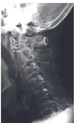
The parameters assessed were: atlanto-axial subluxation, basilar invagination, erosion of the odontoid process of the axis, and subaxial instability. Atlanto-axial subluxation was considered to be present when the distance between the an-terior arch of the atlas and the odontoid process of the axis
was larger than 3 mm (Fig. 1).10,11
Basilar invagination was assessed using the
Redlund-Johnell and Pettersson method12, which involves drawing a
line from the posterior margin of the hard palate to the inferi-or cinferi-ortical surface of the occipital bone on a lateral radiograph
and measuring the distance between that line and the centre of the lower end plate of C2 vertebral body. The normal values of that line are 34 mm or more in males and 29 mm or more in females. This method is appropriate for the assessment of basilar invagination because it avoids the use of measure-ments involving the tip of the odontoid process as reference,
as it is frequently eroded in individuals with RA (Fig. 2).12
Subaxial instability was diagnosed when vertebral
slip-page was greater than 3 mm (Fig. 3).13,14
The participants’ demographic data, along with duration, age at onset of disease, clinical proi le (i.e., presence of nod-ules, associated interstitial lung disease, secondary Sjögren’s syndrome, peripheral neuropathy, and eye involvement), and medication use, were collected from the clinical records. To establish the presence of secondary Sjögren’s syndrome, the
Fig.1 – Lateral radiograph (hyperfl exion) showing increased space between the posterior margin of the anterior arch of the atlas and the anterior surface of the odontoid process of the axis, indicative of atlanto-axial subluxation.
Fig. 2 – Redlund-Johnell’s method for identifying basilar invagination. On a lateral radiograph, a line is drawn from the posterior margin of the hard palate (A) to the inferior cortical surface of the occipital bone (B). The distance from the centre of the lower end plate of C2 vertebral body (C) and line A-B is measured on a line parallel to the longitudinal axis of the odontoid process. The normal values of that distance are 34 mm or more in males and 29 mm or more in females.
Fig. 3 – Lateral cervical spine radiographs in the neutral position (A), hyperextension (B), and hyperfl exion (C). The C4 vertebral body exhibits anterior slippage of 3.5 mm relative to the C5 vertebral body only on the radiograph performed in hyperfl exion (C), indicating subaxial instability.
McGregor line
classii cation criteria formulated by the American-European
consensus group15 were used. The participants were asked
about any clinical complaints they had regarding the cervical region (i.e., pain, stiffness, paresthesia, and weakness of the upper limbs).
The data were grouped into frequency and contingency tables; the measures of central tendency used were medi-ans and interquartile ranges in cases of non-parametric data and means and standard deviations in cases of parametric data. Associations between nominal data were assessed using Fisher’s exact test, and the Mann-Whitney test and unpaired
Student’s t-test were used in cases of numerical data. The
level of signii cance was established as 5%, and analyses were performed using the Graph Pad Prism version 5.0 software.
Results
Descriptive analysis of the investigated population
Among the 80 individuals included in the study, 10 (12.5%) were male, and 70 (87.5%) were female; 18 (22.5%) were Afro-descendants, and 62 (77.5%) were Caucasian. The average age of the sample was 55.4 ± 11.9 years (26-82 years), the average duration of disease was nine years (1-29 years), and the age at disease onset ranged from 17 to 75 years (average of 45.0 ± 12.6 years).
Eleven (13.7%) participants exhibited interstitial lung dis-ease on chest radiograph or tomography, nine (11.2%) ex-hibited subcutaneous nodules, two (2.5%) had scleritis, one (1.25%) had vasculitis, and one (1.25%) had peripheral neu-ropathy. Nineteen (23.7%) participants exhibited secondary Sjögren’s syndrome. Fifty-four participants tested positive for rheumatic factor (RF), 19 (25.0%) for antinuclear factor (ANF), and 22 (70.9%) for the cyclic citrullinated peptide anti-body (anti-CCP). Regarding treatment, 66 (82.5%) participants used methotrexate, 39 (48.7%) used antimalarial agents, 37 (46.2%) used glucocorticoids, 27 (33.7%) used lel unomide,
nine (11.2%) used anti-tumour necrosis factor (TNF)-α agents,
and four (5%) used azathioprine. Ten (12.5%) participants used four DMARDs, 24 (30.0%) used three, 31 (38.7%) used two, and 15 (18.7%) used only one.
The prevalence rates of the various types of radiographic changes observed in the cervical spine are depicted in Figure 4. The distance between the anterior arch of the atlas and the anterior surface of the odontoid process in the individuals with atlanto-axial luxation varied from 3.5 to 5.5 mm (average of 4.3 ± 0.7 mm), and the distance between the odontoid pro-cess and the posterior arch of the atlas varied from 14.0 to 23.0 mm (average of 20.4 ± 2.1 mm). This alteration was noted on the lateral radiograph in the neutral position in only one of the 12 participants with atlanto-axial luxation. The distance from the anterior arch of the atlas to the odontoid process exhibited a median difference of 2.7 mm between the radiographs ac-quired in the neutral position and neck hyperl exion.
The measure of vertebral slippage in the six participants with basilar invagination varied from 20.0 to 28.0 mm (aver-age of 25.6 ± 2.8 mm). Vertebral slipp(aver-age in the 13 participants with subaxial instability varied from 3.5 to 5.0 mm (median of 4.0 mm).
No participant exhibiting radiographic alterations report-ed clinical complaints that could be attributreport-ed to them.
The association between radiographic changes in the cervical spine and the clinical and laboratory profi les
The results of the comparison of the group of participants with some type of cervical spine misalignment with the re-mainder of the participants are described in Table 1. The prevalence of radiographic changes in the cervical spine was higher in the participants with longer disease duration and with earlier disease onset.
Discussion
The sample assessed in this study exhibited a high preva-lence of cervical spine abnormalities (31%). In agreement with
the literature,1 basilar invagination was the least frequent
al-teration. Interestingly, all of the participants in this case se-ries were clinically asymptomatic, and the literature indeed stresses that silent development is characteristic of basilar
invagination.2 Therefore, clinicians must actively evaluate
pa-tients for this pathology.1,2
Therefore, routine follow-up of patients with RA must in-clude radiographs of the cervical spine, which should be per-formed in other positions in addition to the neutral position; otherwise, many alterations might not be identii ed.
Accord-ing to Kauppi et al.,10 50% of subluxations are undiagnosed
when radiographs are taken in the neutral position only. In this case series, atlanto-axial luxation would have been di-agnosed in only one participant if radiographs had been per-formed in only the neutral position. There are no dei nite guidelines in the literature for the interval between radio-graphic assessments. Although the results of this study do not allow for any conclusions to be drawn in that regard, they indicate that individuals with longer disease duration or ear-lier disease onset should be monitored more carefully.
Cervical myelopathy usually appears many years after the
onset of atlanto-axial subluxation.1,4 This delay is believed to
be due to the accumulated effects of repeated microtrauma on an unstable cervical spine over the course of many years,
any kind of misalignment 35
30
20
10
0 25
15
5
atlanto-axial subluxation
basilar invagination
subaxial instability
odontoid erosion
which results in both neuronal and glial cell death and spinal
cord atrophy.1,4,16 Microtrauma seems more relevant for the
genesis of myelopathy than ischaemic injury.5 Once
myelopa-thy manifests itself, the clinical state of deteriorates rapidly,
and the prognosis becomes poorer.1
In one study including 37 individuals with RA and
cervi-cal myelopathy,17 19 participants died; 15 deaths occurred six
months after the onset of symptoms. In that same case se-ries, all of the individuals who had not received cervical collar treatment and half the individuals who had received
treat-ment died within 12 months. In another study18 including
nine individuals with myelopathy and subjected to conserva-tive treatment, all of the participants died within 12 months; the cause of death was attributed to spinal cord compression in four cases.
In the study conducted by Neva et al.6 of Finnish patients
with RA who died, review of the clinical records revealed that cervical spine abnormalities had been diagnosed in only 38 out of 853 individuals and that cervical spine deformities were severe enough to be a potential cause of death in 17 cas-es. Despite these i ndings, according to the ofi cial death cer-tii cates, cervical spine disorder was not the cause of death of any of those individuals. The data indicate that cervical my-elopathy is often not given proper consideration.
There is no consensus on the treatment of cervical spine instability in patients with RA, while the available opinions and recommendations on early and prophylactic surgical
sta-bilisation are exclusively based on retrospective studies.4,19
As a rule, conservative treatment is performed in asymptom-atic individuals, while indications for surgical intervention include intractable pain, neurologic disorders, involvement of the vertebral artery, and high signal intensity in the
spi-nal cord on T1-weighted magnetic resonance imaging.20,21
Although conservative treatment is only used in the milder cases, close monitoring to detect the onset of cervical spine instability is mandatory, especially in the individuals sub-jected to manipulation of the cervical spine, such as patients
requiring orthopaedic surgery.22
Some authors19,23 have reported associations between
cervical spine subluxation and some features of RA, such as positive RF and the presence of subcutaneous nodules. In the present study, neither these nor other clinical features exhib-ited such associations, except for earlier age at onset and lon-ger disease duration. Association with a lonlon-ger-duration ECD
disease has already been reported in the literature,17 although
contradictory i ndings have been described.24 The
disagree-ment relative to associations with positive RF, the presence of subcutaneous nodules, and disease duration might possibly be accounted for in more aggressive RA treatments resulting from novel data on its physiopathology. As has already been mentioned, that type of treatment tends to modify the
natu-ral history of RA, including its effects on the cervical spine.8,25
To conclude, in this study, a sample of individuals with RA exhibited high prevalence rates of asymptomatic disorders of cervical spine alignment. These disorders were more frequent in individuals with longer disease durations.
Confl icts of interest
The authors declare that there is no conl icts of interest.
R E F E R E N C E S
1. Casey ATH, Crockard HA. The cervical spine. In: Firestein GS, Panayi GS, Wollheim FA (eds). Rheumatoid Arthritis. 2.ed. London: Oxford University Press, 2006; p.475-84.
2. Kaupasi MJ, Barcelos A, da Silva JAP. Cervical complications of rheumatoid arthritis. Ann Rheum Dis. 2005;64:355-8.
3. Neva MH, Kotaniemi A, Lehtinen JT, Belt EA, Kauppi M. Atlanto-axial disorders in rheumatoid arthritis associate with the destruction of peripheral and shoulder joints, and decreased bone mineral density. Clin Exp Rheumatol. 2003;21:179-84.
4. Wolfs JFC, Kloppemburg M, Fehlings MG, von Tulder MW, Boers M, Peul WC. Neurologic outcome of surgical and Table 1 – Comparison of the demographic, clinical, and serological proi les of patients with and without cervical spine misalignment.
With some luxation n = 26/80 = 32.5%
Without luxation n = 54/80 = 67.5%
P
Age (years) 26-75
mean 53.9 ± 13.9
34-82 mean 56.0 ± 10.9
0.46
Disease duration (years) 2-29
mean 11.0 IQR 7.5-16.5
1-27 mean 7,0 IQR 4.0-12.0
0.02
Age at disease onset (years) 17- 63
mean 40.5 ± 13.4
23-75 mean 47.0 ±11.8
0.03
Gender 4 males
22 females
6 males 48 females
0.72
Ethnicity 21 Caucasian
5 Afro-descendants
41 Caucasian 13 Afro-descendants
0.77
Nodules 2/26 (7.6%) 7/54 (12.9%) 0.71
Lung i brosis 4/26 (15.3%) 7/54 (12.9%) 0.74
Secondary Sjögren’s 5/26 (19.2%) 14/54 (25.9%) 0.28
Rheumatoid factor 15/26 (57.6%) 39/53 (73.5%) 0.19
Antinuclear factor 4/26 (15.3%) 15/50 (30.0%) 0.10
Cyclic citrullinated peptide antibody 8/13 (61.5%) 14/18 (88.8%) 0.43
conservative treatment of rheumatoid cervical spine subluxation: a systematic review. Arthritis Rheum. 2009;61:1743-52.
5. Wasserman BR, Moskovicich R, Razi AE. Rheumatoid arthritis of cervical spine. Bull NYU Hosp Joint Dis. 2011;69:136-48. 6. Neva H, Myllykangas-Luosujärvi R, Kautiainen H, Kauppi M. Mortality associated with cervical spine disorders: a population-based study of 1666 patients with rheumatoid arthritis who died in Finland in 1989. Rheumatology. 2001;40:123-7.
7. Corbett M, Dalton S, Young A, Silman A, Shipkley M. Factors predicting death, survival and functional outcome in a prospective study of early rheumatoid disease over i fteen years. J Rheumatol. 1993;32:717-23.
8. Neva MH, Kauppi MJ, Kautiainen H, Luukkainen R, Hannonen P, Leirisalo-Rapo M, et al. Combination drug therapy retards the development of rheumatoid atlanto-axial subluxation. Arthritis Rheum. 2000;43:2397-401.
9. Arnett FC, Edworthy SM, Bloch DA, McShane DJ, Fries JF, Cooper NS, et al. The American Rheumatism Association 1987 revised criteria for the classii cation of rheumatoid arthritis. Arthritis Rheum. 1988;31:315-24.
10. Kauppi M, Neva MH. Sensitivity of lateral view cervical spine radiographs taken in the neutral position in atlanto-axial subluxation in rheumatoid diseases. Clin Rheumatol. 1998;17:511-4.
11. Komusi T, Munro T, Harth M. Radiological review: the rheumatoid cervical spine. Semin Arthritis Rheum. 1985;14:187-95.
12. Redlund-Johnell I, Pettersson H. Radiographic measurements of the craniovertebral region. Designed for evaluation of abnormalities in rheumatoid arthritis. Acta Radiol Diagn (Stockh). 1984;25:23-8.
13. Eijk IC, Nielsen MM, van Soesbergen RM, Haumburger HL, Kertens PJSM, Dijkmans BAC, et al. Cervical spine involvement is rare in early rheumatoid arthritis. Ann Rheum Dis. 2006;65:973-4.
14. Souza CP, Deli no HLA. Radiographic study of cervical spine alterations and its clinical correlation in patients with rheumatoid arthritis. Acta Ortop Bras. 2005;13:38-41.
15. Vitali C, Bomardieri S, Jonsson R, Moutsopoulos HM, Alexander EL, Carson SE, et al. Classii cation criteria for Sjögren’s syndrome: a revised version of the European criteria proposed by the American European consensus group. Ann Rheum Dis. 2002;61:554-8.
16. Henderson FC, Geddes JF, Crockard HA. Neuropathology of brain stem and spinal cord in end stage rheumatoid arthritis: implications for treatment. Ann Rheum Dis. 1993;52:629-37.
17. Marks JS, Sharp J. Rheumatoid cervical myelopathy. Q J Med. 1981;50:307-19
18. Meijers KA, van Beusekom GT, Luyendijk W, Duijfjes F. Dislocation of the cervical spine with cord compression in rheumatoid arthritis. J Bone Joint Sur (Br). 1974;56B:668-80. 19. Halla JT, Hardin JG. The spectrum of atlanto-axial facet
joint involvement of rheumatoid arthritis. Arthritis Rheum. 1990;33:325-9.
20. Schmitt-Sody M, Kirchhoff C, Buhmann S, Metz P, Birkenmaier C, Troullier H, et al. Timing of cervical spine stabilization and outcome in patients with rheumatoid arthritis. Int Orthop. 2008; 32: 511–516
21. Christensson D, Saveland H, Rydholm U. Cervical spine surgery in rheumatoid arthritis: A Swedish nation-wide registration of 83 patients. Scand J Rheumatol. 2000;29:314-9. 22. Neva MH, Häkkinen A, Mäkinen H, Hannonen P, Kauppi M,
Sokka T. High prevalence of asymptomatic cervical spine subluxation in patients with rheumatoid arthritis waiting for orthopaedic surgery. Ann Rheum Dis. 2006;65:884-8. 23. Rasker JJ, Cosh JA. Radiological study of cervical spine and
hand in patients with rheumatoid arthritis of 15 years duration: an assessment of the effects of corticosteroids treatment. Ann Rheum Dis. 1978;37:529-35.
24. de Souza MC, de Ávila Fernandes E, Jones A, Lombardi I Jr, Natour J. Assessment of cervical pain and function in patients with rheumatoid arthritis. Clin Rheumatol. 2011;30:831-6.