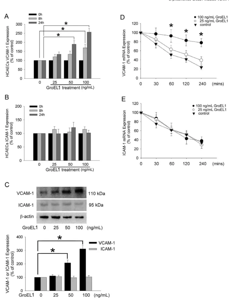
GroEL1, from Chlamydia pneumoniae, induces vascular adhesion molecule 1 expression by p37(AUF1) in endothelial cells and hypercholesterolemic rabbit.
Texto
Imagem

Documentos relacionados
O uso de extratos óleos essenciais, embalagens ativas, atmosferas modificadas, irradiação gama, marinadas, entre outros, como pode-se observar na tabela 1, são
molecule expression and lymphocyte adhesion to endothelial cells: effect of nitric oxide. Exp
Figure 3 - Expression of intercellular adhesion molecule (ICAM)-1 on mesenteric endothelial cells (p ANOVA o 0.001) and the serum levels of corticosterone (p ANOVA=0.01)
Regressemos ao terreno da estética, para observar que a ideia de plasticidade, mesmo nesse seu sentido mais lato, é impossível de pensar fora da tal relação problemática entre arte
OBJECTIVES: To examine older people’s preferences for self-involvement in end-of-life care decision-making in scenarios of mental capacity (competency) and incapacity, and to
Mecha- nisms of eosinophil adherence to cultured vascular endothelial cells: eosinophils bind to the cytokine- induced endothelial ligand vascular cell adhesion mol- ecule-1 via
The present work shows the expression, of the adhesion molecules PECAM-1, ICAM-1, E-selectin, TSP and VCAM, on bovine umbilical vein endothelial cells, stimulated with plas- ma
Glycosylphosphatidylinositol toxin of Plasmodium up- regulates intercellular adhesion molecule-1, vascular cell adhesion molecule-1, and E-selectin expression in vascular
