Links Pathogenesis and Dauer Formation
Victor L. Jensen1,2, Karina T. Simonsen3, Yu-Hui Lee2, Donha Park2, Donald L. Riddle1,2*
1Department of Medical Genetics, University of British Columbia, Vancouver, British Columbia, Canada,2Michael Smith Laboratories, University of British Columbia, Vancouver, British Columbia, Canada,3Department of Biochemistry and Molecular Biology, University of Southern Denmark, Odense, Denmark
Abstract
The DAF-16/FOXO transcription factor is the major downstream output of the insulin/IGF1R signaling pathway controllingC. elegansdauer larva development and aging. To identify novel downstream genes affecting dauer formation, we used RNAi to screen candidate genes previously identified to be regulated by DAF-16. We used a sensitized genetic background [ eri-1(mg366); sdf-9(m708)], which enhances both RNAi efficiency and constitutive dauer formation (Daf-c). Among 513 RNAi clones screened, 21 displayed a synthetic Daf-c (SynDaf) phenotype withsdf-9. One of these genes,srh-100, was previously identified to be SynDaf, but twenty have not previously been associated with dauer formation. Two of the latter genes,lys-1
andcpr-1, are known to participate in innate immunity and six more are predicted to do so, suggesting that the immune response may contribute to the dauer decision. Indeed, we show that two of these genes,lys-1andclc-1, are required for normal resistance toStaphylococcus aureus.clc-1is predicted to function in epithelial cohesion. Dauer formation exhibited bydaf-8(m85),sdf-9(m708), and the wild-type N2 (at 27uC) were all enhanced by exposure to pathogenic bacteria, while not enhanced in a daf-22(m130) background. We conclude that knockdown of the genes required for proper pathogen resistance increases pathogenic infection, leading to increased dauer formation in our screen. We propose that dauer larva formation is a behavioral response to pathogens mediated by increased dauer pheromone production.
Citation:Jensen VL, Simonsen KT, Lee Y-H, Park D, Riddle DL (2010) RNAi Screen of DAF-16/FOXO Target Genes inC. elegansLinks Pathogenesis and Dauer Formation. PLoS ONE 5(12): e15902. doi:10.1371/journal.pone.0015902
Editor:Alejandro Aballay, Duke University Medical Center, United States of America
ReceivedSeptember 8, 2010;AcceptedNovember 30, 2010;PublishedDecember 31, 2010
Copyright:ß2010 Jensen et al. This is an open-access article distributed under the terms of the Creative Commons Attribution License, which permits unrestricted use, distribution, and reproduction in any medium, provided the original author and source are credited.
Funding:This work was supported by the Canadian Institute of Health Research Operating Grant to D.L.R.#MOP-79458 (www.cihr.ca), the Natural Science and Engineering Research Council Scholarship to V.L.J. (www.nserc-crsng.gc.ca), and the Michael Smith Foundation for Health Research Scholarship to V.L.J. (www. msfhr.org). The funders had no role in study design, data collection and analysis, decision to publish, or preparation of the manuscript.
Competing Interests:The authors have declared that no competing interests exist.
* E-mail: [email protected]
Introduction
TheC. elegansdauer larva is a facultative diapause and dispersal stage that develops in response to adverse environmental stimuli such as high temperature, high population density or limited food [1]. Mutations in genes affecting the signal transduction pathways controlling the developmental switch may result either in constitu-tive dauer formation in favorable environments (dauer-constituconstitu-tive, or Daf-c) or a lack of dauer formation in adverse environments (dauer-defective, or Daf-d) [2]. Though there are nearly 30 identified dauer formation (daf) genes inC. elegans, there may be many more genes that have minor effects on the known pathways that are not detectable as single mutants [3–5]. The major pathways involved in dauer formation are the transforming growth factor
b(TGF-b), insulin/insulin-like (IIS) and guanylyl cyclase pathways [6]. Transcriptional targets of the DAF-3/Smad [7], DAF-16/ FOXO [8] and DAF-12 [9] transcription factors are the effectors for parallel processes that execute the dauer/non-dauer switch.
Some of the genes involved in dauer formation function within neurons, and affect neurosensory perception or neuropeptide secretion [10–14]. The dauer pheromone and the competing food signal both require proper sensory perception to elicit a response [15]. Genes shown to be involved in dauer formation include a guanylyl cyclase, G-proteins and genes required for proper amphid cilia formation [10,16,17].
Neural tissue in C. elegans has been previously shown to be refractory to gene expression knockdown by RNAi [18]. This effect can be reduced with mutants that affect the RNAi process includingeri-1, a gene that encodes a siRNAase [18]. This mutant shows a weak Daf-c phenotype when treated with RNAi targeted for the strong Daf-c genesdaf-2anddaf-19. Here we use a strain that containseri-1 as a double mutant with the synthetic dauer formation (SynDaf) mutantsdf-9, a phosphatase-dead phosphatase [4,19,20]. The genetic data suggest that sdf-9 interacts directly with the DAF-2 insulin receptor to stabilize its phosphorylated state, thereby increasing insulin signaling [20]. Although sdf-9(m708)has little or no Daf-c phenotype as a single mutant, it strongly enhances most Daf-c mutants, and results in a synthetic Daf-c phenotype with other genes [4,19,20]. The eri-1; sdf-9
double mutant proved itself useful for assaying enhanced dauer formation resulting from gene knockdown via RNAi.
subsequent dispersal as a defensive response to pathogens in the environment.
Results
RNAi Screen for Enhanced Dauer Formation
As proof of concept for the use oferi-1(mg366); sdf-9(m708) as a sensitized genetic background to detect SynDaf mutations, we tested the effect ofakt-1RNAi on this strain. AKT-1 is involved in transmitting the signal from the DAF-2 receptor to the DAF-16/ FOXO transcription factor [25]. Anakt-1knockout has no Daf-c phenotype as a single mutant, but forms 82% dauer larvae as a double mutant withsdf-9[4]. Theakt-1RNAi treatment resulted in a median constitutive dauer formation of 44% compared to 6% for the control RNAi.
For our screen, we chose genes that were putatively repressed four-fold by DAF-16 activity (in a daf-2 background) from two microarray analyses [23,24], as well as those identified to be direct targets by chromatin immune-precipitation [26]. We chose repressed genes because they are down regulated upon entry into dauer (when DAF-16 is active) and RNAi also represses expression. From the RNAi library [23] we obtained clones corresponding to 513 identified target genes. Sixty-nine of these genes (13%) were obtained from two of our three sources. None were found in all three. Since DAF-16 is a major regulator of dauer formation, we hypothesized that many of its target genes may have small effects on dauer formation, detectable only in a sensitized genetic background.
21 SynDaf Genes
For the primary screen a qualitative assessment of dauer formation was completed for each target gene. 131 of the 513 RNAi clones were judged by visual inspection to result in increased dauer formation (compared to the control), and these were kept for further assessment (for complete target list see Table S1). These included clones that appeared to have only slightly higher dauer formation. In subsequent quantitative screens we required a target gene RNAi treatment to reproduce higher dauer formation significantly (p,0.05) for three consecutive independent trials. In the three retests, we counted each population (dauer and non-dauer larvae) and compared it to the control, if a clone failed to repeat once it was deemed to be negative. Thirty-one genes remained after a first quantitative pass, twenty-three after a second and twenty-one remained after a third and final re-test. Average percent dauer formation for each of the 21 target genes is given in Table 1 (actual counts included in Table S2).
Whereas 69 of the 513 candidate target genes were found in two of the three sources, [23,24,26], eight of the 21 positives were among these 69. The probability that this was random had a p-value (x2
test) of 0.001. Hence, genes from multiple sources were enriched among the 21 positives (Table 1). Nevertheless, most of the positives originated from only one of the three sources.
Each of the three source studies [23,24,26] identified gene classes that were enriched in each of their own data sets. The most enriched protein domain in both the 21 positives we report (Table 1) and the 513 target genes are the CUB (or CUB-like) domain (C1r/C1s, Uegf, Bmp1) [27]. It has been suggested that CUB-domain proteins function in innate immunity due to the organization of their genes in large clusters, the similarity of CUB domains to immunoglobulins and their localization at the cell surface [28,29]. In addition, a CUB domain protein has been identified in a recent RNAi screen for sensitivity to Pseudomonas auruginosaPA14 infection and arsenic stress [30].
Genes Known to Affect Dauer Formation or Insulin Secretion
Several genes identified in our screen function in pathways that have already been associated with dauer formation. This includes one gene that has already been identified as SynDaf, srh-100
[26,31]. SRH-100 is a predicted olfactory G-protein coupled receptor (GPCR) [32]. Detection of this gene shows that our screen can replicate previous results.
A previously unreported SynDaf gene, ccb-1, encodes the b -subunit of the L-type calcium channel, a protein involved in insulin secretion in mammals [33]. Calcium signaling inC. elegans
has been shown to affect dauer formation and insulin secretion [12]. It is likely that loss ofccb-1results in lower insulin output, which has been previously shown in other insulin secretion mutants to result in a SynDaf phenotype [12].
Genes with Unknown Function
Most of the 21 SynDaf genes we identified have predicted protein domains but no assigned functions (Table 1). Five have been shown to interact withdafgenes. C53A3.2 encodes a HAD-superfamily hydrolase and was shown to have a synthetic small brood-size phenotype with daf-18/PTEN [34]. skr-8, a Skp1 homolog that is part of the proteasomal E3 ubiquitin ligase complex, has been shown to be regulated by DAF-12 [35] as well as DAF-16. Three genes (ZK896.5, F35E12.9 and dct-5) are differentially regulated in TGF-b mutants during dauer entry as measured by microarray analysis [5].
Three of the 21 genes have been previously shown to suppress the tumorous gonad phenotype of gld-1 mutants in an RNAi screen of DAF-16 targets [36]. dct-5 (DAF-16-controlled tumor suppressor) encodes a zinc finger transcription factor [36],dct-14
encodes a heat shock protein possibly involved in germ cell apoptosis, anddct-17encodes a protein with CUB and inorganic phosphatase domains. This overlap between the gld-1 -tumor-suppressor genes and the SynDaf positives in this study suggest that these overlapping genes could be involved in the IIS pathway. Finally, three genes, F44D12.8, C24G6.6, and F59B1.2, were SynDaf under our conditions but they have no previously identified involvement in any biological process. F44D12.8 encodes a serine arginine repeat nuclear matrix protein (SRRM), which may function in alternative splicing or mRNA stability [37]. C24G6.6 encodes a flavin-containing amine oxidase and may function in neurotransmission. F59B1.2 encodes a protein with no known or predicted domains.
Innate Immunity Genes
The most notable trend within our list of 21 SynDaf genes is that eight genes have a connection to innate immunity (Table 1). Four genes encode proteins that contain CUB domains and are members of large clusters of paralogs. Several genes in these clusters are induced upon infection [38,39,28], so we include these in our list of immunity genes that are SynDaf. Recently, it has been shown that several CUB-like genes are induced upon infection withYersinia pestis[40]. A total of seven of the eight innate immunity related genes found in our screen, including three CUB domain proteins, dct-17,clc-1, cpr-1 andlys-1are reported to be induced upon infection [38,39].
pathogenic bacteria has not been previously reported for either of these two genes, but LYS-1 over-expression has been shown to confer resistance toSerratia marcescens[42]. It is predicted thatclc-1, which encodes a claudin-like protein, plays a role in epithelial cohesion [43]. It is possible that the epithelial layers inC. elegans
become more permeable toS. aureusas a result ofclc-1RNAi. Dauer Formation on Pathogenic Bacteria
To determine if pathogenesis affects dauer formation, we challenged C. elegans with different pathogenic bacteria. We selected the pathogensPseudomonas aeruginosaPA14 [44],S. aureus,
Agrobacterium tumefaciens and S. marcescens, all of which have been previously tested withC. elegans[42,45,46]. The strains we used reduced survival (compared to the standard laboratory foodE. coli
OP50) similarly to the previous reports (data not shown). We also used Bacillus subtilis, because it had been previously shown to increase the survival ofC. eleganscompared toE. coliOP50 [21].
We first challenged the relatively weak daf-8(m85ts) Daf-c mutant with all the bacterial strains at an intermediate temperature (22.5uC), except for PA14 which we tested at 15uC,
a permissive temperature for daf-8(m85). The percent dauer formation seen fordaf-8 increased on all three pathogens tested compared to OP50, but was reduced on B. subtilis (Table 2). Similarly,sdf-9(m708) formed,20% dauer larvae on OP50 (at
26uC) and 2% onB. subtilis, but formed more than twice as many dauer larvae onA. tumefaciens orS. marcescensand three times as many onS. aureus(Table 3).
We tested N2 for its response to pathogens at 27uC (a condition that induces,5–10% dauer larvae on OP50) to ensure the effect
we observed was not unique to Daf-c mutants [11]. The same trend seen with the two weak Daf-c mutants was repeated in N2 withA. tumefaciens and S. marcescenssignificantly enhancing dauer formation (Table 4). We conclude that part of the C. elegans
response to a pathogenic environment is to enter the dauer stage at greater frequency.
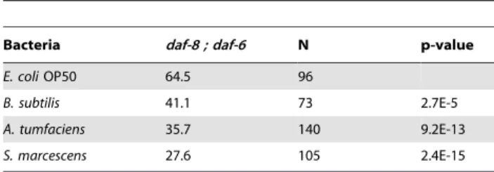
We performed epistasis analysis to determine which part of the dauer signaling pathway affects pathogenesis. We surmised that olfactory sensation might be involved becauseC. elegansis able to discriminate between bacteria [47]. To test this we used the daf-8(m85); daf-6(e1377)double mutant that can form dauer larvae Table 1.Set of 21 SynDaf Genes.
Gene Predicted Functiona Average Dauer Larvae Percent
±S.E. Combined N
Controls
GFP Negative control 5.661.3 1926
akt-1 Dauer signaling kinase, positive control 43.8611.2 1561
Previously Identified SynDaf Gene
srh-100 Predicted olfactory G-protein coupled receptor 37.8612.8 417
New SynDaf Genes
C53A3.2 p-Nitrophenyl phosphatase (Synthetic
small brood size withdaf-18)
26.7611.2 842
skr-8 skp1 protein (Regulated by DAF-12) 26.960.7 420
dct-5 zinc finger transcription factor 12.462.1 780
cyp-35A3 Cytochrome P450 CYP2 subfamily 35.1622.9 289
C24G6.6 Flavin-containing amine oxidase 38.8616.0 329
lase-1 Aminoacylase ACY1 64.1613.7 467
unc-84 Transmembrane protein with a SUN domain 28.067.9 430
ccb-1 Beta subunit of dihydropyridine
sensitive L-type calcium channel
24.764.1 631
dct-14 DNAk, heat shock protein 33.261.1 509
E02C12.8 CHK kinase like, like SRC kinase 21.067.6 927
F59B1.2 Gene 23.768.4 708
F44D12.8 SRRM1 (serine arginine repeat nuclear matrix protein) 33.5613.9 560
Innate Immunity Related New SynDaf Genes
F35E12.9 CUB domain 31.569.7 276
F35E12.10b CUB domain 22.0
612.3 645
ZK896.5b CUB domain 35.4
610.7 436
dct-17b CUB domain and inorganic phosphatase 18.564.9 769
clc-1b Claudin 47.4
67.0 378
lys-1b Lysozyme 19.1
66.5 565
cpr-1b Cysteine proteinase, cathepsin L 31.863.8 433
F52E1.5b Homology to chondroitin proteoglycan 12.5
64.0 842
Actual counts and p-values listed in Table S2.
aPredicted functions are based on previous research and Wormbase annotations. bUpregulated upon infection [38,39].
constitutively (due to the daf-8 mutation), but is defective in chemosensory behavior due to daf-6with improper formation of the sensory channel, preventing the olfactory neurons from contacting the environment [48,49]. While thedaf-8single mutant (which has normal olfactory behavior) responded to pathogenic bacteria by forming a higher percentage of dauer larvae (Table 2), thedaf-8;daf-6double mutant formed fewer dauer larvae on the pathogenic bacteria (Table 5). This indicates that olfactory sensation is required for the increase in dauer formation on pathogenic bacteria.
Our initial observation of increased infection causing higher dauer formation involved RNAi tests using the same bacterial strain (HT115) for control and sample. Hence, the dauer stimulus must not originate from the bacteria, but instead from the worms themselves. To test if the dauer pheromone served as an olfactory cue, we used thedaf-22(m130)mutant that is unable to produce the pheromone [50,51]. It has been reported that the expression of
daf-22increases upon infection with PA14 [38]. Interestingly, daf-22was required for the increase in dauer formation. While a daf-8(e1393) unc-13(e51) strain formed more dauer larvae on pathogenic bacteria, a daf-8(e1393) unc-13(e51); daf-22(m130)
mutant did not (Table 6). In these tests, the unc-13 mutation (which does not affect dauer formation) served to prevent the strain from avoiding the pathogen.
It was previously reported thatdaf-22mutants are able to form dauer larvae at a frequency similar to N2 at 27uC [52]. We compareddaf-22dauer formation on pathogens at 27uC with that of N2. Whereas N2 formed more dauer larvae on the pathogenic bacteria, the daf-22 mutant did not, forming only a few dauer larvae on the laboratory food OP50 and none on the pathogens tested (Table 4). Finally, we used thepdaf-7::GFP reporter gene that exhibits decreased expression with increased pheromone concentrations [53]. Indeed, GFP expression in L2 larvae decreased markedly after exposure to PA14 (Figure S1). Taken together this indicates that increased dauer pheromone production is a mechanism for increased dauer formation in response to bacterial pathogenesis.
Discussion
Mutations insdf-9have been independently isolated three times as enhancers ofunc-31,akt-1anddaf-2mutants [4,19,20]. Because
sdf-9 enhances the phenotype of most Daf-c mutants tested, we utilized it as a sensitized background for identifying new SynDaf genes. Of 20 previously unreported SynDaf genes, three have been shown and five are predicted to play roles in innate immunity. Five genes have been previously linked to insulin or TGF-bsignaling. For example,skr-8is regulated by DAF-12 [35]. It is possible that some of our selected 513 target genes may not be SynDaf with sdf-9, similar to akt-2or the Eak genes [4], but still have a SynDaf phenotype with other mutants. Acknowledging this limitation, our screen nevertheless allowed for the detection of a new set of SynDaf genes and identification of a novel input into the dauer developmental decision.
It is not surprising to see an enrichment of target genes identified in two of the three sources in our positive gene set. There is not a strong consensus among the three gene sets we used
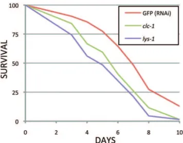
Figure 1. Survival of RNAi treated adults onS. aureus.clc-1and
lys-1RNAi treatment increased pathogen sensitivity compared to the RNAi control (GFP). One of two independent tests is shown. The TD50 (time required for 50% of the nematodes to die) forlys-1was 4.7 days (p,0.0001) and forclc-1was 5.4 days (p = 0.001) compared to 6.8 days for the GFP RNAi control. p-values were calculated using the log-rank test.
doi:10.1371/journal.pone.0015902.g001
Table 2.Percent dauer formation ofdaf-8(m85)on pathogenic bacteria at 22uC.
Bacteria daf-8 N p-value
E. coliOP50 65.3 101
B. subtilis 12.1 107 6.3E-31
A. tumfaciens 84.1 195 3.7E-8
S. marcescens 79.4 102 2.8E-3
E. coliOP50a 0 86
P. aeruginosaPA14a 70.6 68 5.9E-9
aThese tests were carried out at 15 uC. doi:10.1371/journal.pone.0015902.t002
Table 3.Percent dauer formation ofsdf-9(m708)on pathogenic bacteria at 26uC.
Bacteria sdf-9 N p-value
E. coliOP50 19.6 97
B. subtilis 2.6 76 2.0E-4
A. tumfaciens 49.4 79 2.6E-11
S. marcescens 42.2 90 6.3E-08
S. aureus 60.0 40 1.2E-10
doi:10.1371/journal.pone.0015902.t003
Table 4.Percent dauer formation of N2 anddaf-22(m130)on pathogenic bacteria at 27uC.
Bacteria N2 N p-valuea daf-22 N p-valuea
E. coliOP50 11.7 231 3.9 246
B. subtilis 0.78 129 1.1E-4 0 131 0.021
A. tumfaciens 27.6 98 1.0E-6 0 133 0.020
S. marcescens 30.6 111 5.2E-10 0 95 0.049
ap-values given are relative to OP50 sample for each genotype.
[23,24,26], with about 13% (69/513) overlap, There are 67 genes in common between the two microarray experiments and two between the ChIP [26] and the McElweeet al. [23] microarray data, based on our filtering criteria. Eight of the 21 positives were present in the two microarray sources.
We designed our screen to detect genes downstream of DAF-16 that increased dauer formation as a result of reduced activity. Althoughdaf-2mutants show increased pathogen resistance, our target gene set includes many down-regulated innate immunity genes. Despite down-regulation of such genes by DAF-16 the two microarray studies also include many up-regulated DAF-16 innate immunity targets [23,24]. This explains the increased resistance of
daf-2mutants to pathogens despite a number of down-regulated defense genes. Also, many of the innate immunity genes that are down regulated by DAF-16 in our positive gene list are up-regulated in response to other pathogens, which may represent a pathogen-specific response [38,39].
CCB-1 and possible feedback regulation of insulin signaling
Since ccb-1 was detected in our screen, we conclude that its activity normally inhibits dauer formation. It encodes the b -subunit of the L-type calcium channel, which may modulate the sensitivity of the channel [33]. This gene is thought to be a direct target of DAF-16 because it was identified using DAF-16 ChIP, and its promoter contains a DAF-16 binding site [26]. It is possible that DAF-16 regulates the expression ofccb-1to modulate calcium signaling, which has been linked to insulin secretion in mammals and worms [12,33]. The interaction between DAF-16 andccb-1
may be part of a feedback mechanism to reduce insulin secretion during the dauer development. The IIS pathway acts to inhibit DAF-16, but once DAF-16 activity reaches a critical threshold, it could antagonize insulin secretion to stabilize the dauer de-velopmental decision.
Germ line anddctgenes
Three dctgenes were found in our screen. These are putative DAF-16 targets that are gld-1 (Germ Line Defective) tumor suppressors [36]. Whendctexpression is reduced the endomitotic tumors that grow within the germ lines of gld-1 mutants are reduced. Germ line proliferation is actively suppressed in dauer larvae [54], so it is reasonable that the mechanisms governing cell proliferation in adults and dauer larvae may overlap. However, it is not clear why reduction ofdctactivity would trigger constitutive dauer formation in our screen. It is as though inhibition of mitotic progression (e.g., in response to starvation) feeds back to reduce TGF-b and/or insulin signaling and favor dauer arrest, but the point of feedback regulation is not known.
Other genes that regulate both germ line proliferation and dauer formation have already been identified, including AKT-1 and DAF-18/PTEN [54,34]. In our positive gene set, C53A3.2 and skr-8 have been previously shown to have a synthetic small brood size phenotype withdaf-18, an indication of poor germ line proliferation [34,55]. Although it is well known that dauer formation arrests germ line proliferation, these results suggest that the converse may also be true.
Immunity Related Genes
Eight of the 21 positives have been previously implicated in innate immunity, four of which contain the CUB (or CUB-like) domain. The CUB domain consists of ab-barrel with similarity to immunoglobulins, and is predicted to be extracellular [38]. Three of the four CUB domain proteins found in our screen are induced by infection, as are the four remaining innate immunity genes [38,39].
The lys-1 lysozyme is an antimicrobial peptidoglycan N -acetylmuramoylhydrolase that has been shown to protect against infection inC. elegans[42]. Thecpr-1, F52E1.5 andclc-1genes are also predicted to be protective genes because they are induced upon infection [38,39,56]. We propose that under the conditions of the RNAi screen, targeting of these innate immunity genes increases the animal’s susceptibility to, or perception of, infection by the E. coli food [21,57,58]. As a response to this increased sensitivity to infection, the developing larvae may be predisposed to dauer dispersal. This leads to the hypothesis that it is the process of pathogenesis that stimulates increased dauer formation. Indeed, we have shown that pathogenic bacteria enhance dauer formation, and this requires the dauer pheromone.
Two genes,lys-1andclc-1, were required for normal resistance toS. aureus. The remaining six genes may not affect pathogenesis by S. aureusunder our conditions for various reasons, including redundancy among gene families or pathogen-specific interactions. Over-expression oflys-1had been previously shown to increase survival on S. marcescens [42]. LYS-1 is a putative lysozyme, an antimicrobial protein, so we expected that loss oflys-1might make the worm sensitive to infection in spite of possible redundancy with
lys-2. Reduction in survival has not been previously shown for lys-1, but the conditions and pathogens used were different [42].
clc-1encodes a claudin-like protein, and its expression has been seen to be induced upon infection [39,59]. Claudins are predicted to function in epithelial cohesion, indicating that loss of clc-1
function may cause the epithelial layer to loosen. InC. elegans,clc-1
RNAi was reported to increase the permeability of the pharynx to a high molecular mass dye, TRITC-dextran [43]. Thus, increased CLC-1 in response to infection could strengthen the epithelial layers to resist pathogenesis. Indeed, we have shown that survival ofC. elegansis significantly reduced when treated withclc-1RNAi followed by exposure toS. aureusfrom the L4 stage.
Table 5.Percent dauer formation ofdaf-8(m85); daf-6(e1377)
on pathogenic bacteria at 22uC.
Bacteria daf-8 ; daf-6 N p-value
E. coliOP50 64.5 96
B. subtilis 41.1 73 2.7E-5
A. tumfaciens 35.7 140 9.2E-13
S. marcescens 27.6 105 2.4E-15
doi:10.1371/journal.pone.0015902.t005
Table 6.Percent dauer formation ofdaf-8(e1393) unc-13(e51)
anddaf-8(e1393) unc-13(e51); daf-22(m130)on pathogenic bacteria at 20uC.
Bacteria
daf-8
unc-13 N p-value
daf-8 unc-13;
daf-22 N p-value
E. coliOP50 39.2 245 40.5 116
B. subtilis 34.4 93 0.35 33.7 86 0.20
A. tumfaciens 61.9 160 4.1E-09 39.2 79 0.82
S. marcescens 54.5 101 0.0017 35.4 96 0.31
Mechanism for Pathogenic Input into Dauer Formation DAF-2 and DAF-16 have been previously linked to innate immunity.daf-2mutants are resistant to infection [21]. DAF-16 is required for the increased resistance ofdaf-2, just as it is for the longevity and dauer formation phenotypes [21,60,61]. We have shown that the production of dauer pheromone is required for pathogen induced dauer formation with the requirement ofdaf-22
for the dauer induction. Olfactory sensation is also required for the increase in dauer formation, probably through the sensation of dauer pheromone. Worms infected with PA14 increase expression ofdaf-22, a gene that encodes a pheromone biosynthetic enzyme [38,50], and by reducing expression ofdaf-7, an indicator of higher pheromone levels in the environment [53]. Taken together, the data indicate that when C. elegans encounters a pathogenic environment it increases pheromone production to elicit dauer formation. At higher concentrations purified components of the dauer pheromone were found to be a chemo-repellent [62] suggesting that increased dauer pheromone could deter other worms from entering the toxic environment. Pheromone deposited by 100 worms over 60 minutes decreases the response to a chemo-attractant [63].
We rule out starvation as the cause of increased dauer formation on pathogenic bacteria. It is well known that food limitation increases dauer formation [1,64] and that C. eleganscan display avoidance to pathogenic bacteria on plates [47,65]. The AWB ciliated chemosensory neurons are required for this avoidance [47]. Chemosensory function is required for the pathogen induced dauer formation because no increase is observed in a chemosensory mutant, daf-6(Table 5). However, in the unc-13background we show increased dauer formation on pathogenic bacteria, which is suppressed by thedaf-22mutation (Table 6).daf-22single mutants also show no increase in dauer formation on pathogenic bacteria (Table 4) despite normal chemosensory behavior [63]. This indicates that decreased nutrition is not part of the mechanism of pathogen induced dauer formation.
Animals may use cues to recognize infection in other individuals [66]. The original observations included changes in feather brightness or songs of songbirds affecting mate selection. A bird could select for those with genetic resistance to a pathogen by avoidance of potential mates that are infected [66]. Bullfrog tadpoles receive a chemical cue from infected tadpoles, and they spend less time in the presence of those tadpoles to mitigate the risk of infection [67]. We suggest that the dauer pheromone can work in a similar way, as it is used for avoidance and sexual attraction as well as dauer formation [62].
The benefit of dauer formation in a pathogenic environment could accrue from three dauer traits. First, dauer larvae do not feed, which should convey resistance to enteric infection. Second, the dauer stage is used for dispersal, permitting flight from local concentrations of pathogenic bacteria. Third, dauer larvae have a stronger cuticle [1], which could also defend against attachment or entry of pathogenic bacteria [57].
We have identified 21 SynDaf genes, each of which provides insight into dauer formation. Some genes fall into pathways and processes that have already been associated with dauer formation, whereas others suggest a new input into dauer formation, pathogenesis. Indeed, we show that pathogenic bacteria do enhance dauer formation possibly through increased pheromone production. We have explored the connection between dauer formation and suppression of germ line proliferation as well as innate immunity. Our screen is defined by the 513 putative DAF-16 target genes we used. A genome-wide screen should detect additional environmental inputs for dauer formation that do not require DAF-16.
Methods
Gene Target Selection and RNAi Screen
DAF-16 target genes were selected from two microarray studies, 336 from one [23] and 250 from another [24]. The targets chosen were at least four-fold down regulated in a daf-2 background [23,24]. An additional 87 targets were selected from a DAF-16 ChIP study [26]. Only the target genes that were in the Arhinger RNAi library were kept [68]. Bacterial cultures in the library that did not grow after three attempts were also not included, leaving a total of 513 target genes (full target list in Table S1). RNAi clone that targets srh-100 is listed in the RNAi library as srh-99 (or C46E10.7). An RNAi experiment that targets eithersrh-100or srh-99 will likely knockdown both due to high nucleotide identity (88%). For simplicity, we list the target/positive in this paper as
srh-100because it is the primary target of the RNAi experiment. Theeri-1(mg366); sdf-9(m708)strain was constructed by crossing
sdf-9males witheri-1 hermaphrodites. The double mutants were selected in the F2 generation by PCR tests for the deletion ineri-1
and the transposon insert in sdf-9 [18,20]. The screen was performed by first spotting (in duplicate) 50ml of each RNAi clone (thawed from an overnight liquid culture frozen at280uC in 30% glycerol) onto 10 ml NG agar plates containing 100 mg/ml ampicillin and 1 mM IPTG, followed by overnight incubation at room temperature. The clones containing sequences specific for GFP andakt-1were used for the negative and positive controls, respectively. These controls were run with each test.
eri-1; sdf-9was exposed to RNAi for two generations. The two initial 60 mm plates per clone were inoculated with 2–3 L2 or L3 larvae and incubated at 20uC. On the seventh day of incubation, five F1 gravid adults were transferred to fresh RNAi plates (two per clone), made as above, and allowed to lay eggs for approximately 3 hours at room temperature. The plates were then transferred to 25.5uC, where the transferred adults become sterile [18], until the 10th day when the populations were scored for percentage dauer larvae. For the initial screen the two plates per clone were checked visually for an increase in constitutive dauer formation by comparison with the negative (GFP RNAi) and positive (akt-1) controls. For subsequent re-screening the dauer larvae (identified visually in the Zeiss Stemi SV 11 stereomicro-scope at 6606magnification by the presence of dauer alae and radial constriction of the body) and non-dauer larvae were counted. RNAi clones were only re-tested if they produced a significant difference (p,0.05,x2
test). To be kept as a positive, a clone had to show a significant difference in three independent trials with the smallest sample size.30 and largest being.100 (actual counts included in Table S2). Positive clones were confirmed by PCR using a T7 transcriptional start site primer followed by digestion by two different restriction enzymes.
Classification and Comparison of Positives
Function was inferred from previous work, including the ‘‘Gene Summary’’ page on Wormbase [37], as stated for biological process enrichment assessment. Assessing GO term enrichment was completed by using the Wormbase BioMart function (WormMart) to retrieve identifiers for all genes [37,69]. These identities were put into DAVID to identify enriched GO terms [70]. The number of genes overlapping in the target and positive gene sets were compared by ax2test and the p-value reported.
Dauer Formation,daf-7Expression, and Adult Survival on Pathogenic Bacteria
Bacterial strains used were P. aeruginosa PA14, A. tumefaciens
HT115, and E. coli OP50. Fresh overnight cultures of each bacterial strain were spread on each plate to cover approximately half of the plate surface. Bacteria were not spread to the edges in order to minimize the number of dauer larvae crawling off the plate. To assay percent dauer formation, hypochlorite-purified eggs [71] were spotted on to two 60 mm plates for each bacterial strain. Dauer and non-dauer larvae were then counted as the first non-dauers reached egg-laying age. Bacterial strains were compared for nematode pathogenesis as previously described [21] by daily assay of the percent survival on each bacterial strain. PA14 plates were made as described above for thepdaf-7::GFP expression analysis. L2 larvae were placed on PA14 or OP50 control plates and were assayed for GFP expression after 4 hours. GFP worms were imaged on a Zeiss Axioskop with a Qimaging Retiga 2000R camera.
For the survival assays with the eight pathogen-related RNAi treatments, rrf-3(pk1426) was treated with each of the RNAi expressing bacteria for two generations to maximize silencing. F2 larvae were synchronized by hypochlorite treatment followed by hatching overnight in M9 buffer. The synchronized L1’s were then put on a 50-50 mixture of RNAi bacteria for the pathogen related gene and for cdc-25.1to sterilize them for the survival assay. L4 larvae were transferred from the RNAi plates to NG plates seeded with S. aureus and scored daily for survival. GraphPad Prism software was used to calculate significance using the log-rank test and to calculate the TD50 for each strain for each of two replicates. Figure 1 was created in Microsoft Excel.
Supporting Information
Figure S1 Reduction indaf-7expression on PA14.In (A), the native GFP expression on the standard laboratory foodE. coli
OP50 from a daf-7 promoter driving expression of GFP. The expression of daf-7 is much reduced after are exposure to the strong pathogen PA14, as seen in (B). Images were taken with a 1006objective and 106ocular lenses, eight hours after L2 larvae were transferred to either OP50 or PA14 from OP50 plates. (TIF)
Table S1 Full list of target RNAi clones.
(DOCX)
Table S2 Dauer and Non-dauer worm counts for posi-tive genes.
(DOCX)
Acknowledgments
We thank the Caenorhabditis Genetics Center (funded by the National Institutes of Health - National Center for Research Resources) for strains, Dr. Donald Moerman for the RNAi clones, and Nigel O’Neil and Marco Gallo for constructive discussions.
Author Contributions
Conceived and designed the experiments: VLJ DLR. Performed the experiments: VLJ KTS YL DP. Analyzed the data: VLJ KTS DP. Contributed reagents/materials/analysis tools: VLJ DLR. Wrote the paper: VLJ DLR.
References
1. Cassada RC, Russell RL (1975) The dauer larva, a post-embryonic de-velopmental variant of the nematodeCaenorhabditis elegans. Dev Biol 46: 326–42. 2. Riddle DL, Swanson MM, Albert PS (1981) Interacting genes in nematode
dauer larva formation. Nature 290: 668–671.
3. Tewari M, Hu PJ, Ahn JS, Ayivi-Guedehoussou N, Vidalain PO, et al. (2004) Systematic interactome mapping and genetic perturbation analysis of aC. elegans TGF-beta signaling network. Mol Cell 13: 469–82.
4. Hu PJ, Xu J, Ruvkun G (2006) Two membrane-associated tyrosine phosphatase homologs potentiateC. elegansAKT-1/PKB signaling. PLoS Genet 2: e99. 5. Liu T, Zimmerman KK, Patterson GI (2004) Regulation of signaling genes by
TGFbeta during entry into dauer diapause inC. elegans. BMC Dev Biol 4: 11. 6. Jensen VL, Gallo M, Riddle DL (2006) Targets of DAF-16 involved in Caenorhabditis elegansadult longevity and dauer formation. Exp Gerontol 41: 922–927.
7. Patterson GI, Koweek A, Wong A, Liu Y, Ruvkun G (1997) The DAF-3 Smad protein antagonizes TGF-beta-related receptor signaling in theCaenorhabditis elegansdauer pathway. Gene Dev 11: 2679–2690.
8. Ogg S, Paradis S, Gottlieb S, Patterson GI, Lee L, et al. (1997) The Fork head transcription factor DAF-16 transduces insulin-like metabolic and longevity signals inC. elegans. Nature 389: 994–999. doi:10.1038/40194.
9. Antebi A, Yeh WH, Tait D, Hedgecock EM, Riddle DL (2000)daf-12encodes a nuclear receptor that regulates the dauer diapause and developmental age inC. elegans. Gene Dev 14: 1512–1527.
10. Thomas JH, Birnby DA, Vowels JJ (1993) Evidence for parallel processing of sensory information controlling dauer formation inCaenorhabditis elegans. Genetics 134: 1105–17.
11. Ailion M, Thomas JH (2003) Isolation and characterization of high-temperature-induced dauer formation mutants inCaenorhabditis elegans. Genetics 165: 127–44.
12. Speese S, Petrie M, Schuske K, Ailion M, Ann K, et al. (2007) UNC-31 (CAPS) is required for dense-core vesicle but not synaptic vesicle exocytosis in Caenorhabditis elegans. J Neurosci 27: 6150–62.
13. Ailion M, Inoue T, Weaver CI, Holdcraft RW, Thomas JH (1999) Neurosecretory control of aging inCaenorhabditis elegans. Proc Natl Acad Sci 96: 7394–7.
14. Alcedo J, Kenyon C (2004) Regulation of C. elegans longevity by specific gustatory and olfactory neurons. Neuron 41: 45–55.
15. Golden JW, Riddle DL (1984) The Caenorhabditis elegans dauer larva: de-velopmental effects of pheromone, food, and temperature. Dev Biol 102: 368–78. 16. Bell LR, Stone S, Yochem J, Shaw JE, Herman RK (2006) The molecular identities of theCaenorhabditis elegansintraflagellar transport genesdyf-6,daf-10 andosm-1. Genetics 173: 1275–86.
17. Swoboda P, Adler HT, Thomas JH (2000) The RFX-type transcription factor DAF-19 regulates sensory neuron cilium formation inC. elegans. Mol Cell 5: 411–21.
18. Kennedy S, Wang D, Ruvkun G (2004) A conserved siRNA-degrading RNase negatively regulates RNA interference inC. elegans. Nature 427: 645–9. 19. Ohkura K, Suzuki N, Ishihara T, Katsura I (2003) SDF-9, a protein tyrosine
phosphatase-like molecule, regulates the L3/dauer developmental decision through hormonal signaling inC. elegans. Development 130: 3237–48. 20. Jensen VL, Albert PS, Riddle DL (2007)Caenorhabditis elegansSDF-9 enhances
insulin/insulin-like signaling through interaction with DAF-2. Genetics 177: 661–6.
21. Garsin DA, Villanueva JM, Begun J, Kim DH, Sifri CD, et al. (2003) Long-lived C. elegans daf-2mutants are resistant to bacterial pathogens. Science 300: 1921. 22. Honda Y, Honda S (1999) Thedaf-2 gene network for longevity regulates oxidative stress resistance and Mn-superoxide dismutase gene expression in Caenorhabditis elegans. FASEB J 13: 1385–1393.
23. McElwee J, Bubb K, Thomas JH (2003) Transcriptional outputs of the Caenorhabditis elegansforkhead protein DAF-16. Aging Cell 2: 111–21. 24. Murphy CT, McCarroll SA, Bargmann CI, Fraser A, Kamath RS, et al. (2003)
Genes that act downstream of DAF-16 to influence the lifespan ofCaenorhabditis elegans. Nature 424: 277–83.
25. Paradis S, Ruvkun G (1998)Caenorhabditis elegansAkt/PKB transduces insulin receptor-like signals from AGE-1 PI3 kinase to the DAF-16 transcription factor. Gene Dev 12: 2488–2498.
26. Oh SW, Mukhopadhyay A, Dixit BL, Raha T, Green MR, et al. (2006) Identification of direct DAF-16 targets controlling longevity, metabolism and diapause by chromatin immunoprecipitation. Nat Genet 38: 251–7. 27. Bork P, Beckmann G (1993) The CUB domain. A widespread module in
developmentally regulated proteins. J Mol Biol 231: 539–545. doi:10.1006/ jmbi.1993.1305.
28. Thomas JH (2006) Analysis of homologous gene clusters inCaenorhabditis elegans reveals striking regional cluster domains. Genetics 172: 127–43.
29. Shivers RP, Youngman MJ, Kim DH (2008) Transcriptional responses to pathogens inCaenorhabditis elegans. Curr Opin Microbiol 11: 251–6.
30. Nandakumar M, Tan MW (2008) Gamma-linolenic and stearidonic acids are required for basal immunity inCaenorhabditis elegansthrough their effects on p38 MAP kinase activity. PLoS genet 4: e1000273.
31. Lee SS, Kennedy S, Tolonen AC, Ruvkun G (2003) DAF-16 target genes that controlC. eleganslife-span and metabolism. Science 300: 644–7.
duplications and deletions and intron gains and losses. Genome Res 10: 192–203.
33. Davalli AM, Biancardi E, Pollo A, Socci C, Pontiroli AE, et al. (1996) Dihydropyridine-sensitive and -insensitive voltage-operated calcium channels participate in the control of glucose-induced insulin release from human pancreatic beta cells. J Endocrinol 150: 195–203.
34. Suzuki Y, Han M (2006) Genetic redundancy masks diverse functions of the tumor suppressor gene PTEN duringC. elegansdevelopment. Gene Dev 20: 423–8.
35. Shostak Y, Van Gilst MR, Antebi A, Yamamoto KR (2004) Identification ofC. elegansDAF-12-binding sites, response elements, and target genes. Gene Dev 18: 2529–44.
36. Pinkston-Gosse J, Kenyon C (2007) DAF-16/FOXO targets genes that regulate tumor growth inCaenorhabditis elegans. Nat Genet 39: 1403–9.
37. Rogers A, Antoshechkin I, Bieri T, Blasiar D, Bastiani C, et al. (2008) WormBase 2007. Nucleic Acids Res 36: D612–7.
38. Troemel ER, Chu SW, Reinke V, Lee SS, Ausubel FM, et al. (2006) p38 MAPK regulates expression of immune response genes and contributes to longevity inC. elegans. PLoS Genet 2: e183.
39. Shapira M, Hamlin BJ, Rong J, Chen K, Ronen M, et al. (2006) A conserved role for a GATA transcription factor in regulating epithelial innate immune responses. Proc Natl Acad Sci 103: 14086–91.
40. Bolz DD, Tenor JL, Aballay A (2010) A conserved PMK-1/p38 MAPK is required inCaenorhabditis eleganstissue-specific immune response toYersinia pestis infection. J Biol Chem 285: 10832–10840. doi:10.1074/jbc.M109.091629. 41. Simmer F, Tijsterman M, Parrish S, Koushika SP, Nonet ML, et al. (2002) Loss
of the putative RNA-directed RNA polymerase RRF-3 makes C. elegans hypersensitive to RNAi. Curr Biol 12: 1317–9.
42. Mallo GV, Kurz CL, Couillault C, Pujol N, Granjeaud S, et al. (2002) Inducible antibacterial defense system inC. elegans. Curr Biol 12: 1209–14.
43. Asano A, Asano K, Sasaki H, Furuse M, Tsukita S (2003) Claudins in Caenorhabditis elegans: their distribution and barrier function in the epithelium. Curr Biol 13: 1042–6.
44. Tan MW, Mahajan-Miklos S, Ausubel FM (1999) Killing ofCaenorhabditis elegans byPseudomonas aeruginosaused to model mammalian bacterial pathogenesis. Proc Natl Acad Sci 96: 715–20.
45. Couillault C, Ewbank JJ (2002) Diverse bacteria are pathogens ofCaenorhabditis elegans. Infect Immun 70: 4705–7.
46. Sifri CD, Begun J, Ausubel FM, Calderwood SB (2003)Caenorhabditis elegansas a Model Host for Staphylococcus aureus Pathogenesis. Infect Immun 71: 2208–2217. doi:10.1128/IAI.71.4.2208-2217.2003.
47. Zhang Y, Lu H, Bargmann CI (2005) Pathogenic bacteria induce aversive olfactory learning inCaenorhabditis elegans. Nature 438: 179–184. doi:10.1038/ nature04216.
48. Perens EA, Shaham S (2005) C. elegansdaf-6encodes a patched-related protein required for lumen formation. Dev Cell 8: 893–906. doi:10.1016/j. devcel.2005.03.009.
49. Albert PS, Brown SJ, Riddle DL (1981) Sensory control of dauer larva formation in Caenorhabditis elegans. J Comp Neurol 198: 435–451. doi:10.1002/ cne.901980305.
50. Butcher RA, Ragains JR, Li W, Ruvkun G, Clardy J, et al. (2009) Biosynthesis of theCaenorhabditis elegansdauer pheromone. Proc Natl Acad Sci 106: 1875–1879. doi:10.1073/pnas.0810338106.
51. Golden JW, Riddle DL (1985) A gene affecting production of theCaenorhabditis elegansdauer-inducing pheromone. Mol Gen Genet 198: 534–536.
52. Ailion M, Thomas JH (2000) Dauer Formation Induced by High Temperatures inCaenorhabditis elegans. Genetics 156: 1047–1067.
53. Ren P, Lim C, Johnsen R, Albert PS, Pilgrim D, et al. (1996) Control ofC. elegans Larval Development by Neuronal Expression of a TGF-beta Homolog. Science 274: 1389–1391. doi:10.1126/science.274.5291.1389.
54. Narbonne P, Roy R (2006) Inhibition of germline proliferation duringC. elegans dauer development requires PTEN, LKB1 and AMPK signalling. Development 133: 611–9.
55. Sonnichsen B, Koski LB, Walsh A, Marschall P, Neumann B, et al. (2005) Full-genome RNAi profiling of early embryogenesis inCaenorhabditis elegans. Nature 434: 462–9.
56. Wong D, Bazopoulou D, Pujol N, Tavernarakis N, Ewbank JJ (2007) Genome-wide investigation reveals pathogen-specific and shared signatures in the response ofCaenorhabditis elegansto infection. Genome Biol 8: R194. 57. Darby C (2005) Interactions with microbial pathogens. WormBook. pp 1–15. 58. Garigan D, Hsu AL, Fraser AG, Kamath RS, Ahringer J, et al. (2002) Genetic
analysis of tissue aging inCaenorhabditis elegans: a role for heat-shock factor and bacterial proliferation. Genetics 161: 1101–12.
59. Ren M, Feng H, Fu Y, Land M, Rubin CS (2009) Protein Kinase D Is an Essential Regulator of C. elegans Innate Immunity. Immunity 30: 521–532. doi:10.1016/j.immuni.2009.03.007.
60. Larsen PL, Albert PS, Riddle DL (1995) Genes that regulate both development and longevity inCaenorhabditis elegans. Genetics 139: 1567–83.
61. Kenyon C, Chang J, Gensch E, Rudner A, Tabtiang R (1993) AC. elegans mutant that lives twice as long as wild type. Nature 366: 461–464. doi:10.1038/ 366461a0.
62. Srinivasan J, Kaplan F, Ajredini R, Zachariah C, Alborn HT, et al. (2008) A blend of small molecules regulates both mating and development inCaenorhabditis elegans. Nature 454: 1115–1118. doi:10.1038/nature07168.
63. Matsuura T, Sato T, Shingai R (2005) Interactions betweenCaenorhabditis elegans Individuals during Chemotactic Response. Zool Sci 22: 1095–1103. doi:10.2108/zsj.22.1095.
64. Hu PJ (2007) Dauer. WormBook. pp 1–19. doi:10.1895/wormbook.1.144.1. 65. Pradel E, Zhang Y, Pujol N, Matsuyama T, Bargmann CI, et al. (2007)
Detection and avoidance of a natural product from the pathogenic bacterium Serratia marcescensbyCaenorhabditis elegans. Proc Natl Acad Sci 104: 2295–2300. doi:10.1073/pnas.0610281104.
66. Hamilton WD, Zuk M (1982) Heritable true fitness and bright birds: a role for parasites? Science 218: 384–387.
67. Kiesecker JM, Skelly DK, Beard KH, Preisser E (1999) Behavioral reduction of infection risk. Proc Natl Acad Sci 96: 9165–9168.
68. Kamath RS, Fraser AG, Dong Y, Poulin G, Durbin R, et al. (2003) Systematic functional analysis of theCaenorhabditis elegansgenome using RNAi. Nature 421: 231–7.
69. Smedley D, Haider S, Ballester B, Holland R, London D, et al. (2009) BioMart– biological queries made easy. BMC Genomics 10: 22. doi:10.1186/1471-2164-10-22.
70. Dennis G, Sherman BT, Hosack DA, Yang J, Gao W, et al. (2003) DAVID: Database for Annotation, Visualization, and Integrated Discovery. Genome Biol 4: P3.