Duarte Lima Martins
Purification of Complex
Biopharmaceuticals with New Processes,
Advanced Analytics and Computer-Aided
Process Design Tools
Dissertação para obtenção do Grau de Mestre em Biotecnologia
Orientadora : Cristina Peixoto, PhD,
Senior Scientist,
ACTU, IBET/ITQB - UNL
Co-orientador : Professor José Paulo Mota,
Full Professor,
Requimte/CQFB, FCT - UNL
Júri:
Presidente: Prof. Doutor Carlos A. Salgueiro
Arguente: Professor João G. Crespo
Vogal: Doutora Cristina Peixoto
Purification of Complex Biopharmaceuticals with New Processes, Advanced Analytics and Computer-Aided Process Design Tools
Copyright © Duarte Lima Martins, Faculdade de Ciências e Tecnologia, Universidade Nova de Lisboa
A Faculdade de Ciências e Tecnologia e a Universidade Nova de Lisboa têm o direito, perpé-tuo e sem limites geográficos, de arquivar e publicar esta dissertação através de exemplares impressos reproduzidos em papel ou de forma digital, ou por qualquer outro meio conhecido ou que venha a ser inventado, e de a divulgar através de repositórios científicos e de admitir a sua cópia e distribuição com objectivos educacionais ou de investigação, não comerciais, desde que seja dado crédito ao autor e editor.
à minha família
Acknowledgements
I would like thank some people who supported and contributed to the work presented in this MSc thesis, particularly:
To Dr. Cristina Peixoto, my supervisor, for her scientific guidance, remarkable work ethic, confidence and encouragement. I’m deeply thankful for the outstanding people manager that you are, ensuring the highest quality of this work at all stages.
To Professor Paulo Mota, my co-supervisor, for his enthusiasm and enlightening talks on chromatography.
To Professor Paula Alves, for being the demanding and caring head of the ACTU and iBET always pushing the standards; and of course for the opportunity to develop my MSc thesis work at such a stimulating research group.
To Professor Manuel Carrondo, for his inspiring role model of leadership and scientific work.
To Sartorius Stedim Biotech, especially to Dr. Tobias Schleuss, Dr. Franziska Jonas and Dr. Louis Villain, for providing the materials, support and advice throughout this work; and for the opportunity to attend the European Downstream Technology Forum at Sartorius College.
To the funding from Fundação para a Ciência e a Tecnologia (PTDC/EBB-BIO/119501/2010) for supporting the work presented in this thesis.
To Pier, for being a great colleague always up for interesting discussions about purification, his pragmatic view on biopharmaceutical industry and for all the laughs!
To Carina Silva, for the patience and experience shared when teaching me all about adenovirus production and handling.
To all my colleagues of the virus lab at ACTU, for all the help as well as for the fun moments; you guys made wearing two lab coats and two gloves fun!
To all former and current colleagues at the ACTU, especially Nuno, Fabiana, Marco, Carina, João and Marcos, for all the assistance, advice, companionship and good work envi-ronment.
À Lídia pela compreensão e ânimo em todos os momentos, pelo exemplo de trabalho e resiliência. Por completares o outro lado, um obrigado muito especial.
Aos meus pais que acreditaram sempre em mim e me deram a liberdade de fazer o que gosto. Ao meu irmão por ser um exemplo extraordinário de carácter e determinação. Sem o vosso apoio este trabalho não seria possível.
Abstract
Viruses are highly efficient vectors that have been used for vaccination and gene therapy applications. However, their complexity renders downstream process particularly challenging since devices and strategies especially designed for virus purification are still lacking or need further optimization. After an introduction to the challenges of virus purification and the current strategies being employed, this dissertation presents the study of three different stages of the downstream process: clarification, ultrafiltration and chromatography.
A novel clarification procedure based on diatomaceous earth was evaluated. Small-scale batch incubations led to the identification of Divergan RS – a synthetic non-charged material – as the most promising candidate for integration in a scalable filtration set-up.
Ultrafiltration was addressed with the evaluation of cassette and hollow fiber modules. The results obtained show that cassette module with cut-offs in the 500 kDa range and highly hydrophilic materials enable complete recovery of infective Adenovirus while reducing process time in half when compared with the best hollow fibers. Despite the encouraging results with Adenovirus, the experiments using Retrovirus resulted in low yields and possible optimization strategies were identified.
Membrane technology was also evaluated as an alternative to the packed-bed chromatog-raphy columns. By using a scale-down 96-well device, the impact of ligand density, membrane structure and feed conductivity were evaluated for the purification of Adenovirus by ion ex-change chromatography. The hydrogel-grafted membrane with ligand density of 2.4µmol cm-2 operated in bind/elute mode shown the best compromise between yield and purity.
Overall, this thesis contributed to the advancement of virus purification field by exploiting innovative technologies.
Keywords: virus purification; clarification; ultrafiltrattion; membrane chromatography; biopharmaceuticals; innovative technologies.
Resumo
Os vírus são vectores altamente eficientes que tem sido aplicados para vacinação e terapia génica. Contudo, a sua complexidade torna o processo de purificação particularmente de-safiante uma vez que materiais e estratégias desenhadas especificamente para purificação de vírus são inexistentes ou precisam de ser optimizados. Após uma introdução sobre os desafios relativos à purificação de vírus e estratégias atualmente usadas, é apresentado o estudo de três etapas diferentes do processo de purificação: clarificação, ultrafiltração e cromatografia. Foi estudado um inovador processo de clarificação que usa terras de diatomáceas. Os ensaios em pequena escala identificaram Divergan RS – um material sintético não carregado – como o candidato mais promissor para a integração no passo de clarificação.
Relativamente à ultrafiltração, foram testados vários módulos de cassete e de fibras ocas. Os resultados obtidos indicam que os módulos de cassete com umcut-off próximo de 500 kDa e material altamente hidrofílico permitem recuperar 100 % dos Adenovírus infecciosos e ao mesmo tempo reduzir o tempo de processamento para metade relativamente aos módulos de fibras ocas. Apesar dos resultados positivos obtidos com Adenovírus, os ensaios com Retrovírus resultaram em baixos rendimentos e as possíveis causas foram identificadas.
A tecnologia de membrana foi estudada como alternativa à de cromatografia de leito com-pactado. Através um dispositivo de 96 poços, o impacto da densidade de ligando, da estrutura da membrana e da condutividade da carga foram avaliados na purificação de Adenovírus por cromatografia de troca iónica. A membrana modificada com hidrogel e uma densidade de ligando de 2.4µmol cm-2 resultou no melhor compromisso entre rendimento e pureza.
Na globalidade, esta tese contribuiu para o avanço na área da purificação de vírus através do estudo de tecnologias inovadoras.
Palavras-chave: purificação de vírus; clarificação, ultrafiltração; cromatografia de mem-brana; biofármacos; tecnologias inovadoras.
Contents
I Introduction 1
1 Purification of complex biopharmaceuticals 3
1.1 Complex biopharmaceuticals . . . 3
1.1.1 The challenges of complex biopharmaceuticals . . . 3
1.1.2 The Adenovirus - a stable proteic capsid particle . . . 6
1.1.3 The Retrovirus - a labile enveloped particle . . . 7
1.2 Current scalable DSP strategies . . . 8
1.2.1 Harvest . . . 9
1.2.2 Clarification . . . 10
1.2.3 Concentration . . . 11
1.2.4 Intermediate purification . . . 12
1.2.5 Polishing . . . 13
1.3 Scope of the thesis . . . 13
II Materials and Methods 15 2 Materials and Methods 17 2.1 Cell lines, culture media and virus strains . . . 17
2.2 Virus production . . . 18
2.2.1 Ad5 stock production . . . 18
2.2.1.1 CsCl gradient purification . . . 18
2.2.2 Ad5 bioreactor production . . . 18
2.2.3 RV production . . . 19
2.3 Virus purification . . . 19
2.3.1 Harvest and Clarification . . . 19
2.4 Body feed filtration . . . 19
2.5 Ultrafiltration studies . . . 20
2.6 Membrane chromatography . . . 23
2.7 Analytical methods . . . 23
2.7.1 Total virus particles quantification . . . 23
2.7.2 Infectious virus particles titration . . . 23
2.7.3 DNA quantification . . . 24
2.7.4 Protein analysis . . . 24
III Results and Discussion 27 3 Body Feed Filtration 29 3.1 Virus recovery . . . 29
3.2 Impurity removal . . . 31
3.3 Discussion . . . 32
4 Ultrafiltration 33 4.1 R&D prototypes . . . 33
4.1.1 Hydraulic permeability . . . 33
4.1.2 Virus recovery . . . 34
4.1.3 Impurity removal . . . 35
4.1.3.1 Protein clearance . . . 35
4.1.3.2 DNA clearance . . . 36
4.1.4 Productivity analysis . . . 36
4.1.5 Flux Recovery . . . 37
4.2 Commercial/pilot production devices . . . 38
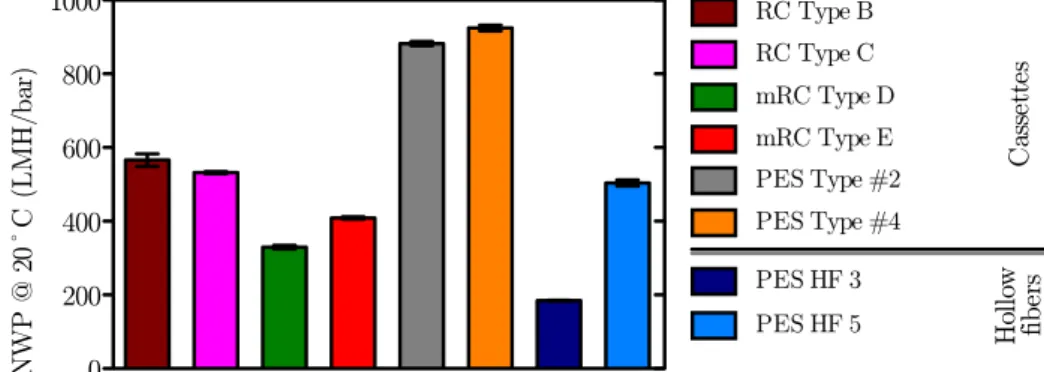
4.2.1 Hydraulic permeability . . . 38
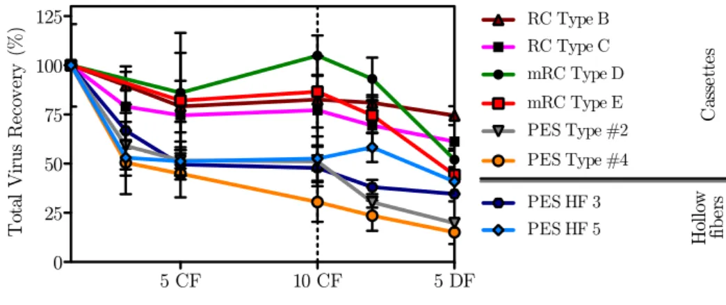
4.2.2 Virus recovery . . . 39
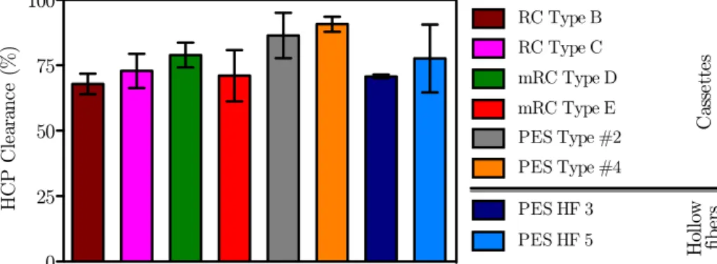
4.2.3 Impurity removal . . . 40
4.2.3.1 Protein clearance . . . 40
4.2.3.2 DNA clearance . . . 41
4.2.4 Productivity analysis . . . 42
4.2.5 Flux Recovery . . . 42
4.3 Discussion . . . 43
5 Membrane chromatography 47 5.1 Hydrogel-grafted membranes . . . 47
5.1.1 Virus recovery . . . 47
5.1.2 Impurity removal . . . 48
5.1.2.1 Protein clearance . . . 48
5.1.2.2 DNA clearance . . . 49
5.2 Directly grafted membranes . . . 50
5.2.1 Virus recovery . . . 50
5.2.2 Impurity removal . . . 51
5.2.2.1 Protein clearance . . . 51 5.2.2.2 DNA clearance . . . 51 5.3 Discussion . . . 52
IV Conclusions 55
6 General Discussion and Conclusion 57
6.1 Debottlenecking the DSP rigth from the beginning . . . 57 6.2 Future work and outlook . . . 58
Appendix 73
List of Figures
1.1 Structure of the Adenovirus by cross-section representation. . . 6
1.2 Structure of the Retovirus by cross-section representation. . . 7
2.1 Schematic representation of the hydrogel-grafted and directly grafted membrane. 23 3.1 TP recovery after incubation with different filter aids. . . 30
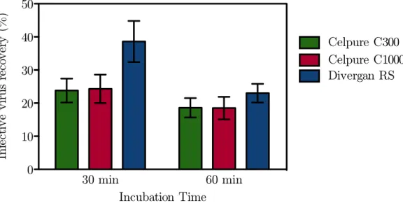
3.2 IP recovery after incubation with different filter aids. . . 30
3.3 DNA clearance after incubation with different filter aids. . . 31
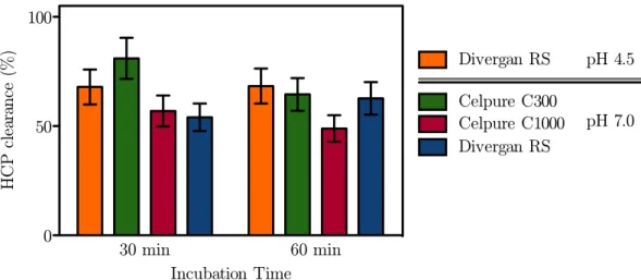
3.4 HCP clearance after incubation with different filter aids. . . 32
4.1 NWP20¶C for the different R&D UF prototypes . . . 33
4.2 TP recovery for the different R&D UF prototypes . . . 34
4.3 HCP clearance for the different R&D UF prototypes . . . 35
4.4 SDS-PAGE comparative analysis of the different R&D UF prototypes . . . . 36
4.5 DNA clearance for the different R&D UF prototypes . . . 36
4.6 Permeate flux and throughput capacity of each R&D UF prototypes . . . 37
4.7 Flux recovery for the different R&D UF prototypes . . . 38
4.8 NWP20¶C for the different UF devices . . . 38
4.9 TP recovery for the different UF devices . . . 39
4.10 HCP clearance for the different UF devices . . . 40
4.11 SDS-PAGE comparative analysis of the different UF devices . . . 41
4.12 DNA clearance for the different UF devices . . . 42
4.13 Permeate flux and throughput capacity of each UF devices . . . 43
4.14 Flux recovery for the different UF devices . . . 44
5.1 TP recovery for the hydrogel-grafted membrane. . . 48
5.2 Protein clearance for the hydrogel-grafted membrane. . . 49
5.3 DNA clearance for the hydrogel-grafted membrane. . . 49
5.4 TP recovery for the directly grafted membrane. . . 50
5.5 Protein clearance for the directly grafted membrane. . . 51
5.6 DNA clearance for the directly grafted membrane. . . 52
5.7 Experimental design space for the hydrogel-grafted membranes. . . 52
A1 Amount of the different filter aids used in dry state. . . 75
A2 Volume filed by the wet filter aid with 50 mL of virus bulk. . . 75
List of Tables
1.1 Specifications for biotechnological products. . . 5
2.1 Membrane characteristics and feed flow rates used. . . 22
4.1 IP recovery for the different R&D UF prototypes . . . 34
4.2 IP recovery for the different UF devices . . . 40
List of Symbols
ACRONYMS
Ad5 Adenovirus serotype 5
ATCC american type culture collection BFF body feed filtration
CEX cation exchange chromatography cGMP current good manufacturing practices CIP cleaning-in-place
D-PBS Dulbecco’s phosphate-buffered saline DE diatomaceous earth
DF diafiltration
DMEM Dulbecco’s Modified Eagle Medium EMA European Medicines Agency
FDA Food and Drug Administration GaLV gibbon ape leukemia virus GFP green fluorescent protein
HEPES 2-[4-(2-hydroxyethyl)piperazin-1-yl]ethanesulfonic acid
HF hollow fiber
HPV human papillomavirus mAb monoclonal antibody MF microfiltration
MLV murine leukemia virus MWM molecular weight marker PES polyethersulfone
pI isoelectric point
PS polysulfone
PVDF polyvinylidene fluoride
RC regenerated cellulose
RV retrovirus
SDS-PAGE sodium dodecyl sulphate polyacrylamide gel electrophoresis SEC size exclusion chromatography
SEM standard error of the mean TFF tangential flow filtration
TRIS tris(hydroxymethyl)aminomethane UF ultrafiltration
VLP virus-like particle
GREEK SYMBOLS
˙
γw shear rate (s-1)
η dynamic viscosity (Pa s) σ conductivity (mS cm-1)
τ throughput capacity (L m-2h-1)
ROMAN SYMBOLS
A area (m2)
C concentration
h flow channel height in a cassette module (m) J flux (LMH ≡L m-2 h-1)
K hydraulic permeability (m)
n number of fibers in a hollow fiber module Q volumetric flow rate (mL min-1)
r radius (m)
Re Reynolds number ( – )
t time (min)
v linear velocity (m s-1)
w flow channel width in a cassette module (m) Y recovery yield (%)
g gravitational acceleration at the Earth’s surface (9.81 m s-2)
Abs absorbance (AU)
CF concentration factor ( – )
Da molecular mass (Dalton≡ g mol-1)
DNA deoxyribonucleic acid (µg mL-1) HCP host cell protein (µg mL-1)
IP infectious particles (particles mL-1)
NWP20¶C normalized water permeability corrected for 20¶C (LMH bar-1)
P pressure (bar)
rpm rotationsper minute (min-1)
T temperature (¶C)
TCF temperature correction factor ( – ) TMP transmembrane pressure (bar) TP total particles (particles mL-1) WCW wet cell weight (g L-1)
SUBSCRIPTS AND SUPERSCRIPTS
f feed
i initial
p permeate
r retentate
Part I
1
Purification of complex
biopharmaceuticals
1.1
Complex biopharmaceuticals
Biopharmaceuticals are structured and highly specific biomolecules capable of targeting unmet medical needs where other drugs have failed. The success of these novel bio-based pharmaceuticals has been reshaping medical therapies over the past decades[1]. Nowadays there are numerous biopharmaceuticals (both approved and under development) with a wide range of sizes, levels of complexity and medical indications[2,3]. Some examples of approved biotherapeutics are the Factor VIII (blood factor), tissue plasminogen activator (antico-agulant/thrombolytic), insulin and insulin-derived products, erythropoietins, interferon-α, pegaptanib (nucleic acid-based anti-angiogenic medicine), monoclonal antibodies (mAbs), human papillomavirus (HPV) virus-like particle (VLP) vaccine, inactivated influenza virus vaccines and adeno-associated viral (AAV) gene therapy vector (Glybera®)[2,4].
Among the above-referred biomolecules virus-based biopharmaceuticals hold a great promise to redefine modern medicine in different fields such as vaccination[5], gene therapy[4] and cancer treatment[6]. However these biopharmaceuticals represent also a series of the challenges both product- and process-wise since their are not as well studied and developed as other less complex biopharmaceuticals already established.
1.1.1 The challenges of complex biopharmaceuticals
Virus and virus-like particle are considerably larger than an insulin protein or a mAb and their size can range from 20 nm (parvovius) to 300 nm (Measles and Mumps virus),
1. PURIFICATION OF COMPLEX BIOPHARMACEUTICALS 1.1. Complex biopharmaceuticals
with different possible morphologies (i. e. the rod-shape of Baculovirus)[7,8]. The highly repetitive tri-dimensional structure is fundamental for immunization in the case of vaccines. Additionally,post-translational modifications, like glycosylation, impact the immunogenicity and antigenicity of the vaccine[9,10]. For gene therapy applications the vector quality is crucial[11].
As a result of this complexity, animal cell lines have to be used often for production of these virus-based biopharmaceuticals. The new virus vaccines are now produced in cell-based systems. The traditional production with fertilized eggs has several drawbacks since it is a work-intensive process, is dependent on egg supply, is not fast enough in the case of a global pandemics and might cause anaphylactic reactions due to egg’s proteins[1,7,12]. Vaccines recently developed and under development make use of continuous mammalian cell lines such as the African green monkey kidney (VERO) cells[13,14], the human lung fibroblast (MRC-5) cells[15], Madin-Darby Canine Kidney (MDCK) cells[16], human embryonic retina-derived PER.C6®cells (Crucell)[17–19] and human embryonic kidney (HEK) 293[19,20].
The VLP-based vaccines might be produced by a wide range of hosts from mammalian cell lines to transgenic plants, however only VLPs produced in yeast and animal cell lines were approved by regulatory authorities[5,8]. The recombinant hepatitis B vaccines are produced almost exclusively in yeast systems (Saccharomyces cerevisiae,Pichia pastorisandHansenula polymorpha) with the exception of chinese hamster ovary (CHO) cells, which is the only
mammalian cell line used. S. cerevisiae is also used for production of the Merck & Co’s HPV-VLP[21]whereas the GSK’s HPV-VLP is produced with the baculovirus-insect cell (B-IC) system[22]. These expression systems seem to have a favorable compromise between high productivity/yield and capability to performpost-translational modifications[5].
Contrarily to VLPs, the gene therapy vectors are less flexible in terms of production system and the system chosen is dependent on the virus used. Ad-based vectors are produced in a complementing cell line (see Section 1.1.2), such as the 293 and the PER.C6® cell
lines[19,23]. In the case of AVV vectors, like UniQure’s Glybera® – recently approved by
EMA –, these can be produced in mammalian cell lines as well as with the B-IC system[24,25]. The current retrovirus production systems rely on genetically engineered virus packaging cell lines (see Section 1.1.3), usually human-derived cell lines[26,27].
As biopharmaceuticals are intended for therapeutic human use there are stringent guide-lines put in place by the regulatory agencies (e. g. EMA or FDA). The Table 1.1 reports the quality attributes evaluated by the regulatory authorities[28,29].
The analyticals for virus-based biopharmaceuticals must be highly specific and robust, however often new analytical assays must be developed and validated, specially for innovative therapeutic biomolecules. Additionally, the lack of well-characterized and accepted standards adds up to the challenges of developing new analytical assays for such specific products[1,31]. The final goal is to obtain a biopharmaceutical of highpurity(process-related impurities,
like HCP and DNA, within regulatory specifications), high potency(highest concentration
feasible of the relevant active component) and high quality(low concentration of product-derived impurities relatively to the desired product, e. i. virus capsid proteins or non
functional virions).
1. PURIFICATION OF COMPLEX BIOPHARMACEUTICALS 1.1. Complex biopharmaceuticals
Table 1.1: Specifications for biotechnological products[28–30].
Attribute Specification
Appearance and description
Color
Physical state Clarity/turbidity (Qualitative statement)
Identity
Multiple tests might be required: Physicochemical
Biological immunichemical (Highly specific but might be qualitative) Purity
Product-related impurities
Degradated product Truncated forms Molecular variants Aggregates
Process-related impurities
Host cell protein (HCP) Host cell DNA
Media components/ancillaries Enzymes/chemicals
Leachables
Potency Cell-based testAnimal-based testor
Quantity Protein massPotency (if applicable)or
Safety
Sterility
Adventitious virus Endotoxins/Pyrogens Mycoplasma
General pH
Osmolarity
Two highly relevant representatives of these virus-based biopharmaceuticals are aden-ovirus and retraden-ovirus.
1. PURIFICATION OF COMPLEX BIOPHARMACEUTICALS 1.1. Complex biopharmaceuticals
1.1.2 The Adenovirus - a stable proteic capsid particle
TheAdenoviridaefamily is composed of over 100 virus, 57 serotypes of which are capable
of infecting humans[32].
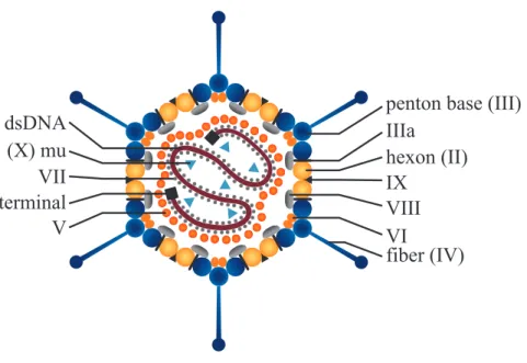
The adenovirus serotype 5 (Ad5) was the first adenovirus discovered when its icosahedral shape was reveled by Horne in 1959[33]. Since then it has been extensively studied, used as virus model and nowadays is widely characterized. The Ad5 is a nonenveloped double-stranded DNA virus with a molecular weight of 150-170 MDa (90-100 nm in diameter)[32,34,35], has a pI of approximately 4.5 and thus is negatively charged at physiological pH[36]. The virus is composed of 11 different proteins (Figure 1.1), 7 are the structural proteins (II, III, IIIa, IV, VI, VIII and IX) which compose the icosahedral virus capsids and the remaining 4 proteins (V, VII, mu and terminal protein) are packed inside the capsid together with the virus DNA to form the core[32,37,38].
The virus capsid is composed of twelve hexon trimers (polypeptide II - 107.9 kDa for Ad5) in each of the 20 facets and twelve pentons in each of the twelve vertices (Figure 1.1). Each penton is a protein composed of one pentamer, the penton base (polypeptide III - 63.3 kDa for Ad5), and one trimer, the fiber (polypeptide IV - 61.6 kDa for Ad5) which is responsible for the adsorption to the cell surface via the CAR (Coxsackie/Adenovirus receptor) and αv-integrins. The remaining (minor) coat proteins are involved in virus stability, correct assemble and disassemble of the virion[32,38,39].
V
terminal
VII
(X) mu
dsDNA
VIII
VI
fiber (IV)
IX
IIIa
hexon (II)
penton base (III)
Figure 1.1: Structure of the Adenovirus by cross-section representation. The coat proteins are listed on the right side and the core proteins and DNA are on the left side.
The application of Ad5 goes beyond fundamental research and its use as biotechnology product is very significant, either as gene therapy vector[40], oncolytic virus[41]or recombinant vaccines[42,43]. The best know examples are the use of recombinant p53-Ad5 approved in China for cancer therapy[44,45] and the Merck & Co’s candidate HIV Ad5-based vaccine
1. PURIFICATION OF COMPLEX BIOPHARMACEUTICALS 1.1. Complex biopharmaceuticals
(although clinical tests failed for Ad5-based vaccine, currently different, rarer Ad serotypes are being evaluated)[46,47].
However to use Ad-based therapies safely, these virus are genetically modified to render replication-incompetent vectors (or conditionally replication-competent virus for oncolytic therapy). To accomplish this several genes of the Ad5 genome were removed or mutated. Initially the genes targeted were involved in viral DNA transcription, inhibition of cell apop-tosis, avoiding host immune response and viral DNA replication[32]. More recently, helper-dependent Ad (also named gutless or high-capacity) were developed. Since these vectors have no viral DNA, a helper virus has to be used to provide all structural proteins[23,32,48,49].
For the production of replication-incompetent adenoviral vectors trans-complementing
cell lines must be used in order to provide the deleted functions. Several cell lines have been used for Ad production, with the 293 and the PER.C6® being the most used. Since the
Ad-producing cell lines can adapted to grow in suspension, stirred-tank bioreactors have been used with or without the aid of microcarriers[19,23].
1.1.3 The Retrovirus - a labile enveloped particle
TheRetroviridae is a family of viruses characterized by their RNA genome and its
retro-transcription into DNA prior to protein synthesis. Notable species of this family include the murine leukemia virus (MLV), belonging to thegamma-retrovirus genus (herein referred to as retrovirus (RV)) and the human immunodeficiency virus (HIV) which belongs to the
Lentivirus genus[50]. The RV is an enveloped, spherical, slightly pleomorphic particle with a
diameter of 100-120 nm (Figure 1.2). The virion structure is divided in three major parts, the
RT PR
RNA IN
CA NC MA
SU Env
Gag
TM
Lipid bilayer
Figure 1.2: Structure of the Retovirus by cross-section representation.
envelope, the proteic capsid and the viral genome. The envelope that covers the the proteic capsid is composed by a bi-lipidic layer and 100-300 Env glycoproteins (69.8 kDa) consisting of transmembrane (TM, 19.9 kDa) and surface (SU, 47.9 kDa) subunits. The SU subunit interacts with the cell membrane receptors to promote the fusion of the envelope with the cell membrane during the infection process. Given its function, the Env protein (and the virus
1. PURIFICATION OF COMPLEX BIOPHARMACEUTICALS 1.2. Current scalable DSP strategies
envelope) is of great relevance of highly infective retroviral preparations. The Gag protein (60.7 kDa) interacts both with the Env proteins by its N-terminal matrix domain (MA) and the inner space of the virus capsid with through its C-terminal nucleocapsid (NC) domain. Inside this proteic structure, besides the ssRNA molecule, there are also the PR (responsible from virus’ protein cleavages during assembling, budding and maturation), the RT (which does the reverse-transcription of the viral RNA) and the IN (responsible for integrating the pro-viral DNA) proteins[51,52].
Retroviral vectors are highly advantageous for gene therapy due to their high transduction efficiency of replicating cells, low immunogenicity and ability to integrate the proviral cDNA (with the therapeutic gene) into the host genome, rendering a long-term effect[48]. Moreover, RV are the second most used gene therapy vector in clinical trials worldwide (the first being adenovirus)[53].
Since retrovirus have a modular structure they can be pseudotyped, i. e. the env region
of the genome can be replaced by the env region of another, more favorable, retrovirus. The possibility to engineer the vector allowed to change, restrict ou broaden vector tropism and to modulate its immunogenicity. Among several Env proteins used, the MLV-based retrovirus pseudotyped with gibbon ape leukemia virus (GaLV) Env has been one of the most studied[26,27].
As earlier described for Ad5, also retroviral vectors being developed for human therapeutic purposes must be replication-incompetent. This is accomplished by providing the transgene of interest together with the packing signal in a different transcriptional unit(s) than the packing functions (env and gag-pro-pol). Current RV generations have three different transcriptional units (transgene,gag-pro-polandenv) with reduced sequence homology, thus reducing greatly the frequency of replication-competent RVs[26,27].
For retrovirus production the packing functions (i. e. structural proteins) can be supplied
either by a plasmid transfection (transient production) or by a packing cell line engineered to express constitutively those functions (stable production). While transient production is only suitable for small scale research purposes, the stable production includes the long process of developing a high titer packing cell line but yields a continuous low-variability production suitable for clinical lots. Currently, human cell lines, namely HEK-derived, are preferred to the murine cell lines for the development of retrovirus-producing cells[26,27,54].
The RV-packing cells are generally cultured in static systems due to their anchorage-dependent feature. High capacity static system such as the Cell Factory® have been reported
for clinical production of RV under current good manufacturing practices (cGMP)[55–57]. These kind of strategies enable a limited but rapid and reliable scale-up and might provide enough material for pre-clinical and early clinical phases.
1.2
Current scalable DSP strategies
The biopharmaceutical process is no longer seen as the sum of upstream with downstream processes and there is a effort to integrate and design together production and purification. Despite this, the downstream process is still crucial to obtain the final intended product; also the substantial developments achieved in the upstream process were not matched by small
1. PURIFICATION OF COMPLEX BIOPHARMACEUTICALS 1.2. Current scalable DSP strategies
improvements in the downstream. Furthermore, the cost of purification can go from 50 % up to 70 % of the total process[58,59]. Meaning that careful choice of operations, materials and equipment should be made to in order to keep the whole process cost-effective. Besides cost, the scalability of the DSP developed is a key issue. Robustness is also a required feature so that small variations in upstream processing as well as in previous downstream operations do not impact the whole bioprocess performance and final product quality.
The following section describe the state-of-the-art regarding virus purification including novel technological advances, emerging industry trends and rational process development approaches for different stages of the DSP train. Some examples are the greater integra-tion between upstream and downstream, high density cell systems, continuous processing, scale-down tools closed systems, modulatiry, numbering up instead of scale-up (for niche biopharmaceuticals) and disposable technologies[60–64].
1.2.1 Harvest
The first steps of the purification train are heavily influenced by the bioreactor bulk fea-tures, namely cell density, cell viability or if the product is secreted for the culture supernatant or if the cells have to be lysed.
In cases such as Ad5 production, is usually chosen to recover both the intra- and extra-cellular virus faction and thus an additional step is performed for virus release[19,23]. The cell lysis can be performed using different methods such as freeze-thaw, detergents, French Press, homogenizer or sonication[65]. Among these, non-ionic detergents especially Triton™X-100 have been preferred[18,19,65–67]. Incubating the bulk with detergent is efficient, fast, robust, easily scalable, cost-effective and does not require any investment in equipment, however removal of this additive has to be confirmed.
Although cell lysis enables the recovery of intra-celullar virus it also releases the host cell DNA and protein which should be removed from the final product. Host cell DNA is of special concern because it increases substantially bulk viscosity but also due to regulatory requirements. The DNA acceptable levels set by the regulatory authorities are generally between 10 ng and 10 pgper dose, depending on type of product, medical indication, pro-duction host and administration route. Therefore, the majority of scientific manuscripts published and patents disclosed refer an incubation step with nuclease (e. g. Benzonase®)
either simultaneously with cell lysis or after clarification[18,19,65–68]. For enveloped virus, like RV or baculovirus, cell lysis is not required since the virus are secreted by the producer cells – budding process. Therefore, the use of nuclease might be avoided for enveloped virus DSP due to its high costs[54,69].
Selective precipitation has been suggested as an alternative to nuclease treatment to remove host cell DNA[17,70–72]. Cationinc detergents (namely domiphen bromide) are able to precipitate DNA as well as Ad particles (both negatively charged and mildly hydrophobic). However fine adjustment of precipitant concentration enables up to 90 % DNA removal with more than 90 % Ad recovery[70]. Selective DNA precipitation has the advantage of being inexpensive, easily scalable (precipitated DNA can be removed by depth filter) and suitable for high cell density processes[17,71].
1. PURIFICATION OF COMPLEX BIOPHARMACEUTICALS 1.2. Current scalable DSP strategies
1.2.2 Clarification
The goal of clarification is to recover and stabilize the product while removing solids, cells/cell debris and process impurities (DNA and HCP). Centrifugation and membrane fil-tration are the two common choices for the initial clarification.
Centrifugation is an operation widely used at laboratory scale for research purposes both to clarify the cell culture fluid (CCF) and to obtain high purity viral preparations (by CsCl gradient ultracentrifugation). The centrifugal separation is based on the solutes’ different densities meaning that clarification is generally a fast process. However, centrifugation has several issues like the high maintenance costs, the lack of reliable scale-down model, does not remove soluble impurities and safety risks must be considered (for instance the Ad5 forms aerosols)[19,68,73]. Despite the earlier described limitations, continuous flow centrifugation are quite common in current industrial-scale operations for mAbs clarification where biore-action yields very high cell density CCFs (107 cell mL-1 and above). However in these cases the clarification also comprises a depth filter operation to achieve higher removal of soluble impurities (DNA and HCP) and low turbidity[74–77].
Microfiltration (MF) is an alternative to centrifugation which can be easily scaled up and implemented in a cGMP setting. Microfiltration membranes have pore sizes in the 0.1–10µm range and can be classified as depth filters or "regular" filter depending on whether the solids are retained throughout filter matrix/medium or only at its surface, respectively. Addition-ally, depth filter might include filter aids,e. g. diatomaceous earth (DE), to modify its struc-ture and charge[78]. Depth filters have been used for a wide variety of VLPs and viruses (en-veloped and non-en(en-veloped) with recovery yields of approximately 90 %[66,67,69,79,80]. Depth filters are usually preferred for feed streams with high biomass contents, sometimes lysed CCF, due to their ability to remove solids and impurities by different mechanisms (size ex-clusion, hydrophobic and electrostatic interactions)[81]. Carefull selection of the filter (pore size, filter material, number and sequence of filter units) and operation parameters (inlet flow rate, pressure drop, conductivity and pH) must be assessed in order to avoid virus losses and to allow solids and soluble impurities retention throughout the whole depth of the fil-ter medium rather than only at the filfil-ter surface. Often the trade-off between feed flow rate/throughput and filter capacity/impurity removal should be evaluated[77,82].
Membrane microfiltration has also been performed either in normal (dead-end) or tangen-tial flow filtration (TFF)[79,83]. Microfiltration is usually operated at low pressures, especially TFF (less than 0.7 bar), and is better suited for low cell density CCFs[74]. However, mem-brane fouling or shear stress-induced cell lysis can become an issue[84].
More recently, a new microfiltration separation was purposed where a filter aid is mixed with the CCF instead of being incorporated in the depth filter units[85,86]. The CCF and filter aid (diatomaceous earth, layered double hydroxides or synthetic materials) are mixed and fed to a microfilter. This novel filtration would permit an enhanced adsorption during mixing and the formation of highly porous and highly permeable cake (body feed) supported by the filter aid, similarly to what is done in the brewing industry where diatomaceous earth is used to clarify beer[87]. Several patents applications claim that the body feed filtration (BFF) would avoid membrane fouling, enable lower pressure operation for longer times at
1. PURIFICATION OF COMPLEX BIOPHARMACEUTICALS 1.2. Current scalable DSP strategies
higher flow rates without compromising impurity removal. One paper recently published[88] referrers the use of DE for clarification of a poliovirus CCF, however the performance of this method was not disclosed and its potential remains unexplored.
1.2.3 Concentration
Large-scale process can produce high volumes of CCF (up to 20 kL for mAbs[89]) which must be concentrated 10–100 times to be further purified by chromatographic steps. Ul-trafiltration (UF) is a pressure-driven separation which employs anisotropic membranes with molecular weight cut-offs (MWCO) ranging from 0.5 to 1000 kDa[84]. (The MWCO is defined by the solutes’ molecular mass which are 90 % retained by the membrane.) These membranes can be constructed with different polymers like regenerated cellulose (RC), polysulfone (PS), polyethersulfone (PES) and polyvinylidene fluoride (PVDF), although RC and modified RC displays better trade-offbetween low (unspecific) protein binding, mechanical strength and resistance to cleaning procedures (chemical agents and temperature)[74,90,91]. Ultrafiltration membrane are generally composed by two main layers, a thick macroporous structure pro-vides mechanical strength while a thin skin layer is responsible for membrane selectivity and permeability[74].
Ultrafiltration is usually performed in TFF mode where the cross flow at the membrane surface creates a "sweeping action" that avoids or lessens concentration polarization and gel layer formation. UF processes are usually performed at constant transmembrane pressure (TMP, see equation 2.5, page 20), however constant permeate flux or constant permeate pressure operation are also feasible[74]. Ultrafiltration has been widely used both in concentartive mode as well for buffer exchange (diafiltration, DF) step and is present in almost every virus DSP described in the literature[79,83,92–97] and patents disclosed[18,71,98]. The membranes used in virus UF have MWCO in the range of 100–750 kDa allowing for high virus recovery (70–100 %). The TMP generally used is between 0.5 and 1.4 bar but the optimum cross flow can vary greatly due to different structural stability of virus; enveloped virus are more labile than non-enveloped virus and thus more prone to shear-induced damage[83,97]. The membrane modules might be constructed in different arrangements, for example flat sheet cassette and hollow fibers. However the majority of the reports published referrers the use of hollow fibers (HF) modules for virus processing[7,99] due to the fact that HF modules provide wider flow paths resulting in lower shear rates[7,84]. Shear rate is proportional to the linear velocity of the feed and inversely proportional to the flow channel diameter or height (see equation 2.9, page 21 for further details). The flow channel geometry influences the shear rate two ways: firstly, the wider flow channel result in lower linear velocity for the same volumetric flow rate; and secondly, the wide flow channel results itself in lower shear rates. Although UF cassettes results in greater shear rates, these modules provide shorter processing times as the flow channel hydrodynamics are more effective than HF in avoiding concentration polarization and gel layer formation[7,84].
Besides UF, rediscovered techniques like aqueous two-phase systems[100,101] and precipi-tation[102–104] have been suggested for early-stage operations in mAb DSP, however its ap-plicability and efficacy remains unclear for virus-based biopharmcaeuticals[105].
1. PURIFICATION OF COMPLEX BIOPHARMACEUTICALS 1.2. Current scalable DSP strategies
1.2.4 Intermediate purification
The purification step(s) employs high resolution unit operations capable of removing impurities closely related with the product, being chromatography the best example. Purifi-cation scientists have relied mainly on ion-exchange and size exclusion chromatography to process virus particles[23,26,92,106], although other techniques like affinity[92,99], hydrophobic interaction (HIC)[107,108] and multimodal[67] have been suggested.
Ion-exchange (IEX) chromatography is the method of choice for capture step. The virus-containing feed is loaded in the chromatographic matrix under low or moderate conductivity and recovered upon a conductivity increase of the mobile phase. The functional groups Q (quaternary amine) and DEAE (diethylaminoethyl) are common choices for this bind/elute (or positive) mode chromatography[23,99]. Accurate process development enables highly se-lective AEX chromatography capable of discriminate between whole virus and virus capsid proteins[109] or different adenovirus serotypes[110,111].
Despite being widely used, packed-bed chromatography has several disadvantages: the pressure drop is high and increases during operation due to matrix deformation or pore obstruction; scale-up requires changes in the column geometry making scale-up problematic; the mass transfer is slow since it is limited by pore diffusion; the capacity is limited to the ligand available at bead surface since the small pores (10–400 µm) will exclude the virus particles. Additionally, the diffusion-limited transport greatly reduces binding capacity at high flow rates (20–500 column volumes per hour)[112–114].
Membrane chromatography, in which a macroporous membrane serves as support to bound functional ligands, has the potential to overcome the limitations of the traditional chro-matography resins[114]. Monoliths – a single polymer block with a network of functionalized flow paths highly interconnected – have also been suggested as an alternative for packed-bed chromatography[115]. The nearly-convective mass transport enables operation at flow rates up to 500 membrane volumes per hour without decreasing binding capacity – reducing pro-cessing times. The wide porous (up to 3 µm) allow lower pressure drops and compact design compared with packed-bed chromatography reduce hardware requirements and facility foot-print. Scale-up is greatly facilitated due to the modular design of chromatography supports. Since membranes adsorbers are single-use devices, column (re)packing, cleaning and valida-tions are avoided. The advantages referred above enable savings with the consumables and labor costs[62,116,117], despite some limitations regarding membrane chromatography. While multilayer membrane modules mitigated the distorted inlet flow, uneven membrane thick-ness and uneven pore size of the first membrane adsorbers some issues still remain. The major drawback of membrane adsorbers is lower binding capacity compared with packed-bed columns due to the lower surface-to-volume ratio[113,114]. AEX membrane adsorbers were first developed as flow-though (negative mode) chromatography for polishing step in mAbs DSP to capture virus, DNA and HCP[118]. However when used for virus purification, the product recovery (desorption) and removal of closely related species (e. g. DNA) is limited.
Consid-ering these drawbacks optimization of ligand density and membrane surface tri-dimensional structure were suggested to overcome the referred limitations[119].
Another different option to improve chromatography is the use of radial columns, where
1. PURIFICATION OF COMPLEX BIOPHARMACEUTICALS 1.3. Scope of the thesis
the chromatographic resin is packed in a cylinder form. The advantage of this columns lie in the radial flow design which enables a small bed length while attaining high column volumes. This feature results in low and uniform pressure drops across the packed-bed allowing for increased flowrates (and productivity), small equipment foot-print and linear scale-up since column geometry is kept[120,121].
Recently, Genzyme and Genentech’s scientists have developed a continuous process for purification of mAbs and recombinant therapeutic enzymes using HIC and affinity chromatog-raphy[63,64,122]. In the conventional batch operation the chromatographic matrix is loaded until product breakthrough is detected at the outlet (i. e. 5 or 10 % of feed concentration). This means that only a fraction (between 30 and 70 %) of the chromatographic resin capacity is used because breakthrough happens before the matrix gets fully loaded. In the described periodic counter-current chromatography (PCC) process the breakthrough effluent is loaded onto a second column enabling loading the first column until exhaustion (up to static binding capacity). This strategy enables, reduced buffer consumption, shorter processing times and better matrix usage (with concurrent investment and facility footprint savings)[63,64].
1.2.5 Polishing
The final step of purification aims to remove small quantities of impurities and to ex-change the buffer for the final formulation. Ultrafiltration and size exclusion chromatogra-phy (SEC) have been used for this purpose although ultrafiltration is preferred for large-scale manufacture[68,71,106] Buffer exchange by size exclusion chromatography is performed in way such that virus particles are excluded from the resin while smaller size molecules (buffer, HCP and DNA) and ions are retained[117]. However, due to the diffusion-limited fractiona-tion mechanism and compressible nature of SEC resin, low flow rates have to be used thus resulting in low productivity. Furthermore, scale up of SEC is limited by technical and eco-nomic constraints[74,123]. On the other hand, ultrafiltration is easily scalable and allows for buffer exchange of high volumes. Additionally, some components of formulation buffers (e.
g. glycerol or trehalose[124,125]) that increase the viscosity can be incorporated in
diafiltra-tion buffers contrarily to chromatography. However TMP, shear rate, diafiltration number, MWCO and membrane material must be carefully optimized for optimal product recovery and short processing time. Hydrophobic interaction chromatography (HIC) was recently sug-gested to remove trace amounts of HCP and DNA, however the high conductivity required limits its application to virus sensitive to high conductivity[107,108,126].
1.3
Scope of the thesis
Virus are biopharmaceuticals holding a tremendous potential for vaccine and gene therapy applications. Despite recent research efforts have targeted purification of complex biological particles, virus downstream processing is still a challenging field. The product characteristics – virus lability/stability or large size – limit the unit operations that can be used. Further-more, the upstream process employed results in closely related impurities that have to be removed in order to achieve clinical-grade preparations. Purification devices and strategies
1. PURIFICATION OF COMPLEX BIOPHARMACEUTICALS 1.3. Scope of the thesis
especially designed for virus purification are lacking or need further optimization; addition-ally most of the research and development (R&D) has been focused on the chromatographic step – considered the major bottleneck of the purification train. As a result, the literature thoroughly evaluating non-chromatographic operations for virus DSP is scarce. This thesis aims to improve the state-of-the-art virus purification by evaluating essential but less studied separations – clarification and ultrafiltration – as well as other better characterized DSP steps – membrane chromatography.
Clarification by depth filter is a technology easily scalable, capable of removing soluble impurites and suspended solids. However limitations such as low filter capacity and low flow rates have been identified[77,82]. In Chapter 3 a novel clarification process based on body feed filtration (or cake filtration) concept was evaluated. For this purpose different filter aids (DE and synthetic polymer; charged and non-charged) were assessed for clarification of an Ad5 bioreactor bulk.
Ultrafiltration is present in all large-volume virus purification schemes. Virus concen-tration and diafilconcen-tration is usually performed using hollow fiber UF modules. Despite its generalized use, HF are known to be slower and more prone to fouling when compared with cassette modules. Considering the advantages of cassette UF, a study with R&D UF cassette prototypes was performed in the first part of Chapter 4. In the second part of the chapter, a comparative evaluation between commercial HF and late-stage cassette devices is described. Two different virus (Ad5, non-enveloped, and RV, enveloped) were used as models to char-acterize membrane MWCO and filter material and its impact on virus recovery, impurity removal and processing time.
Membrane adsorbers have several advantageous features comparing with the traditional packed-bed column chromatography. However, further improvements are required to increase the adoption of this technology. The last part of this thesis (Chapter 5) reports the use of a scale-down tool to evaluate how conductivity of the mobile phase, ligand density and structure of an IEX membrane affect a chromatographic purification of Ad5.
Overall, this thesis aims to evaluate the potential of enhanced and innovative technologies to improve productivity and reduce cost-of-goods in the whole DSP train.
Part II
2
Materials and Methods
2.1
Cell lines, culture media and virus strains
The 293 cell line (ATCC CRL-1573) was used for adenovirus production as well as infec-tious virus particles (IP) titration. A recombinant replication-defective adenovirus serotype 5 harboring a transgene for the green fluorescent protein (GFP) were used; the virus stock was kindly provided by Professor Stefan Kochanek, University of Ulm, Germany. The recombinant RV was produced in 293-derived cell line gene genetically engineered to provide constitutively the packing functions required for RV assembly as described elsewhere[127]. The RV produced is derived from the murine leukemia virus (MLV) with the Gibbon ape leukemia virus (GaLV) ecotropic envelope and harbors a GFP reporter gene. The 293 T cell line (ATCC CRL-11268) was used for infectious RV titration.
The 293 and 293 T cell lines were grown in cell culture flasks (353112, BD Falcon™, USA) with Dulbecco’s Modified Eagle Medium (DMEM, 41966-052, Gibco®, UK) supplemented
with 10 % (V/V) Fetal Bovine Serum (FBS, 10270-106, Gibco®, UK). For the 293 Flex GFP
the medium used was DMEM without phenol red (31053-036, Gibco®, UK), supplemented
with 10 % (V/V) FBS and 4 mM L-Glutamine (25030-024, Gibco®, USA). The 293 cells
were grown in a humidified atmosphere of 5 % CO2 at 37¶C, whereas the Flex 293 and 293 T were grown in a 8 % CO2. The cells were routinely propagated twice a week using 0.05 % Trypsin-EDTA (25300-104, Gibco®, UK). The cell concentration and viability were
determined by counting cells on a Fuchs-Rosenthal haemocytometer (Brand, Germany) using the dye exclusion method with 0.1 % trypan blue (15250-061, Gibco®, UK).
For the suspension culture system (i. e. bioreactor production, see section 2.2.2) the 293 cells adapted to suspension were grown with the commercially available serum-free cul-ture medium Ex-Cell 293 (14570C, SAFC®, USA) supplemented with 4 mM L-Glutamine
2. MATERIALS ANDMETHODS 2.2. Virus production
(25030-024, Gibco®, USA). The cells were routinely propagated twice a week using an
inocu-lum of 0.5×10+6 cell mL-1. Erlenmeyer shake flasks (431255, CORINING, USA) were used
for suspension cell culture in a humidified atmosphere of 8 % CO2 at 37¶C. Agitation was provided by orbital shaking at 110 rpm.
2.2
Virus production
2.2.1 Ad5 stock production
For the small scale virus production, ten 175 cm2-cell culture flasks (353112, BD Falcon™, USA) with 80-90 % confluence were infected with a previously purified and characterized viral stock using a multiplicity of infection (MOI) of 5.
2.2.1.1 CsCl gradient purification
After 48 h, the infected cells were centifuged at 1 500g for 5 min, ressuspended in lysis
buffer (10 mM TRIS, pH 8.0, 0.1 % (V/V) Triton X-100) and vigorously shaken. The cel-lular debris were removed by centrifugation at 1 000 g for 10 min and the virus-containing
supernatant was collected. This fraction was further purified by two rounds of CsCl ultra-centrifugation (Optima™LE-80K, SW 41 Ti rotor both BECKMAN COULTER, USA). The first round consisted in a discontinuous CsCl ultracentrifugation (1.25/1.45 kg/L density) at 151 000 g for 90 min. The visible virus band was extracted and processed by a continuous CsCl gradient ultracentriguation (1.32 kg/L) at 151 000 g for 18 h. After the second of
ul-tracentrifugation, the virus band collected was purified by SEC (HiPrep™ 26/10 Desalting, 17-5087-01, GE Healthcare Life Sciences, Sweden) in order to change for the formulation buffer (10 mM TRIS, pH 8.0, 2 mM MgCl2, 0.5 M trehalose). The purified virus stock was sterilized with 0.2 µm filter (4554, Pall, USA), aliquoted and stored at -80¶C until further
use.
2.2.2 Ad5 bioreactor production
The Ad5 production was performed in a 5-L working volume bioreactor (Sartorius Ste-dim Biotech, Germany). The dissolved oxygen was controlled at 50 % air saturation by a N2/O2/air mixture delivered by a sparger. The aeration rate was 0.01 vvm (vessel volumes
per minute). The pH-value was controlled at 7.2±0.05 by aeration with CO2 in the gas mix-ture and by base addition (1 M NaHCO3). The temperature was controlled at 37¶C using an external water-filled jacket. Mixing was provided by two 6-segment Ruston impellers with the agitation rate controlled between 60 and 210 rpm.
The bioreactor inoculum was 0.5×10+6cell mL-1, the cell concentration at infection (CCI)
was 1×10+6 cell mL-1and a MOI of 5 was used. The bioreactor was harvested 48 hourspost
infection (hpi).
2. MATERIALS ANDMETHODS 2.3. Virus purification
2.2.3 RV production
For RV production twenty 175 cm2-cell culture flasks (353112, BD Falcon™, USA) were used to expand the 293 Flex GFP cell in the serum-supplement medium. When the cell confluence reached 80-90 % the medium was replaced by 25 mL of serum-free medium. The cell culture supernatant was collected 24 h after medium exchange.
2.3
Virus purification
2.3.1 Harvest and Clarification
After Ad5 bioreactor harvest, the cells were lysed by adding Triton X-100 (X100, SIGMA-ALDRICH®, Switzerland) to a final concentration of 0.1 % (w/w).
Simultane-ously, Benzonase® (101654, Merck Millipore, Germany) was added to a final concentration
of 50 U/mL. The virus-containing bioreactor bulk was incubated at 37¶C for 2 h.
Clarification of Ad5 bulk was performed resourcing to a Sartopore® 2 filter with
0.8 + 0.45 µm pore size (5445306G8 -- OO, Sartorius, Germany). Before filtration the mod-ule was primed with three capsmod-ule volumes of TRIS buffered saline, pH 8.0 (TBS; T6664, SIGMA-ALDRICH®, Switzerland). The virus-containing bulk was loaded to the filter at a
constant flow rare equivalent to 150 LMH using a Tandem 1082 Pump (Sartorius Stedim Biotech, Germany).
Cell lysis and nuclease incubation was not performed for the RV-containing bulk since the these enveloped virus were collected in the cell culture medium. The RV-containing super-natant was clarified using a 0.45 µm vacuum-driven filter (SCHVU05RE, Merck Millipore, Germany). The filter was conditioned with 500 mL of TBS, pH 8.0 before virus microfiltra-tion.
2.4
Body feed filtration
For the screening four filter aids were tested:
Celpure® C300 (Advanced Mineral™, USA)
Celpure® C1000 (Advanced Mineral™, USA)
Celite® (World Maetrials, USA)
Divergan® RS (BASF, Germany)
All the filter aids were kindly provided by Dr. Franziska Jonas (Downstream Process Technologies, Sartorius Stedim Biotech, Germany)
The screening procedure consisted in incubating 10 g of each of the filter aids with 50 mL of Ad5-containing lysed cell bulk at pH 4.5 and pH 7.0 (the bulk used was not treated with Benzonase® nor clarified in any way). Before adding the lysed cell bulk to the glass beakers
with the filter aids a sample was retrieved, the was measured (MM40+, CRISON, Spain) and the turbidity was determined (2100Qis Portable Turbidometer, HACH®, Germany). The
2. MATERIALS ANDMETHODS 2.5. Ultrafiltration studies
mixtures were incubated at 20-22¶C under constant and gentle stirring. After 30 min and
60 min of incubation the stirring was stopped (allowing the filter aid to settle by gravity) a sample was collected.
Upon sample analysis the TP and IP recovery yield (YTP and YIP) were determined accordingly with equation 2.1 and 2.2, respectively.
YVP= CVPV CVP,iVi
(2.1)
YIP= CIPV CIP,iVi
(2.2)
Impurity – i. e., DNA and HCP – clearance (%) were also assessed and calculated as
defined by equation 2.3 and 2.4, respectively.
DNA Clearance= 1− CDNAV CDNA,iVi
(2.3)
HCP Clearance= 1− CHCPV CHCP,iVi
(2.4)
2.5
Ultrafiltration studies
The ultrafiltration devices were kindly provided by Dr. Tobias Schleuss (Membrane R&D, Sartorius Stedim Biotech, Germany). The membranes modules were set up accordingly with the manufactures’ instructions. Briefly, a Tandem 1082 Pump (Sartorius Stedim Biotech, Germany) was used to feed the clarified bulk to the membrane device, the retentate was recycled to the feed container and the permeate was collected separately. The TMP (see equation 2.5) was controlled by adjusting the retentate flow rate using a flow restriction valve. Sartocon Slice 200 Filter Holder was used (#17525 --- 01, Sartorius Stedim Biotech, Germany) for cassette devices. MasterFlex®14 tubing (MasterFlex Group, Germany) with
1.6 mm internal diameter was used. The pressure was monitored at feed inlet, retentate outlet and permeate outlet by in-line pressure transducers (080-699PSX-5, SciLog®, USA).
Retentate temperature was measured with thermocouple sensor (56-4110-05 and PRT-DPM-3T/50, GE Healthcare Life Sciences, Sweden). The feed/retentate and the permeate volumes were monitored using a technical scale (TE4101, Sartorius Stedim Biotech, Germany).
TMP= Pf +Pr
2 −Pp (2.5)
Before any experiment the membranes were thoroughly rinsed with ultrapure water (Grade 1, as defined in ISO 3696) to eliminate trace preservatives. Membrane permeabil-ity was determined by the normalized water permeabilpermeabil-ity corrected for 20¶C (NWP
20¶C,
LMH bar-1
≡ L m-2 h-1 bar-1). The NWP20¶C (equation 2.6) was calculated based on pure
2. MATERIALS ANDMETHODS 2.5. Ultrafiltration studies
water permeate fluxes (JP) measured at five different TMP between 0.4 and 2.0 bar.
NWP20¶C=
K η =
Jp
TMPTCF (2.6)
The NWP was corrected for 20¶C using the temperature correction factor (TCF,
equa-tion 2.7) based on water dynamic viscosity (η)[128].
TCF= ηT¶C η20¶C
(2.7)
This measurement was done before the experiment, after the experiment and after CIP. After conditioning the UF system with diafiltration buffer (20 mM TRIS, pH 8.0, 25 mM NaCl), 450 mL of clarified feedstock containing Ad5 or RV were concentrated 10-fold and then were diafiltered five times. The UF/DF test was performed at a constant pressure of 1.2 bar and at a constant feed flow rate (cross-flow) equivalent to a linear velocity (v) of 0.202 m s-1 (see Table 2.1). For the RV ultrafiltration with Type H membrane it was not possible to achieve the TMP of 1.2 bar while maintaining a recirculation flow rate appropriated for TFF. Therefore, exceptionally in this case the performance test was run at a TMP of 0.9 bar. The linear velocity used was kept constant for all experiments in order to have the same tangential flow/force. Unless stated otherwise, the membranes used for RV were coated before UF experiment with 40 mL of 4 g L-1 human serum albumin (HSA) prepareted in buffer 1. The HSA was recirculated at 120 mL min-1 for 10 min.
Throughout the filtration process samples of the retentate were collected and stored at -80¶C for further analysis.
The permeate flux was recorded by means of gravimetric control and membrane through-put capacity – τ, the amount of feed processed within a period of time (t) using a given membrane area (A) – was determined according to equation 2.8.
τ = Vi
At (2.8)
Also the shear rate at the wall (γw˙ ) under laminar flow conditions (Reynolds number, Re, < 2100 ) for HF and cassette modules were calculated as defined in equation 2.9 and 2.10[129], respectively.
˙ γw =
8v d =
4Q
nπr3 (2.9)
˙
γw = 6v h =
6Q
wh2 (2.10)
The CIP procedure consisted of washing the UF system with 1 M NaOH at a flow rate of 500 mL/min and then a 60-min incubation. After this treatment the system was rinsed with ultrapure water until the outlet stream reached pH 7.
For the Ad5 runs, all procedures were conducted at 20–22¶C, while fro RV runs all
pro-cedures were performed at 8–14¶C.
2. M AT E R IA L S A N D M ET HODS 2.5. Ultr afiltr ation studies
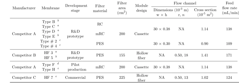
Table 2.1: Membrane characteristics and feed flow rates used.
Manufacturer Membrane Developmentstage Filter material
Filter area (cm2)
Module design
Flow channel Feed
flow rate (mL/min) Dimensions (10-3 m) Cross section
(10-5 m2
)
w×h r, n
Competitor A
Type B b
R&D prototype
RC
200 Cassette
30× 0.38 NA 1.14 138
Type C a
Type D a
mRC Type E b
Type # 2 c
PES 30× 0.30 NA 0.90 109
Type # 4 c
Competitor B HF 3
a
R&D
prototype PES 155 Hollowfiber NA 0.50, 18 1.41 171
HF 5 b
Competitor A Type F
d
Pilot
production mRC 200 Cassette 30 × 0.38 NA 1.14 138
Type H e
Competitor C HF 7 e
Commercial PES 225 Hollow
fiber NA 0.50, 13 1.02 124
RC: regenerated cellulose;
mRC: regenerated cellulose modified with a highly hydrophilic cross-linking; PES: polyethersulfone;
NA: not applicable/not available;
a,b,c,d ande: The precise MWCO was not available. However it was disclosed the relative MWCO within a MWCO range; a,bandcwere, respectively, the prototypes of low, medium and high MWCO considering a 300–1000 kDa range;
d andewere, respectively, the prototypes of low and high MWCO considering a 500–750 kDa range;
w and h are the cassette flow channel width and height, respectively; r andnare the hollow fiber internal radius and fibers number, respectively.
2. MATERIALS ANDMETHODS 2.6. Membrane chromatography
2.6
Membrane chromatography
Membrane sheets and 96-well membrane holder were kindly provided by Dr. Louis Villain (Membrane R&D, Sartorius Stedim Biotech, Germany). The membrane adsorbers func-tionalized with a quaternary amine (Q) had three ligand densities and two different surface structures, hydrogel-grafted and directly grafted (see Figure 2.1). The hydrogel-grafted
mem-(a)
(b)
Figure 2.1: Schematic representation of the two membrane structures: (a) hydrogel-grafted membrane where the Q ligand (red dots) is immobilized in the tentacle-like structure; and (b) directly grafted membrane where the ligand is immobilized directly onto the membrane surface. Experimental design space for the hydrogel-grafted membranes.
brane had ligand densities of 1.7, 2.4 and 3.3µmol cm-2, while the directly grafted membrane had 0.5, 2.5 and 4.5 µmol cm-2. The small-scale device had a bed volume of 23 µL and 500µL of buffer or virus (clarified, concentrated and diafiltered by UF) were used. The chro-matography consisted of three steps: equilibration; virus load, and elution. The equilibration buffer was 50 mM HEPES, pH 7.5 with different NaCl concentrations (0, 50, 100, 150 and 200 mM). The virus feed was diluted 1:2 with concetrated buffer to match the conductivity of the equilibration buffer. Elution was performed with 50 mM HEPES, pH 7.5, 1.0 M NaCl. After depositing the equilibration buffer inside the wells a vacuum of 0.35 bar was applied to displace the liquid across the membrane bed; the same procedure was applied for virus load and elution steps. The scale-down chromatography was performed in duplicate and the samples were stored at -80¶C until further analysis.
2.7
Analytical methods
2.7.1 Total virus particles quantification
Total virus particles concentration and size distribution were measured using the NanoSIGHT NS500 (NanoSIGHT Ltd, UK). The samples were diluted in D-PBS (14190-169, Gibco®, UK) so that virus concentration would be in the 10+8–10+9 particles mL-1– the instrument’s linear range. For each sample three 60-second videos were acquired and particles between 70 and 130 nm were considered.
2.7.2 Infectious virus particles titration
The infectious virus assay for both Ad5 and RV is based on GFP transgene expression upon infection (transfection) and flow citometry analysis.
For Ad5 titration, 293 cells were seeded at 0.25×10+6 cellper well in 24-well flat bottom
plates (42475, Nunc, Denmark). After 24 h, the cells from three wells were trypsinized
![Table 1.1: Specifications for biotechnological products [28–30] .](https://thumb-eu.123doks.com/thumbv2/123dok_br/16671241.742718/29.892.187.718.131.833/table-specifications-for-biotechnological-products.webp)