K I D N E Y B I O P S Y I N L U P U S N E P H R I T I S
:
S E I Z E T H E O P P O R T U N I T Y
Derek M. Fine
Michelle Petri
A B S T R A C T
Lupus nephritis is common in systemic lupus erythematosus (SLE), occurring in up to 60% of affected adults during the course of their disease. The histopathologic manifestations of lupus nephritis are diverse and determine not only the diagnosis and prognosis, but also substantially guide management of this com-plex disease. As the armamentarium available to treat lupus nephritis expands it becomes even more im-perative that the correct diagnosis be made prior to instituting therapy. When deciding whether to pursue kidney biopsy we need to balance the risks of limited diagnostic information, which may include poten-tially preventable progression of renal disease or the avoidable use of a possibly toxic therapy, with the risk of a kidney biopsy. With the advances in imaging technology and biopsy needle mechanics kidney, biop-sies have become safer and generally well tolerated. The combination of safer biopbiop-sies with a greater need than ever for correct diagnosis has made the risk/benefit for performing renal biopsies even more favora-ble in most cases of new kidney involvement in systemic lupus erythematosus.
Key words: Systemic Lupus Erythematosus; Kidney Biopsy; Therapy.
R E S U M O
A nefrite lúpica é comum no Lúpus Eritematoso Sistémico (LES), ocorrendo em cerca de 60% dos adultos afectados por esta doença. As manifestações histopatológicas de nefrite lúpica são diversificadas e deter-minam não só o diagnóstico e prognóstico, mas também a orientação terapêutica desta doença complexa. À medida que o arsenal terapêutico disponível para tratar a nefrite lúpica expande, torna-se cada vez mais imperativo estabelecer um diagnóstico correcto antes de instituir a terapêutica. Quando se decide se se efectua uma biópsia renal é necessário balançar os riscos de uma informação diagnóstica limitada (in-formação que potencialmente pode permitir a prevenção da doença renal ou, pelo contrário, evitar o uso de terapêuticas tóxicas) com o risco de uma biópsia renal. Com os avanços na tecnologia de imagem e na mecânica das agulhas de biópsia as biópsias renais tornaram-se mais seguras e melhor toleradas. A com-binação de biópsias mais seguras com uma maior necessidade de obter um diagnóstico correcto fez com que a relação risco/benefício da biópsia renal ficasse mais favorável na maioria dos casos de envolvimento renal no LES.
Ó R G Ã O O F I C I A L D A S O C I E D A D E P O R T U G U E S A D E R E U M AT O L O G I A - A C TA R E U M P O R T. 2 0 0 5 ; 3 0 : 1 2 1 - 2 7
123
A R T I G O D E R E V I S Ã O
K I D N E Y B I O P S Y I N L U P U S N E P H R I T I S
:
S E I Z E T H E O P P O R T U N I T YDerek M. Fine, MD, Michelle Petri, MD, MPH
Introduction
The role of kidney biopsy in the evaluation of kid-ney disease in patients with systemic lupus erythe-matosus (SLE) has been the subject of much debate over the years. Though some early studies1
conclu-ded that biopsy adconclu-ded little relevant clinical infor-mation, most clinicians now accept the benefits of performing renal biopsies in lupus nephritis.2-4The
timing of such biopsies remains the source of some controversy. Some have recommended the use of renal biopsy in all patients newly diagnosed with SLE,5while others have suggested that biopsy be
performed only selectively in those whose renal in-volvement has failed to respond to therapy.6In this
review I hope to show that biopsy can provide criti-cal information in the diagnosis and management of lupus nephritis and should be considered in any SLE patient with clinical evidence of kidney in-volvement.
Kidney Involvement in SLE
The kidney is a major target organ of SLE. Up to 60% of patients with SLE will develop renal manifesta-tions at some point in their course, with 25-50% presenting with kidney involvement early.7The
clinical presentation of kidney involvement is high-ly variable, ranging from mild asymptomatic pro-teinuria to rapidly progressive glomerulonephritis. Features generally include varying degrees of glomerular involvement with proteinuria – nephrotic in 45-65% of cases7– as well as hematuria
with red cell casts and/or acute renal failure.
Diagnostic utility and rationale of kidney
biopsy in SLE
The histopathologic manifestations of lupus
ne-phritis are classified into several categories origi-nally designated by the World Health Organization (WHO) classification in 1982.8These criteria have
undergone a subsequent revisions,9the most
re-cent of which evolved under the auspices of both the International Society of Nephrology and the Re-nal Pathology Society.10The general structure
in-cludes 6 principal pathologic patterns (classes I--VI), shown in Table I. The system has allowed physicians and pathologists taking care of those with lupus nephritis to reliably compare renal pa-thologic findings in a disease with complex his-topathologic manifestations.
The focal and diffuse forms of lupus nephritis (classes III and IV, respectively) are distinguished from each other only by the percentage of glomeruli involved. These renal lesions generally present with nephritic urine sediments and progressive renal failure. In contrast, the membranous lesion (class V) typically presents with nephrotic-range protei-nuria. Several studies, however, have illustrated the unreliability of diagnoses rendered on the basis of clinical features alone.11-14The kidney biopsy
there-fore becomes an essential tool in the differentiation of the histopathologic classes.
Making a diagnosis on clinical grounds is parti-cularly difficult, for example, when patients pre-sent with low or moderate levels of proteinuria without acute renal failure. Such a presentation could be the result of mesangial lupus, a mild mem-branous lupus or of greatest concern, a prolifera-tive lesion, either with mild activity or in early stages of a more active lesion. Without a biopsy these cannot be distinguished. Though it can be argued that in these cases the more severe forms of disease will progress to a point where a biopsy will be prompted,6at that point there may already be
significant damage to the kidney if an active prolife-rative lupus nephritis were present. With early intervention shown to result in better outcomes, it would be imperative that a kidney biopsy be perfor-med so that an early diagnosis can be made.15-17
Biopsy is therefore indicated even in the presence Department of Medicine,Johns Hopkins University School of Medicine
of apparent mild clinical involvement (Table II). Because the optimal treatment of lupus nephritis varies with the type of glomerular disease (see be-low and Table 1), the provision of definitive diag-nosis by kidney biopsy becomes even more essen-tial.3, 4, 18
It must also be taken into account that the diffe-rential diagnosis of kidney involvement in SLE ex-tends beyond the WHO or ISN classification to in-clude renal thrombotic microangiopathy, usually related to the presence of antiphospholipid anti-bodies, which may be present in 15-90% of SLE pa-tients.19Other non-lupus causes of kidney disease
that may affect a person of similar age or gender (for example, IgA nephropathy, focal segmental glomerulosclerosis or non-steroidal anti-inflam-matory drug induced acute interstitial nephritis) must also be considered.20, 21The differentiation of
these disorders is once again enhanced by perfor-mance of kidney biopsy.
The need for performance of a second biopsy in follow-up and assessment of treatment efficacy re-mains controversial. Failure to respond
adequate-ly to therapy, suggested by persistent proteinuria or worsening renal function, should prompt a re-peat biopsy. By revealing a transition from one histopathologic class to another22(most
common-ly class IV to V while under treatment7), a change
in activity or chronicity of the previous lesion23, or
the presence of non-lupus disease, repeat biopsy may result in a substantial change in treatment. Since histopathologic patterns in lupus fre-quently transform spontaneously,7even in the
presence of an established diagnosis with a mild histopathologic lesion (class I or II) one should re-main vigilant for clinical and laboratory evidence of change in disease activity. Such changes should be assessed with a kidney biopsy.
Histopathology as a Predictor of Outcome
In addition to making a definitive diagnosis, the prediction of outcomes is significantly enhanced K I D N E Y B I O P S Y I N L U P U S N E P H R I T I S: S E I Z E T H E O P P O R T U N I T Y
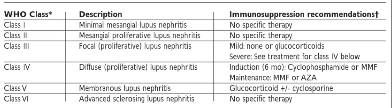
Table I. Classification and Treatment of the Different Forms of Lupus Nephritis
WHO Class* Description Immunosuppression recommendations†
Class I Minimal mesangial lupus nephritis No specific therapy Class II Mesangial proliferative lupus nephritis No specific therapy
Class III Focal (proliferative) lupus nephritis Mild: none or glucocorticoids
Severe: See treatment for class IV below Class IV Diffuse (proliferative) lupus nephritis Induction (6 mo): Cyclophosphamide or MMF
Maintenance: MMF or AZA Class V Membranous lupus nephritis Glucocorticoid +/- cyclosporine Class VI Advanced sclerosing lupus nephritis No specific therapy
* Subclasses omitted (refer to Weening et al.10for full classification)
†Recommendations derived from Rose et al.53
Table II. Indications for Performance of Kidney Biopsy in Lupus Nephritis
Increasing creatinine
Urine protein > 500 mg per 24 hours OR
Urine protein-to-creatinine ratio > 0.5 g protein/g creatinine
Hematuria and/or Red Blood Cell Casts Presence of cellular casts
Table III. Key Points: Advantages of Performing Kidney Biopsy
Early diagnosis = Better outcome
Correct diagnosis means appropriate therapy for severity of disease
Insufficient therapy results in loss of kidney function Inappropriate therapy may result in exposure to
unnecessary toxicity
Histopathologic features allow prognosis determination Allows for comparison at future time point if second
Ó R G Ã O O F I C I A L D A S O C I E D A D E P O R T U G U E S A D E R E U M AT O L O G I A - A C TA R E U M P O R T. 2 0 0 5 ; 3 0 : 1 2 1 - 2 7
125 by information obtained by renal biopsy.24, 25Two
re-cent commentaries on the role of kidney biopsy in lupus nephritis have thoroughly and effectively re-viewed the well recognized relationship between the histological features on biopsy and the clinical course of lupus nephritis.4, 18The WHO
classifica-tion system is well established in predicting outco-mes,26-28as are the NIH histologic activity and
chronicity indices.23, 29-31
The availability of this information is helpful in ensuring that the type, duration and intensity of treatment match the severity of the disease.
Though clinical variables such as elevation of serum creatinine and nephrotic syndrome at pre-sentation, persistent elevations of blood pressure, low hematocrit, hypocomplementemia and pre-sence of anti-DNA antibodies have prognostic va-lue,5,25,29,31-36the histologic information obtained
from biopsies enhances outcomes prediction significantly.24, 25, 37
Current Treatment Options in Lupus Nephritis
With a growing armamentarium of drugs available to treat lupus nephritis, the need for biopsy prior to initiating therapy has become even more pres-sing. The optimal treatment regimen in lupus ne-phritis varies according to WHO class (Table I).7, 38
Patients with the mildest forms of lupus nephritis (WHO class I or II) generally do well without spe-cific intervention, such as blockade of the renin--angiotensin system. In the absence of appropria-te immunosuppressive therapy, however, the pro-liferative forms (Class III and IV) of lupus nephri-tis typically progress to chronic renal failure.39The
benefits of early treatment are well documented.16
Since late and inadequate therapy allows active le-sions to evolve into chronic irreversible ones, there is a tendency to treat all patients with proliferative lesions regardless of severity. In patients with the most aggressive forms of lupus nephritis, aggres-sive therapy is warranted.
What becomes important then is having an ac-curate diagnosis with appropriate determination of severity, as the blind use of these drugs may re-sult in significant toxicity to the patient who other-wise might not have been exposed to a particular drug. The side effects of glucocorticoids are well known and are particularly disliked by patients. The physiologic effects include accelerated atherosclerosis, infections, osteoporosis, avascular
necrosis of bone and induction of diabetes melli-tus. Cyclophosphamide, which is usually given in monthly boluses by the “NIH protocol”, has im-mediate toxicity including nausea, vomiting, hair loss, and fatigue. Major toxicities include cytope-nias, serious infections, hemorrhagic cystitis, ma-lignancy and, of enormous importance to most young female patients, gonadal failure.40Serious
infections are common in SLE and death from in-fection correlates with the recent use of glucocor-ticoids and cyclophosphamide.40, 41
The more recent use of mycophenolate mofetil (MMF) has provided a drug that has equal if not better efficacy compared with cyclophosphamide, with much less toxicity.42-44In addition to risk of
in-fections, particularly viral ones, the toxicities of MMF are more limited and include predominant-ly reversible abdominal pain, diarrhea and leu-kopenia.45-48It is this improved toxicity compared
with most of the other agents that has resulted in the growing use of MMF in the treatment of lupus nephritis and further reinforces the need for accu-rate histopathologic diagnosis.
Patients may be spared all these toxicities if histopathology obtained by biopsy reveals a mild lesion (class I or II), a severely advanced chronic sion (class VI), or the presence of non-lupus le-sions.
Safety of Kidney Biopsy
Kidney biopsy is most frequently performed by percutaneous approach with either ultrasound or CT guidance. These techniques have been establi-shed as safe and effective in obtaining renal parenchyma. With improved imaging and the use of semi-automated biopsy guns, complications are uncommon and those of most concern are related to bleeding. Major complications, those requiring need for blood transfusion or invasive interven-tion, have been reported in 049to 6.4 %50of
biop-sies. Predictors of complications have included high creatinine and low starting hematocrit.51
Pa-tients with SLE may be at risk of bleeding due to concurrent corticosteroid use and platelet dys-function, though this has not been studied. In high-risk patients such as those with ongoing an-ticoagulation needs in the context of antiphos-pholipid antibody syndrome, or those with low platelet counts, alternative biopsy methods have been used such as transjugular kidney biopsy and D E R E K M. F I N E E C O L.
surgical laporoscopic biopsy.52
The only absolute contraindication to percuta-neous kidney biopsy is the inability of the patient to cooperate with the procedure. Relative con-traindications include the presence of a bleeding diatheses, such as those mentioned above, solitary kidney and advanced kidney disease with bilate-rally small kidneys52. In bleeding diatheses, if the
bleeding risk can be minimized before and for a few days after the biopsy, then a percutaneous ap-proach can be used. However, there is still an in-creased bleeding risk for at least 6 weeks after the procedure.52
Conclusion
The goals of management of patients with lupus nephritis are early treatment of disease and pre-vention of end-stage renal disease. In order to achieve these objectives is becomes clear that kid-ney biopsy is essential in the establishment of ear-ly and definitive diagnosis, allowing appropriate type, duration and intensity of treatment that will match the class and severity of the disease defined by both clinical and histopathologic parameters. To be effective, the clinician must be aware of the di-verse clinical and pathologic manifestations of lu-pus nephritis. It is especially important that the clinician recognize early stages of renal involve-ment at which point kidney biopsy should be per-formed without delay. This will allow effective treatment prior to the development of irreversible damage.
References:
1. Fries JF, Porta J, Liang MH. Marginal benefit of renal biopsy in systemic lupus erythematosus. Arch Intern Med 1978; 138: 1386-1389.
2. Gault MH, Muehrcke RC. Renal biopsy: current views and controversies. Nephron 1983; 34: 1-34.
3. Kashgarian M. Lupus nephritis: lessons from the path lab. Kidney Int 1994; 45: 928-938.
4. Grande JP, Balow JE. Renal biopsy in lupus nephritis. Lupus 1998; 7: 611-617.
5. Neumann K, Wallace DJ, Azen C, et al. Lupus in the 1980s: III. Influence of clinical variables, biopsy, and treatment on the outcome in 150 patients with lupus nephritis seen at a single center. Semin Arthritis Rheum 1995; 25: 47-55.
6. Salach RH, Cash JM. Managing lupus nephritis: algo-rithms for conservative use of renal biopsy. Cleve Clin J Med 1996; 63: 106-115.
7. Cameron JS. Lupus nephritis. J Am Soc Nephrol 1999; 10: 413-424.
8. Churg J, Sobin LH. Renal disease:classification and at-las of glomerular diseases. Tokyo, New York: Igaku-Shoin; 1982.
9. Churg J, Bernstein J, Glassock RJ. Renal disease:classi-fication and atlas of glomerular diseases (second edi-tion). Tokyo, New York: Igaku-Shoin; 1995.
10. Weening JJ, D’Agati VD, Schwartz MM, et al. The classi-fication of glomerulonephritis in systemic lupus ery-thematosus revisited. Kidney Int 2004; 65: 521-530. 11. Huong DL, Papo T, Beaufils H, et al. Renal involvement
in systemic lupus erythematosus. A study of 180 pa-tients from a single center. Medicine (Baltimore) 1999; 78: 148-166.
12. Nossent JC, Henzen-Logmans SC, Vroom TM, Huysen V, Berden JH, Swaak AJ. Relation between serological data at the time of biopsy and renal histology in lupus nephritis. Rheumatol Int 1991; 11: 77-82.
13. Gladman DD, Urowitz MB, Cole E, Ritchie S, Chang CH, Churg J. Kidney biopsy in SLE. I. A clinical-mor-phologic evaluation. Q J Med 1989; 73: 1125-1133. 14. Leehey DJ, Katz AI, Azaran AH, Aronson AJ, Spargo BH.
Silent diffuse lupus nephritis: long-term follow--up. Am J Kidney Dis 1982; 2(Suppl 1): 188-196. 15. Houssiau FA. Management of lupus nephritis: an
up-date. J Am Soc Nephrol 2004; 15: 2694-2704.
16. Esdaile JM, Joseph L, MacKenzie T, Kashgarian M, Hayslett JP. The benefit of early treatment with im-munosuppressive agents in lupus nephritis. J Rheuma-tol 1994; 21: 2046-2051.
17. Houssiau FA, Vasconcelos C, D’Cruz D, et al. Early res-ponse to immunosuppressive therapy predicts good renal outcome in lupus nephritis: lessons from long--term followup of patients in the Euro-Lupus Nephritis Trial. Arthritis Rheum Dec 2004; 50: 3934-3940. 18. Mittal B, Rennke H, Singh AK. The role of kidney
biop-sy in the management of lupus nephritis. Curr Opin Nephrol Hypertens 2005; 14: 1-8.
19. Nzerue CM, Hewan-Lowe K, Pierangeli S, Harris EN. «Black swan in the kidney»: renal involvement in the antiphospholipid antibody syndrome. Kidney Int 2002; 62: 733-744.
20. Baranowska-Daca E, Choi YJ, Barrios R, Nassar G, Suki WN, Truong LD. Nonlupus nephritides in patients with systemic lupus erythematosus: a comprehensive clini-copathologic study and review of the literature. Hum Pathol 2001; 32: 1125-1135.
21. Ling BN, Bourke E, Campbell WG, Jr., Delaney VB. Naproxen-induced nephropathy in systemic lupus erythematosus. Nephron 1990; 54: 249-255.
22. Esdaile JM, Joseph L, MacKenzie T, Kashgarian M, Hayslett JP. The pathogenesis and prognosis of lupus nephritis: information from repeat renal biopsy. Semin Arthritis Rheum 1993; 23: 135-148.
23. Moroni G, Pasquali S, Quaglini S, et al. Clinical and prognostic value of serial renal biopsies in lupus nephritis. Am J Kidney Dis 1999; 34: 530-539.
24. Austin HA, 3rd, Boumpas DT, Vaughan EM, Balow JE. Predicting renal outcomes in severe lupus nephritis: contributions of clinical and histologic data. Kidney Int 1994; 45: 544-550.
Ó R G Ã O O F I C I A L D A S O C I E D A D E P O R T U G U E S A D E R E U M AT O L O G I A - A C TA R E U M P O R T. 2 0 0 5 ; 3 0 : 1 2 1 - 2 7
127 25. Austin HA, 3rd, Boumpas DT, Vaughan EM, Balow JE.
High-risk features of lupus nephritis: importance of race and clinical and histological factors in 166 pa-tients. Nephrol Dial Transplant 1995; 10: 1620-1628. 26. Schwartz MM, Lan SP, Bonsib SM, Gephardt GN,
Shar-ma HM. Clinical outcome of three discrete histologic patterns of injury in severe lupus glomerulonephritis. Am J Kidney Dis 1989; 13: 273-283.
27. Kashgarian M. The role of the kidney biopsy in the treatment of lupus nephritis. Ren Fail 1996; 18: 765--773.
28. McLaughlin J, Gladman DD, Urowitz MB, Bombardier C, Farewell VT, Cole E. Kidney biopsy in systemic lupus erythematosus. II. Survival analyses according to biop-sy results. Arthritis Rheum 1991; 34: 1268-1273. 29. Austin HA, 3rd, Muenz LR, Joyce KM, et al. Prognostic
factors in lupus nephritis. Contribution of renal histo-logic data. Am J Med 1983; 75: 382-391.
30. Austin HA, 3rd, Muenz LR, Joyce KM, Antonovych TT, Balow JE. Diffuse proliferative lupus nephritis: identifi-cation of specific pathologic features affecting renal outcome. Kidney Int 1984; 25: 689-695.
31. Esdaile JM, Federgreen W, Quintal H, Suissa S, Hayslett JP, Kashgarian M. Predictors of one year outcome in lu-pus nephritis: the importance of renal biopsy. Q J Med. Nov 1991; 81: 907-918.
32. Arce-Salinas CA, Villa AR, Martinez-Rueda JO, et al. Factors associated with chronic renal failure in 121 pa-tients with diffuse proliferative lupus nephritis: a case-control study. Lupus 1995; 4: 197-203.
33. Donadio JV, Jr., Hart GM, Bergstralh EJ, Holley KE. Prognostic determinants in lupus nephritis: a long-term clinicopathologic study. Lupus 1995; 4: 109-115. 34. Esdaile JM, Levinton C, Federgreen W, Hayslett JP,
Kashgarian M. The clinical and renal biopsy predictors of long-term outcome in lupus nephritis: a study of 87 patients and review of the literature. Q J Med 1989; 72: 779-833.
35. Korbet SM, Lewis EJ, Schwartz MM, Reichlin M, Evans J, Rohde RD. Factors predictive of outcome in severe lupus nephritis. Lupus Nephritis Collaborative Study Group. Am J Kidney Dis 2000; 35: 904-914.
36. Mok CC, Wong RW, Lau CS. Lupus nephritis in Sou-thern Chinese patients: clinicopathologic findings and long-term outcome. Am J Kidney Dis 1999;34: 315-323. 37. Esdaile JM, Mackenzie T, Barre P, et al. Can experien-ced clinicians predict the outcome of lupus nephritis? Lupus 1992; 1: 205-214.
38. Contreras G, Roth D, Pardo V, Striker LG, Schultz DR. Lupus nephritis: a clinical review for practicing nephrologists. Clin Nephrol 2002; 57: 95-107.
39. Appel GB, Cohen DJ, Pirani CL, Meltzer JI, Estes D. Long-term follow-up of patients with lupus nephritis. A study based on the classification of the World Health Organization. Am J Med 1987; 83: 877-885.
40. Petri M. Cyclophosphamide: new approaches for sys-temic lupus erythematosus. Lupus 2004; 13: 366-371. 41. Hellmann DB, Petri M, Whiting-O’Keefe Q. Fatal
infec-tions in systemic lupus erythematosus: the role of op-portunistic organisms. Medicine (Baltimore) 1987; 66:
341-348.
42. Contreras G, Pardo V, Leclercq B, et al. Sequential the-rapies for proliferative lupus nephritis. N Engl J Med 2004; 350: 971-980.
43. Appel GB, Ginzler EM, Radhakrishnan J, et al. Multi-center controlled trial of mycophenolate mofetil as in-duction therapy for severe lupus nephritis [Abstract]. J Am Soc Nephrol 2003; 14: 38A.
44. Chan TM, Li FK, Tang CS, et al. Efficacy of mycopheno-late mofetil in patients with diffuse proliferative lupus nephritis. Hong Kong-Guangzhou Nephrology Study Group. N Engl J Med 2000; 343: 1156-1162.
45. Halloran P, Mathew T, Tomlanovich S, Groth C, Hooft-man L, Barker C. Mycophenolate mofetil in renal allo-graft recipients: a pooled efficacy analysis of three ran-domized, double-blind, clinical studies in prevention of rejection. The International Mycophenolate Mofetil Renal Transplant Study Groups. Transplantation 1997; 63: 39-47.
46. Sollinger HW. Mycophenolate mofetil for the preven-tion of acute rejecpreven-tion in primary cadaveric renal allo-graft recipients. U.S. Renal Transplant Mycophenolate Mofetil Study Group. Transplantation 1995; 60: 225--232.
47. Mycophenolate Mofetil (Cellcept) product informa-tion. Roche Laboratories, 1995.
48. Placebo-controlled study of mycophenolate mofetil combined with cyclosporin and corticosteroids for prevention of acute rejection. European Mycopheno-late Mofetil Cooperative Study Group. Lancet 1995; 345: 1321-1325.
49. Hergesell O, Felten H, Andrassy K, Kuhn K, Ritz E. Safe-ty of ultrasound-guided percutaneous renal biopsy-retrospective analysis of 1090 consecutive cases. Nephrol Dial Transplant 1998; 13: 975-977.
50. Marwah DS, Korbet SM. Timing of complications in percutaneous renal biopsy: what is the optimal period of observation? Am J Kidney Dis 1996; 28: 47-52. 51. Whittier WL, Korbet SM. Renal biopsy: update. Curr
Opin Nephrol Hypertens 2004; 13: 661-665.
52. Stiles KP, Yuan CM, Chung EM, Lyon RD, Lane JD, Ab-bott KC. Renal biopsy in high-risk patients with medi-cal diseases of the kidney. Am J Kidney Dis 2000; 36: 419-433.
53. Rose BD, Schur PH, Falk RJ, Appel GB. Treatment of Lupus Nephritis. In: UpToDate, Rose, BD (Ed). Welles-ley, MA; 2004.
Corresponding Author:
Derek M. Fine, MD
1830 E. Monument Street, Suite 416 Baltimore, MD 21205
Tel: (410) 955-5268 Fax: (410) 955-0485 E-mail: [email protected]
The Johns Hopkins Lupus Cohort is supported by NIH AR43727 and the Johns Hopkins General Clinical Research Center M-1-RR00052