Potential for transmission of Stenocarpella macrospora from inoculated
seeds to maize plants grown under controlled conditions
1Carolina da Silva Siqueira
2*, José da Cruz Machado
2, Ellen Noly Barrocas
3,
Mirella Figueiró de Almeida
2ABSTRACT – Maize seeds infected by Stenocarpella macrospora can cause stalk and ear rot and leaf spot. Transmission
of this pathogen through seeds may vary according to the cultivar, climatic conditions, and virulence of the pathogen among
other factors. The aim of this study was to assess the transmission rate of S. macrospora from seeds of the maize cultivars
C1-RB9308YG and C2-RB9108 using artificially infected seeds grown under two temperatures (20 °C and 25 °C). Seeds were inoculated by the osmotic conditioning method for 24 h (inoculum potential - P1), 48 h (P2), 72 h (P3) and 96 h (P4). After inoculation, 25 seeds were distributed individually in plastic cups with substrate, with 4 replicates per treatment. At the end of
twenty-eight days of daily assessments, all plants were analyzed for the presence of the pathogen by biological methods, and
some were sampled at random and analyzed Bio-PCR. The maximum percentages of dead seeds/seedlings in pre-emergence were 74.5% and 82.5% for P3 and P4, respectively. The highest total rate of transmission of the pathogen under study was 85.8% for seeds of the cultivar C1 at the highest inoculum potential (P4), grown at the temperature of 20 °C.
Index terms: seed pathology, inoculum potential, ear and stalk rot, fungus.
Potencial de transmissão de
Stenocarpella macrospora de sementes
inoculadas para plantas de milho cultivadas em condições controladas
RESUMO - Sementes de milho infectadas por Stenocarpella macrospora podem ocasionar podridão do colmo e da espiga
e manchas foliares. A transmissão deste patógeno por meio de sementes pode variar de acordo com a cultivar, as condições climáticas e a virulência do patógeno, entre outros fatores. O objetivo foi avaliar a taxa de transmissão de S.macrospora a partir de sementes das cultivares de milho C1-RB9308YG e C2-RB9108, infectadas artificialmente e cultivadas sob duas temperaturas (20 °C e 25 °C). A inoculação das sementes foi realizada pelo método de condicionamento osmótico, por 24 (potencial de inóculo - P1), 48 (P2), 72 (P3) e 96 (P4) horas. Após a inoculação, 25 sementes foram distribuídas, individualmente, em copos plásticos contendo substrato, com 4 repetições por tratamento. Ao final de 28 dias de avaliações
diárias, todas as plantas foram analisadas para a presença do patógeno por métodos biológicos e algumas, amostradas ao acaso,
por Bio-PCR. As porcentagens máximas de morte em pré-emergência de sementes/plântulas foram de 74,5% e 82,5%, para P3 e P4, respectivamente. A maior taxa de transmissão total do referido patógeno neste estudo foi de 85,8%, ocorrida no potencial de inóculo mais elevado (P4) em sementes da cultivar C1, cultivadas na temperatura de 20 °C.
Termos para indexação: patologia de sementes, potencial de inóculo, podridão do colmo e da espiga, fungo.
1Submitted on 10/07/2013. Accepted for publication on 03/25/2014. 2Departamento de Fitopatologia, Universidade Federal de Lavras - UFLA, Caixa Postal 3037, 37200-000, Lavras, MG, Brasil.
3Department of Plant and Environmental Sciences, 2360, Taastrup, Denmark. *Corresponding author <kerolpet@gmail.com>
Introduction
Stenocarpella macrospora (Earle) Sutton, causal agent of
stalk and ear rot and leaf spots in maize has been increasing in various maize producing regions in Brazil. Currently, it is quite often detected in seeds, a situation attributed to the increase in the occurrence and intensity of the disease in the
fields (Casa et al., 2006; Mário et al., 2011).
The fungus S. macrospora is necrotrophic, exhibiting a
parasitic phase in the developing plant and saprophytic phase in crop residue. Thus, the pathogen may be found surviving outside the crop season as mycelium inside the seeds, forming pycnidia in
the crop residues on the soil surface (Casa et al., 2003). Therefore,
by the fungus (injured kernels) is unsuitable for consumption and for formulating feeds since they may have the toxin diplodiol (Petatán-Sagahón et al., 2011).
The transmission process of S. macrospora from the seed to the plant occurs through the mesocotyle, reaching the
crown, the roots, and, finally, the base of the stalk. It is a slow
process and may be found in different phases of the crop, from
the beginning to the end (Casa et al., 2006). The association of
the pathogen with seeds ensures direct access of the parasite to the nutrition source at the time of seed germination and seedling emergence. Due to diverse physical and biological factors, in addition to the position of the pathogen within the seed and its intensity, the pattern of transmission through seeds becomes quite variable.
Quantification of the transmission rate of S. macrospora
in maize has not yet been undertaken and according to Casa et
al. (2006), it is a difficult task through the fact of this fungus
being linked to lack of seed germination, to asymptomatic plants, and to the presence of other fungi associated with
the seed, which may interfere in this quantification since a
selective medium for the pathogen is not available.
The aim of this study was to understand and to assess the transmission rate of S. macrospora in maize seeds, considering the interference of aspects like the inoculum potential of the pathogen, the environment temperature, and genotypes under controlled growing conditions.
Materials and Methods
Obtaining and multiplying fungal isolates and health testing
of the seeds used:two isolates of S. macrospora, CMLAPS375
and CMLAPS10, from the mycological collection of the Seed Pathology Laboratory of Federal University of Lavras (Lavras, Minas Gerais, Brazil) were used. The isolates were multiplied in Petri dishes containing the PDA culture medium (20 g of agar, 20 g of dextrose, and 200 g of potato/L) and placed in a BOD chamber at a temperature of 25 ± 2 °C and 12 hours phototperiod. The cultivars used, RB9308YG (cultivar susceptible to S.
macrospora - C1) and RB9108 (cultivar moderately resistant to
S. macrospora - C2), were provided by the Riber Seeds company,
located in Patos de Minas, MG. The health and physiological
testing of the maize seeds were determined according to the
Rules for Seed Testing (Brasil, 2009a) and the Seed Health Analysis Manual (Brasil, 2009b). From those analysis, it was
observed that the seed lots used in this study were not carriers
of S. macrospora. For the cultivar C1 (RB9308YG), incidences
of 28.5% of Fusarium verticillioides and 13% of Penicillium sp.
were detected, and, for C2 (RB9108), an incidence of 25.5% and 11%, for the respective fungi were observed in C1. Germinations
of these lots were 98% normal seedlings for C1 and 96% for C2.
Seed preparation and inoculation: inoculation of the
seeds was carried out by the osmotic conditioning method
(Machado et al., 2012) through which the seeds were kept
in contact with colonies of the fungal isolates for different
exposure times. First, the isolates were cultivated on PDA culture medium in Petri dishes containing mannitol solute with water potential of -1.4 MPa, adjusted by the software SPPM (Michel and Radcliffe, 1995). The seeds of both cultivars were disinfested with 1% sodium hypochlorite for 1 minute,
washed three times with distilled water, and dried in laboratory conditions. After that, the seeds were evenly distributed over
the fungal colonies five days old, for periods of 24, 48, 72 and 96 hours, these times corresponding to the different inoculum potentials of P1, P2, P3 and P4, respectively. Incubation occurred in a BOD chamber with temperature of 25 ± 2 °C
and 12-h photoperiod. For the control treatments, the same periods as mentioned for inoculation were also used, in which
seeds of both cultivars were placed in Petri dishes containing only the PDA medium with the addition of mannitol for
assessment of the possible effect of water restriction on the physiological quality of the maize seeds.
Seed health testing: to obtain the percentage of incidence of
S. macrospora in inoculated seeds, blotter test were performed to
estimate the transmission rate of the fungus from the seeds to the plants in the different inoculum potentials. Thus, the inoculated maize seeds were distributed on a paper substrate soaked with
the OA medium (20 g of agar and 30 g of oatmeal/L), a medium favorable to the formation of pycnidia of the species (Silva and Juliatti, 2005), in 15-cm diameter Petri dishes, with eight replications of 25 seeds per dish. The seeds were then placed in a freezer at -20 °C for 24 hours and then incubated for 7-15 days in a growing chamber with a temperature of 20 ± 2 °C and 12-h photoperiod. At the end of this period, the seeds were examined
individually through a stereoscopic microscope, checking for the incidence of the S. macrospora.
Assessments for determination of the transmission rate:
the seeds were distributed in 200 mL plastic cups containing a
commercial substrate (Multiplanta trop sc 25 kg), sowing one
per cup and a total of 100 cups equally distributed in four trays
(replications). The experiment was conducted in chambers with temperatures adjusted to 20 °C and 25 ± 2 °C and a 12 light-hour photoperiod (daylight NSK T10 40 W 6500 K FL40T10-6 60 Hz)/12 hours of darkness. The emergence
of plants which were symptomatic and asymptomatic of the disease in focus was assessed daily. Fragments of symptomatic
plants were aseptically placed and incubated in Petri dishes containing the PDA medium for confirmation of the presence
after sowing, all the asymptomatic plants were collected
and 2 cm fragments at the height of the root collar (C) and last leaf insertion (LI) were disinfested in 70% alcohol, 1%
sodium hypochlorite, and distilled and sterilized water for 1 minute and dried on sterilized paper. All the fragments were
deposited on Petri dishes containing the PDA medium and incubated at the temperature of 25 °C and 12-h photoperiod. After 7 and 15 days, the fragments were individually assessed
in a stereoscopic microscope for observation of structures characteristic of S. macrospora (Mario and Reis, 2001).
Detection of S. macrospora in at least one of the fragments
examined per plant was sufficient for confirmation of the
transmission of such pathogen from the seed to the plant. A
randomized block experimental design was used in a triple factorial arrangement of 2 x 2 x 4 (2 temperatures, 2 cultivars, and 4 inoculum potentials), with four replications per treatment. Determination of the total transmission rate (T.T.)
of S. macrospora from the seeds to the plants for each type of
exposure time was calculated based on the formula (Teixeira and Machado, 2003):
T.T.(%) = [I.R.(%) / I.S.(%)] * 100
in which:
I.R.(%) = infection rate of S. macrospora in seeds in
pre-emergence up to fragments (C and LI) of maize plants at 28 days of age;
I.S.(%) = incidence of S. macrospora in inoculated seeds, based on the seed health testing.
Confirmation of the presence of S. macrospora in
asymptomatic plants by the molecular technique: at
least 20% of the plants of each treatment in which disease
symptoms were not observed were collected at random and
subjected to analysis in Bio-PCR. For that purpose, 2-cm fragments from the region of the root collar (C) and from the last leaf insertion (LI) of the plants were assessed individually. The Wizard® Genomic DNA Purification Kit (Promega,
Madison, WI) was used for DNA extractions, according to
the protocol recommended by the manufacturer. The primers
P1/P2 described by Xia and Achar (2001), specific for the
genus Stenocarpella, were used to detect the presence of the
fungus in the tissues (Barrocas et al., 2012). Amplification was performed in 25 µL of the reaction containing the PCR buffer (IB buffer - Phoneutria, Brazil – 500 mM KCl, 100 mM Tris-HCl pH 8.4, 1% Triton X-100, MgCl2), dNTPs (2.5 mM of each dNTP), primers (10 µM of each forward or reverse primer), and 5 U/µL of Taq DNA polymerase
(Phoneutria, Brazil), with the addition of 2 µL of the DNA
to make up the total volume. The cycle initially consisted of
95 °C for 3 minutes, denaturation at 94 °C for 30 seconds, annealing at 60 °C for 1 minute, and extension of 72 °C for 1 minute, with final extension of 72 °C for 10 minutes, for a total of 30 cycles. An aliquot of 10 µL was used to analyze the PCR products in 1% agarose gel in TBE buffer, stained
with GelRed® at 150 V for approximately 2 hours. The PCR
products were observed in a UV transilluminator, L-Pix HE equipment (Loccus Biotecnologia, Brasil).
Statistical analysis: statistical analyses were carried
out with the assistance of the Sisvar® program, version
5.3 (Ferreira, 2011). Analyses of variance were carried out
individually for each isolate of Stenocarpella macrospora, in
addition to the control (not inoculated), in the triple factorial
arrangement. For the death in pre-emergence and transmission rate variables with observation of symptomatic and asymptomatic plants, the analyses of variance were corrected
through transformation of the data in square root (data+1).
The mean values among the treatments were compared by
regression, the Tukey test, or Student t test (P ≤ 0.05). For the
total transmission rate, all the percentages of the transmission rates of symptomatic and asymptomatic plants and death in pre-emergence were considered.
Results and Discussion
Analysis of variance for death in pre-emergence and transmission rates of Stenocarpellamacrospora from seeds to
plants exhibited a triple non-significant interaction (p ≤ 0.05)
when the seeds were inoculated with the two isolates of the pathogen.
The presence of the pathogen was not detected in the plants of the control treatment. Therefore, the seeds of these treatments responded only to the possible interference of water restriction from mannitol in their physiological quality, and these results were used for estimation of the percentage of death in pre-emergence caused by the pathogen. The same is undertaken in different studies of the fungus-seed interaction for subtraction of the incidental and consequent
interferences in physiological quality (Araújo et al., 2006; Costa et al., 2003) which, in the present study, were minimal.
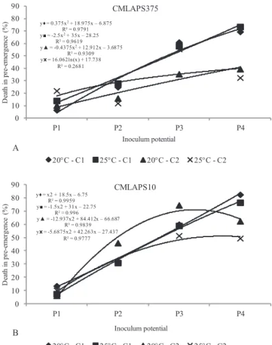
The results obtained in assessment of death in
pre-emergence of maize seeds/seedlings (Figure 1) were higher
than those of the transmission rates of symptomatic and
asymptomatic plants (Figures 2 and 3) for all the inoculum potentials. According to Tanaka and Machado (1985), the quantity of inoculum present in the seed influences germination and transmission of pathogens. When associated with the
y = -2.5x2+ 35x –28.25 R² = 0.9619
y = 0.375x2+ 18.975x –6.875 R² = 0.9791
y = -0.4375x2+ 12.912x –3.6875 R² = 0.9309 yx= 16.062ln(x) + 17.738
R² = 0.2681
0 10 20 30 40 50 60 70 80 90
P1 P2 P3 P4
D eat h in pre -e me rg en ce (%) Inoculum potential CMLAPS375
20C - C1 25C - C1 20C - C2 25C - C2
y = x2 + 18.5x –6.75 R² = 0.9959
y = -1.5x2 + 31x –22.75 R² = 0.996
y = -12.937x2 + 84.412x –66.687
R² = 0.9839 yx= -5.6875x2 + 42.263x –27.437
R² = 0.9777
0 10 20 30 40 50 60 70 80 90
P1 P2 P3 P4
D eat h in pre -e me rg en ce (%) Inoculum potential CMLAPS10
20C - C1 25C - C1 20C - C2 25C - C2
A
B
Figure 1. Assessment of the death of seeds/seedlings in
pre-emergence caused by the fungus Stenocarpella
macrospora in relation to the isolates CMLAPS375
(A) and CMLAPS10 (B) at the inoculum potentials P1 (24 h), P2 (48 h), P3 (72 h), and P4 (96 h), in the cultivars C1 (RB9308YG) and C2 (RB9108), kept at the temperatures of 20 °C and 25 °C.
y= -0.9509x2+ 2.8613x + 5.4763 R² = 0.9998 y = -0.3757x2+ 0.7443x + 6.274
R² = 0.9857
y = -0.8345x2+ 3.5203x + 1.2638 R² = 0.9897
yx= -0.7196x2+ 2.9107x + 3.1876 R² = 0.8741
0 1 2 3 4 5 6 7 8 9
P1 P2 P3 P4
T ra ns m is si on ra te of sy mp to matic p la n ts (%) Inoculum potential CMLAPS10
20C - C1 25C - C1 20C - C2 25C - C2
y= -1.1634x2+ 4.6336x + 2.0099 R² = 0.9664
y= -0.5416x2+ 1.9749x + 2.1783 R² = 0.9287
y = 0.0665x2–1.6358x + 8.1524 R² = 0.9168
yx= -0.2241x2+ 0.4241x + 4.625 R² = 0.6605
0 1 2 3 4 5 6 7 8 9
P1 P2 P3 P4
Tr an sm is sio n rate of sym pt om at ic p la n ts (%) Inoculum potential CMLAPS375
20C - C1 25C - C1 20C - C2 25C - C2
A
B
Figure 2. Transmission rates of Stenocarpella macrospora
in symptomatic plants for the two isolates
(CMLAPS375-A and CMLAPS10-B) at the potentials P1, P2, P3, and P4 (24 h, 48 h, 72 h, and 96 h), cultivars C1 (RB9308YG) and C2 (RB9108), at the temperatures of 20 °C and 25 °C.
The highest percentages of death in pre-emergence
were reached in the P3 and P4 inoculum potentials for both isolates, both temperatures, and both cultivars. In the P3 and P4 potentials for the CMLAPS375 isolate, the maximum percentages were 61% and 73.5%, respectively, for the C1
cultivar, with characteristics of pathogen susceptibility at both growing temperatures, reinforcing the difference of cultivar
response to the pathogen (Mendes et al., 2011). However, for the isolate CMLAPS10, the same result was not observed – the maximum value for P3 was 74.5%, occurring for the two cultivars maintained at 20 °C, and, for P4, the maximum percentage was 82.5% for C1, at the two growing temperatures.
These results in reference to death in pre-emergence for
maize seeds/seedlings infected by S. macrospora reflected the
most recurrent consequence of the activity of this pathogen,
compromising germination of maize seeds (Casa et al., 2006).
In a study with Colletotrichum gossypii var. cephalosporioides
in cotton seeds, Araújo et al. (2006) also observed that higher
temperatures of osmotic conditioning, together with presence of the fungus, led to progressive reduction in seed germination. Another study that corroborates the results obtained in this research was that of common bean seeds inoculated with F.
oxysporum f. sp. phaseoli, in which a proportional reduction
was observed in germination of normal seedlings with a 0 to
144 hour incubation period (Costa et al., 2003).
S. macrospora is a pathogen which most commonly
exhibits symptoms over more advanced vegetative stages of
the plant, culminating in a high incidence at the end of the
crop cycle, which may explain the low transmission rates for symptomatic plants observed in this study (Figure 2). The
format, normally measuring from 1 to 3 cm in length, with a
tan color and, at times, exhibiting darker concentric rings as of the initial point of infection. In addition, these first lesions
could also be seen in the form of small yellowish or tan streaks
that later increased in size, extending in the longitudinal
direction of the leaf, which were able to tear the infected plant
tissue (Casa et al., 2006; Duarte et al., 2009).
The transmission rates for symptomatic plants (Figure 2) were higher in the P1 and P2 inoculum potentials, for both isolates. It was observed that P1 exhibited the highest stand and P4 the lowest stand compared to the other potentials. Therefore, in P4, the lowest occurrence of symptoms in
surviving plants was seen, and this indicates that death in pre- and post-emergence is the main consequence of high inoculum
potentials of the pathogen. For the isolate CMLAPS375, potentials P1 and P2 led to the highest transmission rates for symptomatic plants of 6.8% and 7.1%, respectively, and, for CMLAPS10, of 7.4% (P2 and P3). These results occurred in C1 grown at 20 °C; although in literature the temperature of
25 °C is indicated as one of the most favorable conditions for
the activity of S. macrospora (Casa et al., 2007).
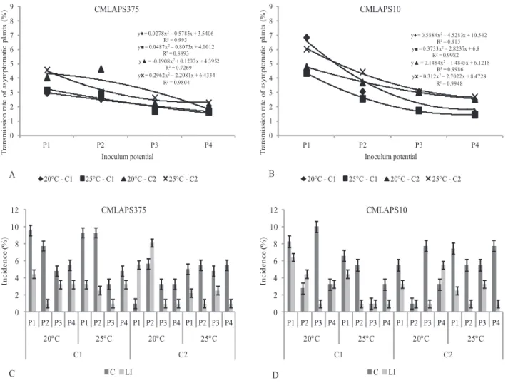
When compared to the transmission rates for asymptomatic
plants, the rates in relation to symptomatic plants were higher,
but they generally occurred at potentials P1 and P2; for the asymptomatic plants, the highest rates occurred at P1 for the cultivar C1 kept in temperature of 20 °C.
In the asymptomatic plants (Figures 3 A and B), incidence
of the fungus on root collar and last leaf insertion, was
observed only in few cases, reflecting the low transmission
rates observed in those treatments. The highest percentages
occurred at the P1 inoculum potential, in which 6.9% was observed for C1 at temperature of 20 °C with the isolate CMLAPS10; which was the isolate responsible for most of
the positive results for latent infection by S. macrospora in the plants analyzed. These results were compiled including
the positive results provided by the Bio-PCR technique,
which allowed detection of S. macrospora in almost all the treatments assessed with asymptomatic plants.
y= 0.0278x2–0.5785x + 3.5406
R² = 0.993 y = 0.0487x2–0.8073x + 4.0012
R² = 0.8893 y = -0.1908x2+ 0.1233x + 4.3952
R² = 0.7269
yx= 0.2962x2–2.2081x + 6.4334
R² = 0.9804
0 1 2 3 4 5 6 7 8 9
P1 P2 P3 P4
Tr a n s m is s io n rat e of a s y m p to m a tic p la n ts (% ) Inoculum potential CMLAPS375
20C - C1 25C - C1 20C - C2 25C - C2
y= 0.5884x2–4.5283x + 10.542
R² = 0.915
y = 0.3733x2–2.8237x + 6.8
R² = 0.9982 y = 0.1484x2–1.4845x + 6.1218
R² = 0.9986
yx= 0.312x2–2.7022x + 8.4728
R² = 0.9948
0 1 2 3 4 5 6 7 8 9
P1 P2 P3 P4
Tr a n s m is s io n rat e of a s y m p to m a tic p la n ts (% ) Inoculum potential CMLAPS10
20C - C1 25C - C1 20C - C2 25C - C2
0 2 4 6 8 10 12
P1 P2 P3 P4 P1 P2 P3 P4 P1 P2 P3 P4 P1 P2 P3 P4
20C 25C 20C 25C
C1 C2 In ci d en ce (% ) C LI 0 2 4 6 8 10 12
P1 P2 P3 P4 P1 P2 P3 P4 P1 P2 P3 P4 P1 P2 P3 P4
20C 25C 20C 25C
C1 C2 In ci d en ce (% ) C LI B A CMLAPS375 CMLAPS10 D C
Figure 3. A and B - Transmission rate of Stenocarpella macrospora in asymptomatic plants. C and D - Incidence of S.
macrospora in the sectioned parts of plants: root collar (C) and the last leaf insertion (LI). C - isolate CMLAPS375
The highest percentage of positive results detected for the
pathogen in the Bio-PCR (Figure 4) occurred for the isolate CMLAPS375, with 8.21% incidence in the C2 plants kept at 20 °C. For the isolate CMLAPS10, the maximum percentage
also occurred in the same cultivar and temperature observed
for CMLAPS375; however, the incidence was 7.78%. It is important to register that positive results in PCR analysis
for the presence of fungi in asymptomatic plants should be considered with caution because the pathogen may be unviable for developing and causing disease.
Considering that the asymptomatic plants presented a higher incidence of the pathogen in the region of the root collar, this may be related to the permanence of the fungus in this part of the plant from which the pathogen under favorable conditions may cause the disease collapse. This
kind of infection is similar what takes place in the field,
where infected plant organs near or below the soil derived from contaminated seeds represent source of inoculum for
the disease development (Casa et al., 2006; Reis et al., 2004).
From the transmission rates observed in this study, it turns out that the asymptomatic plants will be certainly able to transmit the pathogen from the mother plant to the seeds on the ear.
For S. macrospora transmission of the pathogen was
observed (Figure 5), from the seed to the plant, at all the
inoculum potentials and it increased from the lower to the higher potentials, i.e., the highest rates of total transmission
were reached at P3 and P4 for both isolates, both temperatures and both cultivars. At the highest inoculum potential, P4, the highest rates, 76.4% and 85.8%, occurred for CMLAPS375 and CMLAPS10, both being for C1 (RB9308YG) at the two
growing temperatures. For this cultivar, high rates of total
transmission were observed in all the treatments, confirming the
susceptibility of this genotype. Taking into consideration the
lowest rate of total transmission of this pathogen, 15%, which occurred with the isolate CMLAPS10 at potential P1 and cultivar C2 (RB9108), at the temperature of 20 °C, it was observed that
even in the less favorable conditions for the pathogen, this
interaction should be considered of great significance from the epidemiological point of view. For both potentials, P2 and P3, the values of total transmission rates were around 50% for both
isolates, both temperatures and both cultivars.
The total transmission rates observed were directly proportional to and progressive with the inoculum potentials, i.e., the longer the period of time the seed remained in contact with S.
macrospora, the higher infection of seed tissues and, consequently,
the higher transmission of the pathogen to the plants from the
seed. The same was observed by Botelho et al. (2013) in a study
on Sclerotinia sclerotiorum in common bean seeds, in which
there was an increase of the disease when the inoculum potential, obtained by the variation of the time of inoculation, increased from
36 to 96 hours of contact of the seeds with the pathogen in two cultivars assessed in the study. Barrocas et al. (2014) also verified
this proportionality as a result of increases in the inoculum potentials in cotton seeds inoculated with Colletotrichum
gosypii var. cephalosporioides. However, in another study
with maize seeds and the fungus Acremonium strictum,
Teixeira and Machado (2003) observed that the infection
rate, assessed in the above ground part of plants at 28 days
of age, was higher with an increase in the time of exposure
0 1 2 3 4
5 6 7
8 9 10
P1 P2 P3 P4 P1 P2 P3 P4 P1 P2 P3 P4 P1 P2 P3 P4
20C 25 C 20C 25 C
C1 C2
In
ci
d
en
ce
(%
)
CMLAPS375 CMLAPS10
A
M E
CP2
CP1 43 44 45 389 390 391 - 397
B C 25C D 20C E
Figure 4. A – Detection of the isolates CMLAPS375 and CMLAPS10 of Stenocarpella macrospora at the
inoculum potentials P1-24 h, P2-48 h, P3-72 h, and P4-96 h by the Bio-PCR technique undertaken
in sections of asymptomatic plants of the cultivars C1-RB9308YG and C2-RB9108 developed at the
temperatures of 20 °C and 25 °C. B - positive control of the Bio-PCR (CP1 and CP2) and water (E). C – some positive results of plant samples kept at 25 °C (43 and 45). D – positive results of plant samples kept at 20 °C (389, 391, and 397). E - marker.
In relation to the parts of the asymptomatic plants assessed
(Figures 3 C and D), root collar (C) and region of the last leaf insertion (LI), it was possible to observe a higher occurrence
of the fungus in the root collar region, for both isolates, a
maximum of 10.04% incidence of CMLAPS10 at P3 potential and 9.59% of CMLAPS375, at P1. With regard the incidence
of S. macrospora at the last leaf insertion, the maximum
of the seeds to the pathogen (from 0 to 120 hours), but the
transmission rates were statistically equals for the times of 24,
72 and 120 hours, differing only in the zero hour time.
Conclusions
Total transmission of S. macrospora from maize seeds to
plants may reach high rates, maximum of 85.8%.
Transmission of S. macrospora by maize seeds may occur in an asymptomatic manner under certain circumstances
reaching rate of 6.9%.
Death in pre-emergence and total transmission rates are directly proportional to the inoculum potentials of S.
macrospora in maize seeds.
Acknowledgments
To the CNPq, CAPES, and FAPEMIG for financial
support to carry out this study.
References
ARAÚJO, D.V.; POZZA, E.A.; MACHADO, J.C.; ZAMBENEDETTI, E.B.; CELANO, F.A.O.; CARVALHO, E.M.; CAMARGOS, V. N. Influência da temperatura e do tempo de inoculação das sementes de algodão na
transmissibilidade de Colletotrichum gossypii var. cephalosporioides. Fitopatologia Brasileira, v.31, n.1, p.035-040, 2006. http://www.scielo.br/ pdf/fb/v31n1/a06v31n1.pdf
BRASIL. Ministério da Agricultura, Pecuária e Abastecimento. Regras para análise de sementes.Ministério da Agricultura, Pecuária e Abastecimento. Secretaria de Defesa Agropecuária. Brasília: MAPA/ACS, 2009a. 395p. http:// www.agricultura.gov.br/arq_editor/file/2946_regras_analise__sementes.pdf
BRASIL. Ministério da Agricultura, Pecuária e Abastecimento. Manual de Análise Sanitária de Sementes. Ministério da Agricultura, Pecuária e Abastecimento. Secretaria de Defesa Agropecuária. Brasília: MAPA/ ACS, 2009b. 200p. http://www.agricultura.gov.br/arq_editor/file/12261_ sementes_-web.pdf
BARROCAS, E.N.; MACHADO, J.C.; ALVES, M.C.; CORRÊA, C.L. Desempenho de sementes de algodão submetidas à deficiência hídrica e
presença de Colletotrichum gossypii var. cephalosporioides. Bioscience Journal, v.30, n.2, p.421-428, 2014. http://www.seer.ufu.br/index.php/ biosciencejournal/article/view/17993
BARROCAS, E.N.; MACHADO, J.C.; ALMEIDA, M.F.; BOTELHO, L.S.; VON PINHO, E.V.R. Sensibility of the PCR technique in the detection of Stenocarpella sp. associated with maize seeds. Revista Brasileira de Sementes, v.34, n.2, p.218-224, 2012. http://www.scielo.br/pdf/rbs/v34n2/05.pdf
BOTELHO, L.S.; ZANCAN, W.L.A.; MACHADO, J.C.; BARROCAS, E.N. Performance of common bean seeds infected by the fungus Sclerotinia sclerotiorum. Journal of Seed Science, v.35, n.2, p.153-160, 2013. http:// www.scielo.br/pdf/jss/v35n2/03.pdf
CASA, R.T.; REIS, E.M.; ZAMBOLIM, L. Decomposição dos restos culturais do milho e sobrevivência saprofítica de Stenocarpella macrospora e S. maydis. Fitopatologia Brasileira, v.28, n.4, p.355-361, 2003. http://www. scielo.br/pdf/fb/v28n4/17007.pdf
CASA, R.T.; REIS, E.M.; ZAMBOLIM, L. Doenças do milho causadas por
fungos do gênero Stenocarpella. Fitopatologia Brasileira, v.31, n.5, p.427-439, 2006. http://www.scielo.br/pdf/fb/v31n5/01.pdf
y= 0.6375x2+ 16.833x + 9.2685 R² = 0.9903 y = -1.5023x2+ 28.921x –9.6761
R² = 0.9958
y = -13.624x2+ 86.448x –59.302 R² = 0.9811 yx= -6.0951x2+ 42.471x –15.777
R² = 0.982
0 10 20 30 40
50 60 70
80 90 100
P1 P2 P3 P4
To
ta
l
tr
an
sm
is
sio
n
ra
te
(%)
Inoculum potencial
CMLAPS10
20C - C1 25C - C1 20C - C2 25C - C2
y= -3.6355x2+ 39.055x –22.699 R² = 0.9625
y= -0.1179x2+ 20.143x –0.6955 R² = 0.9756
y = -0.5618x2+ 11.4x + 8.8602 R² = 0.9044 yx= -3.1779x2+ 21.966x + 6.5584
R² = 0.2201
0 10 20 30 40
50 60 70
80 90 100
P1 P2 P3 P4
To
ta
l
tr
an
sm
is
sio
n
ra
te
(%)
Inoculum potential
CMLAPS375
20C - C1 25C - C1 20C - C2 25C - C2
A
B
Figure 5. Total transmission rate of Stenocarpella macrospora,
observing the results of seeds/seedlings with
death in pre-emergence, symptomatic plants and
asymptomatic plants. A - isolate CMLAPS375 and B - isolate CMLAPS10, at the inoculum potentials P1 (24 h), P2 (48 h), P3 (72 h), and P4 (96 h), in the cultivars C1 (RB9308YG) and C2 (RB9108), kept at temperatures of 20 °C and 25 °C.
Once S. macrospora comes to the contact with maize seeds and there starts the infection process it may cause high rates of transmission as demonstrated in this study. From the results it was seen that such pathogen
may lead to the death of the seed/seedling or generate
CASA, R.T.; REIS, E.M.; ZAMBOLIM, L.; MOREIRA, E.N. Efeito da temperatura e de regimes de luz no crescimento do micélio, germinação de conídios e esporulação de Stenocarpella macrospora e Stenocarpella maydis. Fitopatologia Brasileira,v.32, p.137-142, 2007. http://www.scielo.br/pdf/fb/
v32n2/07.pdf
COSTA, M.L.N.; MACHADO, J.C.; GUIMARÃES, R.M; POZZA, E.A; ORIDE, D. Inoculação de Fusarium oxysporum f.sp. phaseoli em sementes
de feijoeiro através de restrição hídrica. Ciência e Agrotecnologia, v.27, n.5, p.1023-1030, 2003. http://www.scielo.br/pdf/cagro/v27n5/a08v27n5.pdf
DUARTE, R.P.; JULIATTI, F.C.; FREITAS, P.T. Eficácia de diferentes
fungicidas na cultura do milho. Bioscience Journal, v.25, n.4, p.101-111, 2009. http://www.seer.ufu.br/index.php/biosciencejournal/article/view/6966/4614
FERREIRA, D.F. SISVAR: A computer statistical analysis system. Ciência e Agrotecnologia, v.35, n.6, p.1039-1042, 2011. http://www.scielo.br/pdf/ cagro/v35n6/a01v35n6.pdf
MACHADO, J.C.; BARROCAS, E.N.; COSTA, M.L.N.; GUIMARÃES, R.M.; MACHADO, C.F. Uso da técnica de restrição hídrica ou
condicionamento osmótico em patologia de sementes. Revisão Anual de Patologia de Plantas, v.20, p.1-24, 2012.
MARIO, J.L.; REIS, E.M. Método simples para diferenciar Diplodia macrospora de D. maydis em testes de patologia de sementes de milho. Fitopatologia Brasileira, v.26, n.3, p.670-672, 2001. http://www.scielo.br/ pdf/fb/v26n3/a18v26n3.pdf
MARIO, J.L.; REIS, E.M.; JULIATTI, F.C. Three inoculation methods for
screening corn germplasm to white ear rot resistance. Tropical Plant Pathology,
v.36, n.6, p.362-366, 2011. http://www.scielo.br/pdf/tpp/v36n6/04.pdf
MENDES, M.C.; VON PINHO, R.G.; MACHADO, J.C.; ALBUQUERQUE, C.J.B.; FALQUETE, J.C.F. Qualidade sanitária de grãos de milho com e sem inoculação a campo dos fungos causadores de podridões de espigas. Ciência e Agrotecnologia, v.35, n.5, p.931-939, 2011. http://www.scielo.br/pdf/cagro/ v35n5/a10v35n5.pdf
MICHEL, B.E.; RADCLIFFE, D. A computer program relating solute potential to solution composition for five solutes. Agronomy Journal,
v.87, n.1, p.131-136, 1995. http://www.scielo.br/scielo.php?script=sci_ nlinks&ref=000161&pid=S2317-1537201300020000300013&lng=e
PETATÁN-SAGAHÓN, I.; ANDUCHO-REYES, M.A.; SILVA-ROJAS, H.V.; ARANA-CUENCA, A.; TELLEZ-JURADO, A.; CÁRDENAS-ÁLVAREZ, I.O.; MERCADO-FLORES, Y. Isolation of bacteria with antifungal activity
against the phytopathogenic fungi Stenocarpella maydis and Stenocarpella macrospora. International Journal of Molecular Sciences, v.12, p.5522-5537, 2011. http://www.ncbi.nlm.nih.gov/pmc/articles/PMC3189730/
REIS, E.M.; CASA, R.T.; BRESOLIN, A.C.R. Manual de diagnose e controle de doenças do milho. Lages: Grapel, 2004.144p.
SILVA, A.R.; JULIATTI, F.C. Esporulação de Diplodia maydis e Diplodia macrospora em diferentes meios de cultura. Bioscience Journal, v.21, n.3,
p.127-131, 2005. http://snida.agricultura.gov.br:81/cgibin%5Cwxis.exe?I
-sisScript=Cenagri_Search.xis&method=post&caminho=f:\xitami\webpages\ binagri\bases\&agb=agb&formato=1&quantidade=25&proxdoc=1&inver
-so=on&expressao=Juliatti,%20F.C.
TANAKA, M.A.S.; MACHADO, J.C. Patologia de sementes. Informe Agropecuário, v.11, n.122, p.40-46. 1985.
TEIXEIRA, H; MACHADO, J.C. Transmissibilidade e efeito de Acremonium strictum em sementes de milho. Ciência e Agrotecnologia, v.25, n.5, p.1045-1052, 2003. http://www.scielo.br/pdf/cagro/v27n5/a11v27n5.pdf
XIA, Z.; ACHAR, N. Random amplified polymorphic DNA and polymerase