2016
UNIVERSIDADE DE LISBOA
FACULDADE DE CIÊNCIAS
DEPARTAMENTO DE BIOLOGIA VEGETAL
Impact of Plasmodium infection on the expression of
bradykinin-receptor 2 on trophoblasts
André Filipe Rivais Martins Barateiro
Mestrado em Biologia Molecular e Genética
Dissertação orientada por:
Doutora Luciana Vieira de Moraes
I
Acknowledgments
To the Disease Genetics Group:
I am truly thankful and grateful for the amazing three years that I spent at the Disease Genetics Group, at Instituto Gulbenkian de Ciência (IGC). For the friendship, counselling and support that everyone gave me through the good and bad times.
I want to thank Dr. Carlos Penha-Gonçalves for having me at the group, for all the wise and good advices, and for putting his trust in the work that I developed so far. Additionally, I am also thankful for supporting my future, no matter what I choose.
I want to thank to Teresa for all the cheerful moments and teaching. To Inês for all the patience, friendship and endless discussion about life, in general. To Sónia for all the support and help. To Nádia for being so patient with my meaningful doubts and for teaching me everything that I know about Western Blots. To Rita for the amazing almond pies and support. Last but not the least, to my good friend Yash, for all the endless discussions, for his true friendship and for his desire to take me to Tanzania.
Specially, I want to thank Luciana de Moraes for being such an amazing supervisor and, above everything, an amazing friend. For believing in me, for trusting my work and opinions, for all the things that she gave me, I am truly thankful. I will never be capable to pay off all the effort that she invested on me.
To the IGC:
I want to thank the Flow Cytometry and the Animal House facilities for all advices, teaching and for making my work a lot easier. Additionally, I also would like to thank the IGC community for all the good moments and support.
To Faculdade de Ciências da Universidade de Lisboa (FCUL):
I want to thank Professora Doutora Rita Zilhão for giving me the pleasure of being her student. I am grateful for all the support, and for being such a dedicated and helpful supervisor. For all the thrust and for believing in my true potential, I am truly thankful.
To my friends:
To all my friends, I want to thank them for believing in me every single day and for making me a better person. Yet, I want to make some special considerations for those who were always there, even during the hardest times:
II
To Diogo Joaquim and Abel Santos, for endless conversations, for sharing and support, for all the times that we dreamt together and for making me believe in a better tomorrow.
To Jorge Sampaio, Rita Esteves, Mariana Dias, Rita Dias, Andreia Teixeira and Bárbara Oliveira for giving me the funniest and weirdest days of my life, showing me that true friendship does not choose sizes or origins and for making me believe that this is something that can last for ages, no matter who far the destiny takes us.
To André Carvalho and Bruno Amoroso, for being the best housemates that a person can have. For supporting and helping me every day, for all the conversations and outpourings and for making my life amazing.
To Beatriz Pinheiro, for being my best friend. For making me laugh and for clarifying my mind. For supporting my decisions and for making me a better and stronger person. For all the crying, fights and outpourings, for all the things that we have been through, I am sincerely thankful.
To my family:
To my mother Ana Carvalho, to my father Luís Barateiro and to my brother João Barateiro I want to say that everything that I accomplished is dedicated to you who made my education and personal growth possible. Everything that I am is because of you; it is a fruit of your work, education, wise advices and effort. You always believed that I would be someone in the future and I am going that far because of what you did. Thank you so much for what you did and for being who you are.
Finally, I want to thank to my grandmother Maria dos Anjos and my grandfather João Barateiro, which is not among us anymore, for making my dreams possible. I know that they would like to see or understand my achievements and how far I am going. Such thing is not possible yet, you will always be in my heart. Thank you.
III
Abstract
Pregnant women living in malaria endemic areas are at high risk of developing severe infection that can lead to abortion, preterm delivery, intrauterine growth restrictions and foetal and maternal mortality. Plasmodium, the causing agent of malaria, infects erythrocytes that sequester in the placenta by interaction of Plasmodium specific proteins to receptors expressed on trophoblasts (placental cells). Placental response to infection is characterized by an exacerbated proinflammatory response, coagulopathy and fibrin accumulation, linking placental pathology to microcirculatory defects. This hostile environment impacts foetal development and viability. In vivo studies suggest that trophoblasts might respond to vasoactive factors leading to vasoconstriction or vasodilation. In fact, angiogenic and vasodilatory factors are expressed in trophoblasts; these molecules have a critical role in the maintenance of local homeostasis and normotension in uteroplacental perfusion. Unpublished results from our group showed that mRNA levels of bradykinin-receptor 2 (B2R) gene (Bdkrb2) among other vasoactive factors were downregulated at late gestation in mice infected with P. berghei compared to non-infected animals. Additionally, kininogen levels were also reduced in the peripheral blood and placentas of infected mice. We hypothesized that infection modulates B2R expression in trophoblasts leading to impaired signaling and to vasoconstriction of maternal blood spaces. Using an established placental malaria (PM) mouse model, we assessed percentage and surface expression of B2R on trophoblasts at late gestation (gestational day 18) and on primary trophoblasts co-cultured with
Plasmodium-IEs (infected erythrocytes). The in vitro system was set as follows: trophoblasts
were incubated with P. berghei-IEs or NIEs (non-infected erythrocytes) for 2 hours. In other conditions, trophoblasts were kept in culture for an additional period of 2 hours with medium only after incubation with IEs. The percentage of B2R+ trophoblasts was increased in a subpopulation of trophoblasts in G18 placentas from P. berghei-infected mice. B2R expression levels were reduced after 2 hours of incubation with IEs, an effect that seems to be maintained for an additional period of 2 hours. Furthermore, an increase in the percentage of cells expressing B2R was only observed after the additional 2-hour period of culture. Our results strongly suggest that Plasmodium infection is capable of modulating expression of B2R. This effect might impact the downstream signaling leading to insufficient vasodilation and possibly impaired placental perfusion, which can affect foetal growth and survival.
IV
Resumo
A malária é uma doença que afecta um elevado número de indivíduos em todo o mundo. Todos os anos, milhares de pessoas são diagnosticadas como infectadas, das quais uma grande maioria sucumbe à doença. Durante os últimos anos, grandes esforços têm sido feitos para reduzir a prevalência e incidência da doença, no entanto, o número de casos é ainda alarmante. A grande maioria dos casos ocorre na África sub-Sahariana, onde a mortalidade é elevada, principalmente entre as crianças, podendo também estender-se aos adultos. Os infectados desenvolvem sintomas bastante severos como anemia aguda, complicações respiratórias e malária cerebral, muitas vezes resultando na morte do hospedeiro. Nestes países subdesenvolvidos, as mulheres grávidas pertencem a um grupo de risco sendo altamente susceptíveis à doença e, quando infectadas, podem desenvolver um vasto leque de sintomas desde anemia severa a complicações no crescimento intrauterino. Todos estes sintomas levam a complicações no desenvolvimento fetal, resultando numa elevada taxa de abortos e nascimentos precoces o que terá, futuramente, um impacto na longevidade da criança devido ao reduzido peso e aos problemas no desenvolvimento.
A doença é causada por um protozoário do género Plasmodium, injectado na circulação do hospedeiro quando um mosquito infectado (vector) do género Anopheles toma uma alimentação de sangue. Os esporozoítos, após entrarem em circulação, viajam rapidamente para o fígado onde vão desenvolver-se e multiplicar-se, naquela que é conhecida como a fase não sintomática da doença. Depois de 8 a 12 dias de maturação e multiplicação dentro dos hepatócitos, os parasitas (merozoítos) são libertados em circulação, infectando eritrócitos, iniciando assim a fase sintomática da doença. Na fase eritrocitária, o parasita continua a multiplicar-se e a maturar atingindo o estadio de esquizonte que, devido ao seu elevado número, vai promover a lise dos eritrócitos, com libertação dos merozoítos em circulação, capazes de infectar novos eritrócitos. Alguns parasitas diferenciam-se em gametócitos que são colectados por mosquitos, onde vão realizar a fase sexuada completando, posteriormente, o ciclo de vida. Dentro dos eritrócitos, o parasita é capaz de promover a expressão dos seus antigénios na superfície da célula, sendo muitos deles responsáveis por promover a adesão dos eritrócitos infectados ao endotélio vascular e ao tecido de vários órgãos, num processo designado por sequestração (sequestration). Estes mecanismos de adesão são um factor de virulência do parasita que permitem que este consiga evitar o sistema imunitário e a remoção pelo baço.
Durante a gravidez, a placenta constitui um nicho favorável para a acumulação do parasita, que é capaz de aderir aos trofoblastos (células da placenta de origem fetal). P. falciparum
V
(estirpe de Plasmodium mais virulenta em humanos) liga-se a um glicosaminoglicano, nomeadamente o sulfato de condroitina A, existente em receptores expressos na placenta, por intermédio de uma proteína de adesão designada por var2CSA. A acumulação de parasita na placenta leva à produção de quimiocinas e citocinas pro-inflamatórias que vão levar ao recrutamento de células do sistema imunitário para o local da infecção. A coagulopatia, deposição de fibrina e supressão da sua degradação, em conjunto com o perfil pro-inflamatório vão contribuir para a patologia placentária, promovendo defeitos na microcirculação e no transporte de nutrientes que terão impacto no desenvolvimento e crescimento fetal.
A placenta é um órgão fundamental cujo desenvolvimento e função devem ser preservados. Este órgão é responsável pelas trocas de gases e nutrientes entre a mãe e o feto, conferindo ainda tolerância contra o sistema imunitário materno e protecção física e imunológica contra patogénios, assegurando a sobrevivênvia e o desenvolvimento do feto. A grande maioria das complicações que podem ocorrer na gravidez resultam de insuficiência placentária causada por defeitos na vascularização da placenta que podem levar ao reduzido fluxo de sangue que terá futuras consequências no desenvolvimento fetal. Um trabalho publicado pelo laboratório (de Moraes et al., 2013) mostrou que os trofoblastos são capazes de controlar o fluxo sanguíneo abrindo e fechando espaços de circulação materna sugerindo que estas células provavelmente serão capazes de responder a factores de vasomodulação. Um grande número de trabalhos reportou que os trofoblastos, embora não tendo uma origem vascular, expressam um enorme reportório de factores ligados à angiogénese e à vasomodulação, os quais têm um profundo impacto na placentação, mantendo a homeostasia e a pressão sanguínea normalizada na perfusão utero-placentária. Tendo em conta que a desregulação destes factores leva a um fluxo sanguíneo insuficiente que terá consequências no desenrolar da gravidez, formulámos a hipótese de que a infecção causada pelo parasita terá impacto na regulação da expressão destes factores. Trabalhos anteriores mostraram que os níveis de mRNA do gene Bdkrb2 (gene do receptor da bradicinina 2, B2R) e do gene Nos3 (sintetase de óxido nítrico endotelial, eNOS) estão reduzidos em placentas infectadas, assim como os níveis de quininogéneo (precursor da bradicinina) sugerindo que a infecção afecta mecanismos de vasomodulação ligados à via de sinalização do B2R pela bradicinina.
Os factores quantificados pertencem ao sistema das calicreinas-quininas (kallikrein-kinin
system, KKS), um sistema de sinalização celular pleiotrópico. As quininas (como é o caso
da bradicinina), contêm actividade bioquímica exercida após ligação ao seu receptor. Este tipo de sinalização pode resultar num vasto tipo de respostas desde vasodilatação e permeabilização vascular à resposta inflamatória. As quininas, geradas a partir da
VI
conversão proteolítica do quininogéneo por uma quininase designanda por calicreina, vão exercer determinadas funções biológicas consoante o tipo de receptor a que se ligam. São conhecidos dois receptores associados a esta cascata de sinalização: o receptor do tipo 1 (B1R), cuja expressão é induzida em resposta a alterações do sistema imunitário, participando na resposta inflamatória e na sensação de dor; e um receptor do tipo 2 (B2R), expresso constitutivamente em muitos tipos celulares (maioritariamente em células endoteliais) que, quando activado, promove vasodilatação, permeabilidade vascular e impede a agregação de plaquetas. O B2R, após activação, induz a mobilização de cálcio intracelular que promove a activada da sintetase de óxido nítrico endotelial, produtora de óxido nítrico, um potente vasodilator, exercendo o relaxamento do músculo liso levando ao aumento da vasodilatação e consequente redução da pressão sanguínea (hipotensão). Este factores foram mencionados como tendo a expressão aumentada durante a gravidez, sugerindo a importância da vasodilatação durante o decorrer da gestação. Também a expressão das enzimas e receptores constituintes desta via, foi documentada como estando desregulada nos trofoblastos em doenças em que ocorrem complicações na gravidez. Defeitos na sinalização via B2R podem promover vasocontrição e oclusão dos espaços de circulação materna na placenta, explicando o reduzido fluxo sanguíneo, a insuficiêcia placentária e as restrições no desenvolvimento fetal.
De modo a testar esta hipótese, utilizámos um modelo animal (murganho) de malária placentária para avaliar os níveis de expressão do B2R em trofoblastos isolados de placentas infectadas com P. berghei. A expressão do receptor não foi significativamente alterada em trofoblastos isolados no final da gestação. Verificámos, um aumento significativo na expressão do B2R numa subpopulação de trofoblastos que, naturalmente, já expressava níveis consideráveis do receptor. Tendo em conta que, na placenta, uma enorme quantidade de factores maternos, fetais e do parasita podem afectar os níveis de expressão do receptor, crescemos e purificámos, em cultura, trofoblastos isolados de placentas não infectadas que foram, posteriormente, inoculados com eritrócitos infectados com parasita. As condições de cultura foram as seguintes: trofoblastos co-cultivados com eritrócitos não infectados ou infectados com P. berghei durante duas horas; adicionalmente, para avaliar se a presença do parasita é necessária para induzir a modulação do B2R, criámos outra condição experimental em que as células, após serem infectadas, foram incubadas com meio por um período adicional de duas horas. Estas duas horas de incubação com o parasita foram suficientes para reduzir os níveis de expressão do B2R, o efeito que parece ser prolongado até duas horas após remoção dos eritrócitos infectados. O aumento do número de células a expressar o receptor apenas foi observado durante o período de incubação adicional sem parasita. Os nossos resultados sugerem que a
VII
presença do Plasmodium leva à modulação do B2R e que este efeito pode ter consequências na vasomodulação dos espaços de circulação materna na placenta, reduzindo a vasodilatação, consequentemente promovendo a redução do fluxo sanguíneo, a insuficiência placentária e o reduzido desenvolvimento fetal.
VIII
Index
Introduction ... 1
Malaria epidemiology and biology ... 1
Placental-malaria (PM) ... 3
Placental Development and Function ... 4
Plasmodium infection impact on vasoactive factors ... 7
The bradykinin-signaling pathway ... 9
Materials and Methods ... 11
Results ... 15
Characterization of the placental malaria mouse model ... 15
Impact of Plasmodium infection on B2R expression by trophoblasts in vivo ... 17
Plasmodium infection modulates B2R levels of expression in vitro ... 19
Acute Plasmodium infection lead to a prolonged modulation of B2R expression ... 21
Discussion ... 23
1
Introduction
Malaria epidemiology and biology
Malaria is a disease that affects a concerning number of people worldwide. Despite the tremendous progress made on reducing diseases’ incidence and prevalence, up to 214 million cases were reported last year. In sub-Saharan Africa, malaria is the fourth highest cause of death in children under 5 years but the disease also affects adults and especially pregnant women (WHO, 2015). The disease has a high prevalence in children (Eijk et al., 2015), which are prone to develop severe symptoms from the infection such as severe anaemia, metabolic acidosis, respiratory distress and cerebral malaria (Miller et al., 2002). The disease is caused by a protozoan parasite from the gender Plasmodium, from which P.
falciparum and P. vivax are associated to the more severe pathological cases in humans.
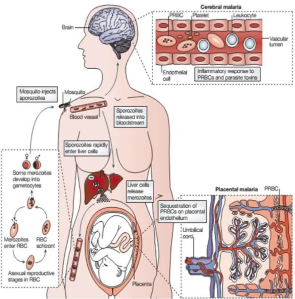
Parasite is transmitted when an infected female mosquito from the gender Anopheles takes a blood meal from a mammalian host (Fig. 1). After being injected subcutaneously, sporozoites travel through the blood to rapidly invade the hepatocytes in the liver, where they develop and multiply into thousands of merozoites (Prudêncio et al., 2006). Abrupt increase in merozoite numbers will eventually lead to hepatocyte rupture with parasite release into circulation, beginning a systemic infection with the onset of the first symptoms. When in circulation, merozoites are capable of invading erythrocytes relying on their highly variable surface receptors to mediate binding (Miller et al., 2002). Once inside erythrocytes, the parasite changes the surface protein repertoire of the cell by expressing its own proteins at the membrane. P. falciparum erythrocyte membrane protein 1 (PfEMP1) is a protein with variable surface antigens (VSA) encoded by an extended var gene family that mediates adhesion of the infected erythrocytes (IEs) to the vasculature and brain endothelium, as well as to the placenta in pregnant women (Ho and White, 1999). This leads to a local accumulation of IEs in a process known as sequestration, which promotes a set of conditions that allows maturation and multiplication of the parasite. In addition, this phenomenon has an impact also in IE survival since it avoids parasite clearance by the spleen (Engwerda et al., 2005). Once it reaches late stage, the fully mature parasite (schizont) leaves the cell due to erythrocyte lysis releasing merozoites, which are capable of engaging circulatory erythrocytes. Gametocytes, that are responsible for ensuring the sexual stage of the life cycle inside the mosquito, are also released from erythrocytes upon lysis, becoming available to be collected by the invertebrate host to fulfill the cycle. Inside the mammalian host, the accumulation of IEs can happen in several tissues as previously mentioned (Ho and White, 1999). Accumulation in the brain (cerebral malaria) and in the placenta (placental malaria) (Fig. 1) may lead to the more severe pathological outcomes of
2
the disease, triggering an exacerbated immunological response that not only contributes to the disease control, rather contributing to the fatalities (Schofield and Grau, 2005).
Figure 1. Plasmodium life cycle and pathological outcomes. Upon taking a blood meal, the infected
Anopheles injects the sporozoites into circulation that rapidly travel to the liver. Hepatocytes fulfil their
nutritional needs and give them shelter until they become fully developed and mature. Maturity results in the burst of hepatocytes and release of merozoites into circulation, which infect erythrocytes, maturing into gametocytes and late stage schizonts. Erythrocytes invasion lead to the onset of the first symptoms and sequestration in the brain and placenta result in pathologies as cerebral malaria (CM) and placental malaria (PM), respectively (Schofield and Grau, 2005).
3
Placental-malaria (PM)
In sub-Saharan Africa approximately 28 million pregnant women are exposed to malaria and are at high risk of developing placental malaria (WHO, 2015). Amongst other factors, susceptibility to infection during pregnancy is highly associated to the placenta, which constitutes a suitable niche for parasite accumulation. The adhesion of P. falciparum-IEs is mediated by a variation of the PfEMP1 protein known as var2CSA (Salanti et al., 2003) that promotes the interaction between the IEs and the glycosaminoglycan chondroitin sulfate A (CSA) (Fried and Duffy, 1996), which is expressed in syncytiotrophoblasts (placental cells). This process of IE sequestration is the leading cause of poor pregnancy outcomes. Abortion, preterm delivery and intrauterine growth restrictions (IUGR) will affect newborn’s longevity, which generally have low birth weight (LBW) upon delivery (Desai et al., 2007).
Major consequences of Plasmodium infection in the placenta are inflammation and impaired microcirculation. Infection induces trophoblasts (foetal-derived placental cells) to produce chemokines, which will act as chemoattractants for proinflammatory cells (Lucchi et al., 2008, 2011; Walter et al., 1982) that are recruited to the site of infection. Proinflammatory cytokines such as IFN-γ (interferon- γ) and TNF-α (tumor necrosis factor α) are secreted (Fried et al., 1998) and these were shown to be linked to placental microcirculatory defects and foetal loss. Knockout mice for IFN-γ had delayed foetal death yet, sustained TNF-α production was shown to induce placental disruption and excessive coagulation (Poovassery et al., 2009). Additionally, infection leads to high fibrin deposition and fibrinolysis resistance (Avery et al., 2012). Together, the cytokine-driven coagulopathy and antifibrinolytic state affect placental microcirculation leading to foetal growth restrictions (FGR). Moreover, the complement pathway molecule C5a, a regulator of inflammatory and immune response, was also shown to be linked to FGR; C5a was shown to be upregulated in PM, affecting angiogenesis and, consequently, foetal development (Conroy et al., 2013). Besides microcirculatory defects it has been suggested that inflammation might also impact metabolic pathways, affecting foetal development. Inflammation in the placenta has been shown to reduce the expression of some amino acid transporters (Boeuf et al., 2013), glucose transport isoform 1 (GLUT-1) (Chandrasiri et al., 2014) and lipid and hormone transporters (Lybbert et al., 2016), which are critical for foetal growth. Additionally, disruption of other metabolic pathways, namely iron homeostasis, might also contribute to increased foetal death in PM (Penha-Gonçalves et al., 2014).
Despite current knowledge on PM pathology, more studies are needed to fully understand this disease and for that, animal models are essential tools that should be explored. Mouse models for the study of PM remain controversial due to the lack of molecular homology
4
between mouse and human strains of Plasmodium (Hviid et al., 2010). However, PM models have been shown to recapitulate several aspects of human PM; infection of BALB/c mice at mid-stage pregnancy with P. berghei impacts foetal development and viability (Neres et al., 2008). P. berghei pre-immune mice showed less severe pathology and were protected after multiple gestations – a model that reproduces features of women living in high malaria endemic areas (Marinho et al., 2009). Several parasite strains where shown to be capable of inducing placental pathology in mice. Infection of C57Bl/6 pregnant mice with P. berghei strains that do not induced cerebral malaria (CM) in B6 mice such as NK65, K173, PM4KO (Rodrigues-Duarte et al., 2012) lead to severe placental pathology suggesting different mechanisms underlying pathology between CM and PM. Moreover, P. chabaudi infection at earlier gestational stage (G10) induces abortion by IFN-γ and TNF-α-driven coagulopathy (Poovassery and Moore, 2009). Recently our lab has developed a var2CSA-based PM model induced by a transgenic P. berghei expressing var2CSA (de Moraes et al., 2016). This model is a tool for in vivo pre-clinical studies to understand var2CSA-mediated pathology. These models will give a tremendous contribution to the understanding of PM pathology, foetal and placental abnormalities.
Placental Development and Function
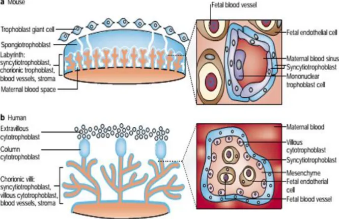
It is imperative to ensure placental development and function, which is critical for the survival and proper development of the foetus. Placenta has a vast role of function guarantying nutrients and gas exchanges (Lager and Powell, 2012), tolerance against maternal immune system (Kanellopoulos-Langevin et al., 2003) and protection against pathogens (Robbins and Bakardjiev, 2012).
Two structurally and functionally distinct units compose this highly complexed and specialized organ: the chorionic plate, constituted by foetally derived villi, which contain the physical structures that separate foetal compartment from maternal blood spaces, and the basal plate that is constituted by the uterine wall that separates placental microenvironment from maternal decidua and myometrium. In humans (Gude et al., 2004) (Fig. 2b), the chorionic villi are branched extension of the foetal chorion mainly composed by a very particular type of foetally derived cells known as trophoblasts. The villi have protrusions that increase the area of contact with the maternal blood, turning physiological exchanges more effective, and those are surround by a multicellular layer called the placental membrane. This membrane is the physical barrier that separates foetal from maternal compartments. It is constituted by a multinucleated layer of syncytiotrophoblast (STB) that mediates all the physiological processes that were previously mentioned. Below it, a layer of mononuclear
5
cytotrophoblast (CTB) can be used to replace syncytium or to promote the anchoring of the villi to the decidua after undergoing a phenotypic change to extravillous cytotrophoblast (EVT). In the core of these chorionic villi, extensive networks of foetal capillary surrounded by endothelium receive nutrients and oxygen transported by trophoblasts, delivering foetal waste products to be secreted into the maternal circulation. At the anchoring villi, CTBs and STBs ensure the attachment of the villi to the uterine wall and the phenotypic switch of the CTBs to EVTs make them capable of invading and remodeling the uterine spiral arteries. Therefore, these cells, after replacing maternal endothelium, they become responsible for controlling placental perfusion by modulation blood flow.
Mice have functionally identical but structurally distinct placentas when comparing with humans (Rossant and Cross, 2001) (Fig. 2a). Mice placentas are arranged in a structure called the labyrinth, which is different from the human chorionic villi. Nonetheless, both are comparable in terms of some cellular populations regarding their features. Early in placental development, trophoblast giant cells (TGCs) acquire an early invasive phenotype that is analogous to the one from EVTs in humans. Invasion and implantation takes place followed by a replacement of maternal endometrium by the chorionallantoic structure that results from fusogenic events between chorion and allantois. Arising from the chorion, two layers of STB and mononuclear trophoblastic cells cover the foetal vasculature that rises from the allantois as it happens in humans. Eroded endometrium is replaced by trophoblasts, which surround maternal sinuses (Adamson et al., 2002) canalizing maternal blood across the placenta. Mononuclear trophoblastic cells are in direct contact with the maternal blood but their functions are still unknown.
Both placental types are haemochorial (Wildman et al., 2006), which means that chorion-derived structures are in direct contact with the blood that is freely circulating in the maternal blood spaces. Despite the structural differences between human and mouse placentas, the similarities between trophoblast populations, the haemochorial structure and hemotrophic nutrition, make the rodent model a very useful tool in the study of placental diseases, being always mindful of how careful one must be regarding the extrapolations that can be made from one model to the other (Malassiné et al., 2003).
6
Figure 2. Comparative anatomy of the mouse and human placenta: structural and cellular comparison. a) Mouse placenta. Structure of the labyrinth and detailed view of maternal-foetal interface.
Bilayer of syncytiotrophoblast (STBs) surrounding maternal sinus separating maternal blood from foetal capillary. Mononuclear trophoblastic cells of unknown function in direct contact with the maternal blood. b)
Human placenta. Chorionic villi are the main structures in human placenta. Detailed picture show the villous
surrounded by a monolayer of STB containing cytotrophoblasts (CTB) beaned it. This membrane separates maternal blood that bathe the villous from foetal vessels. (Rossant and Cross, 2001).
7
Plasmodium infection impact on vasoactive factors
As previously mentioned, trophoblasts are in direct contact with the maternal blood, being responsible for the regulation of blood flow, canalizing the blood into maternal blood spaces. Previous works, using an established placental malaria mouse model have shown that trophoblasts are capable of actively modulate the blood flow in the placenta, rearranging compartments by “opening and closing” maternal blood spaces (de Moraes et al., 2013). These findings suggest that trophoblasts might respond to vasomodulatory factors leading to vasoconstriction or vasodilation of maternal sinuses. It is known that trophoblasts express a vast repertoire of angiogenic and vasomodulatory factors that have a critical role in placentation, maintaining local homeostasis and normotension in uteroplacental perfusion (Valdes et al., 2009). Previous studies in our lab (unpublished) showed dysregulation of the mRNA levels of expression of vasomodulatory factors such as the bradykinin-receptor 2 gene (Bdkrb2, B2R) (Fig. 3A, B and E) and endothelial nitric oxide synthase (Nos3, eNOS) (Fig. 3C, D and F) during infection. In non-infected mice (NI), placental Bdkrb2 transcript levels increased throughout late gestation (Fig. 3A) whereas in infected (INF) ones Bdkrb2 levels were modulated from gestational day 16 (G16) onwards and significantly decreased at G18. (Fig. 3B). Yet, when comparing the mRNA levels between INF and NI placentas for the different time-points, we observed a marked downregulation at G18 (Fig. 3E). The levels of
Nos3 mRNA also increased progressively from G16 onwards in NI placentas (Fig. 3C)
whereas in INF placentas no alterations were observed from G16 to G18 (Fig. 3D). In comparisons between INF and NI placentas, Nos3 mRNA levels were downregulated from G17 onwards (Fig. 3F). Additionally, kininogen levels were decreased in the context of infection both in the peripheral blood (Fig. 3G) and in placental extracts (Fig. 3H), as well as nitric oxide levels (data not shown). These components, which expression and production were altered during infection are members of the bradykinin-signaling pathway that was shown to be linked to vasodilation and are dysregulated in complicated prengancies (Corthorn et al., 2006). As such we hypothesized that infection might impair the transduction signaling through the bradykinin-signaling pathway leading to placenta insufficiency and poor pregnancy outcomes.
8
Figure 3. Plasmodium infection induces dysregulation of Bdkrb2, Nos3 mRNA levels and kininogen levels. Pregnant mice infected at G13 were euthanized at different time-points (G16-G18) from which
placentas were removed for RNA extraction and quantification of the mRNA levels of the Bdkrb2 (B2R) gene (A, B and E) and Nos 3 (eNOS) gene (C, D and F) by qPCR. Fold change to G16 of the mRNA levels of the
Bdkrb2 gene in A) non-infected (NI) and B) infected (INF) placentas. Fold change to G16 of the mRNA levels
of the Nos3 gene in C) NI and D) INF placentas. E) Fold change of the mRNA levels of the Bdkrb2 gene normalized to NI. F) Fold change of the mRNA levels of the Nos3 gene normalized to NI. Additionally, kininogen levels were measured in G) maternal serum and H) placental extracts in NI and INF mice using competition ELISA. * p<0.05, ** p<0.01, *** p<0.001; line refer to mean.
9
The bradykinin-signaling pathway
The bradykinin-signaling pathway belongs to the pleiotropic kallikrein-kinin system (KKS). This complex network is responsible for triggering a broad spectrum of cellular and physiological responses after ligand binding to the respective receptor, being involved in vasomodulation, vascular permeability, inflammation and pain (Bhoola et al., 1992). Briefly, all kinin peptides (i.e. bradykinin) are converted from a kininogen precursor (HK) that is cleaved into active kinins by the action of a peptidase known as kallikrein, which acts freely in the plasma or in the tissue (Fig. 4). Kinins induce their pleiotropic effects via activation of two known receptors: the type 1 (B1R) and the type 2 (B2R), that belong to a family of G protein-coupled receptors (GPCR) (Leeb-Lundberg et al., 2005), which are differentially expressed across the tissue. The B1R has minimal or null expression, which is induced in response to injuries and inflammation (Calixto et al., 2004). In contrast, B2R is constitutively expressed, being predominant in endothelium but less expressed in the smooth muscle (Figueroa et al., 2001). This receptor is expressed mainly at the cell surface but also has cytosolic and nuclear distribution due to internalization and cytosolic/nuclear trafficking (Takano and Matsuyama, 2014). As depicted in Fig. 4, bradykinin (BK), the most important and functionally active kinin, binds to the B2R inducing vasodilation by promoting the release of nitric oxide (NO) (Drummond and Cocks, 1995) by endothelial nitric oxide synthase (eNOS) (Knowles and Moncada, 1994) and the production prostaglandins (PGs) (Cherry et al., 1982). It was previously reported that HK (Brann et al., 2011) and NO (Conrad et al., 1993) production increases during pregnancy suggesting a contribution to maternal vasodilation. Additionally, elements of this signaling cascade, namely B2R and eNOS were shown to be expressed in the placenta (endothelial cells and trophoblasts), and dysregulation was shown to be linked to pathologies with complicated pregnancies as it happens in pre-eclampsia (Corthorn et al., 2006). This disease of unknown cause is related to placental malaria in a way that recapitulates some pathological outcomes caused by the parasite namely foetal growth restriction, hypertension and foetal and maternal mortality (Sibai et al., 2005).
Therefore, our previous unpublished data together with published reports support the hypothesis that Plasmodium infection might affect the bradykinin-signaling pathway leading to microcirculatory defects in the placenta and consequent foetal growth restriction. The present study focused on the evaluation of the expression levels of bradykinin-receptor 2 (B2R) on trophoblasts during infection. As such, we aimed at:
1. Ex vivo approach: Expression of B2R was assessed on trophoblasts isolated from infected placentas at G18. The choice for this time-point is based on features
10
previously observed: reduction of foetal weight, high values of parasitemia and downregulation of the vasoactive factors;
2. In vitro approach: B2R levels were evaluated on trophoblasts co-cultured with P.
berghei-IEs in vitro. Results will help clarify if IEs interaction with trophoblasts leads
to modulation of the B2R expression.
Figure 4. The bradykinin-signaling pathway. Kininogen (HK) is converted in the plasma or in the tissue by
kallikrein into active kinins. Bradykinin (BK) is the most important kinin, mediating several physiological processes throw the activation of the bradykinin-receptor 2 (B2R). Activation of this receptor promotes vascular permeability and avoids platelet aggregation. Additionally, it induces cytosolic calcium (Ca2+) mobilization that stabilizes endothelial nitric oxide synthase (eNOS) activity. The following nitric oxide (NO) production, together with prostaglandins (PGs), will induce vascular relaxation and vasodilation.
11
Materials and Methods
Animals and parasites
Eight week-old BALB/c females and C57Bl/6 males were obtained from the animal facility at the Instituto Gulbenkian de Ciência. Mice were bred and maintained under specific-pathogen free (SPF) conditions. Plasmodium berghei ANKA (Pb-ANKA) parasite constitutively expressing green fluorescent protein (GFP) under the control of the eef1 promoter (15cyl clone) (Janse et al., 2006) was used for all in vitro and in vivo experiments. For parasite expansion, infected-erythrocytes (IEs) from frozen vials were intraperitoneally (i.p.) injected in BALB/c mice and used when parasitemia reached 5-10% measured by GFP detection using flow cytometry. All procedures involving laboratory mice were in accordance with national (Portaria 1005/92) and European regulations (European Directive 86/609/CEE) on animal experimentation and were approved by the Instituto Gulbenkian de Ciência Ethics Committee and the Direcção-Geral de Veterinária (Official National Entity for regulation laboratory animals usage).
Pregnancy mouse model
BALB/c females were mated to C57Bl/6 males (2:1; female: male ratio) and separated in the next day. Females were weighed upon separation considering that day as the gestational day 1 (G1). Females were monitored and a weight gain of 4 – 5 g at G13 was taken as an indicative of pregnancy. Pregnant mice were used for experimental infection or for the isolation and culture of primary trophoblasts. Breedings from this allogenic model give litters with a high number of pups having small variations of weight between them.
Experimental design for evaluation of B2R expression
Ex vivo approach. At G13, females were intravenously (i.v.) infected with 106 IEs of the previously expanded parasite. Weight gain and parasitemia were monitored from G17 to G18. Infected and non-infected pregnant mice were euthanized at G18 from which placentas were collected for trophoblast isolation. Trophoblasts were stained for flow cytometry (FACS) analysis to assess isolates’ purity, by targeting cytokeratin 7 (KRT7) trophoblast marker, and bradykinin receptor 2 (B2R) surface expression.
In vitro approach. Primary trophoblasts were isolated at G18 and were either co-cultured
with Pb-IEs or non-infected erythrocytes (NIEs) after 6-7 days of enrichment. Trophoblasts were removed from culture, stained with anti-KRT7 and anti-B2R fluorescent antibodies and analyzed by FACS.
12
Primary trophoblast isolation
Primary trophoblasts were isolated as described elsewhere (adapted from Pennington et al., 2012). Briefly, pregnant mice were euthanized at G18 by CO2 narcosis and the uterus was carefully removed. Placentas from foetuses with similar weight between them were separated and pooled in groups. After dissection, placentas were coarsely minced inside a petri dish using a proper blade and transferred to a tube containing 1 mg/mL of collagenase IA (Sigma-Aldrich) and 4 U/µL DNase diluted in 25 mL of DMEM medium (Biowest) previously supplemented with 20 mM HEPES (Gibco) and 0.35 g/L sodium bicarbonate (Gibco) (wash buffer). Incubation was done at 37 ºC water bath for 30 – 40 minutes with vigorous pipetting every 10 minutes to digest the minced placentas. Macerated tissue was passed through a 70 µm strainer and washed at 400 x g for 10 minutes at 4 ºC. Cells were then added to a Percoll (Sigma-Aldrich) gradient made as described elsewhere (Nagamatsu
et al., 2004). Pelleted cells were resuspended in 4 mL of 25% Percoll, being gently layered
on the top of 4 mL of 40% Percoll contained inside a 15 mL tube. These layers were done by diluting, in wash buffer, a 90% Percoll solution previously prepared with PBS 10x. 2 mL of PBS 1x was added to the top of the 25% layer, followed by a centrifugation of 800 x g for 20 minutes at 4 ºC (without brake). Cells disposed on the interface between the two Percoll layers were collected, washed with PBS 1x and treated with ACK (ammonium-chloride-potassium buffer) for 5 minutes to lyse erythrocytes. Cells were washed, counted and plated in 24-well plate at a density of 2 x 10⁵ cells/well. Cells were cultured in DMEM complete medium containing 10% FBS (Gibco), 1% HEPES (Gibco), 1% sodium pyruvate (Gibco), 1% penicilin and streptomicin (Gibco), 1% non-essential amino acids (BioWhittaker), 1% glutamine, 0.1% β-mercaptoethanol (Gibco) and 0.1% gentamicin (Sigma). Cells were cultured for 1 week for enrichment of trophoblasts and washed every other day to remove non-adherent cells.
Parasite synchronization
P. berghei-IEs were synchronized as described elsewhere (Janse et al., 2006) to obtain late
stage schizont. Briefly, IEs were expanded in non-pregnant BALB/c mice during approximately one week reaching a parasitemia between 5 – 10%. Infected mice were sacrificed and immediately bleed by cardiac puncture using heparin to avoid clothing. Collected blood was transferred to a tube containing approximately 5 mL of RPMI (Gibco) supplemented with 20% FBS (Gibco) and 0.1% neomycin (Sigma-Aldrich) (synchronization medium) and centrifuged at 450 x g for 8 minutes. Pelleted erythrocytes were resuspended in 50 mL of synchronization medium and transferred to a 75 cm2 flask followed by the addition of a mixture of gas containing 5% CO2, 5% O2 and 90% N2 for 90 seconds at a 1.5 –
13
2 bar pressure. IEs were incubated overnight in a shaking incubator set for 50 rpm at 36.5 ºC. On next day, in the morning (approximately 18 hours after incubation), IEs-containing schizonts were sorted by passing suspension throw a 25 LS column using magnetic-associated cell sorting (MACS) technique. Column was washed with PBS 1x and cell suspension was passed throw it directly from the flask. Column was washed 6 times with PBS 1x and IEs were eluted using DMEM complete medium. Cells were washed at 450 x g for 8 minutes to pellet the IEs-containing schizont and then resuspended in complete culture medium for counting and further use.
In vitro assays
A number of 2 x 10⁵ synchronized Pb-IEs or NIEs were added to trophoblast cultures (1:1 ratio; erythrocytes-trophoblasts). NIEs used as a control were freshly collected from the tail of a non-infected mouse and centrifuged at 500 x g for 5 minutes to remove any plasma component that could influence the response of cultured trophoblasts. Pelleted NIEs were resuspended in complete medium, counted, and added to cultures. Experiments were conducted using different conditions: 1) Trophoblasts were incubated with IEs/NIEs for a period of 2h; 2) Trophoblasts were cultured with IEs/NIEs for 2h, washed with ACK to remove non-phagocytosed IEs/NIEs and then incubated with complete medium for an additional period of 2h. After incubation period, trophoblasts were detached from wells with 100 µL of Accutase (eBioscience) for 15 – 20 minutes at 37 ºC. Complete medium was added to neutralize the Accutase activity and cells were then aspirated to a FACS plate and stained with fluorescent antibody markers for KRT7 and B2R.
Flow Cytometry
Primary trophoblasts freshly isolated or detached from culture plates were stained to assess purity and the expression of the B2R by flow cytometry. Stainings were performed in FACS 96-well plates with U-shaped bottom and between 2 – 4 x 10⁵ cells were used per staining condition. Cells were firstly incubated with 100 µL of Fc Block (2.4G2 ab specific for FcII/III) diluted in PBS 1x containing 2% FBS and 0.1% sodium azide (NaN3) (FACS buffer) for 15 minutes at 4 ºC and then washed at 550 x g for 1 minute at 4 ºC. Pelleted cells were incubated with 50 µL of the anti-B2R Cy5 antibody (1:100) (Bioss Antibodies) for 30 minutes at 4 ºC. Cells were washed 2 times and resuspended in 100 µL of Fixation/Permeabilization solution (BD Biosciences) for 20 minutes at 4 ºC. Fixed cells were washed 2 times with Perm/Wash solution 1x (BioLegend), which kept them permeable for intracellular staining. After washing, cells were incubated with 50 µL of Perm/Wash 1x containing the anti-KRT7 (cytokeratin 7) PE antibody (1 µg per 2 – 4 x 10⁵ cells) (Santa Cruz Biotechnology) for 30
14
minutes at 4 ºC. Ultimately, cells were washed 2 times with Perm/Wash 1x and then with FACS buffer prior to cytometry analysis. Cells were resuspended in 100 – 400 µL of FACS buffer and acquired using the LSR Fortessa X-20 cytometer (BD Biosciences). Specific combinations laser (nm) - filter were used to detect the different fluorescence: 561 nm - 586/15E to detect PE, 640 nm – 640/30 to detect Cy5 and 488 nm – 530/30 to identify autofluorescent events.
Statistical Analysis
Data are displayed as scattered dot-plot with line at mean (normal distribution) or median (non-normal distribution), or as single dots representing mean ± SD. Unpaired T test and Mann-Whitney test were performed using GraphPad Prism 6.0 software. Comparisons between data with probability value of p<0.05 were considered significant.
15
Results
Characterization of the placental malaria mouse model
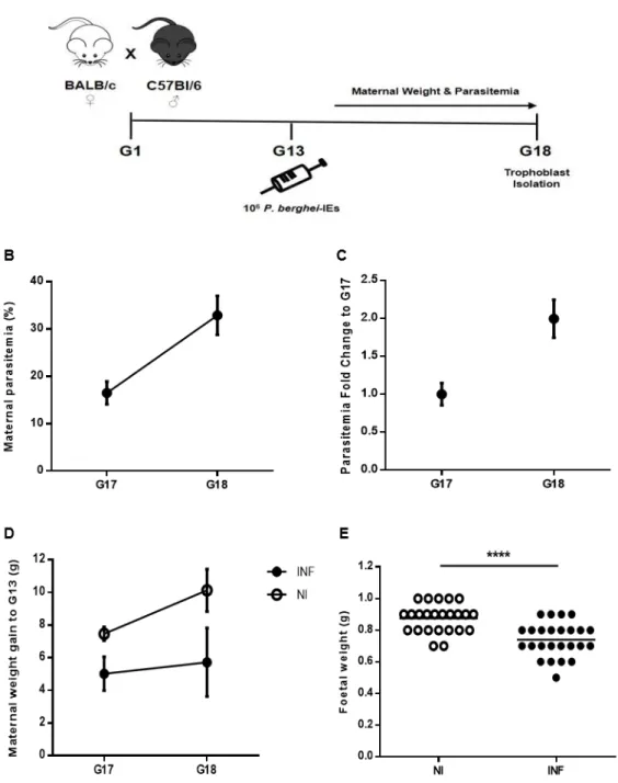
In our placental malaria mouse model (Fig. 5A) mice with confirmed pregnancies at G13 were infected with 106 P. berghei-IEs. Maternal weight gain and parasitemia were monitored during the following days until G18, when mice were euthanized for trophoblast isolation. Maternal weight increases throughout pregnancy and weight gain at late gestation (from G17 to G18) reflects that phenomenon. Non-infected mice gain an average of 7.467 ± 0.416 g at G17 and 10.133 ± 1.301 g at G18, approximately gaining 3 g within this 24-hour window (Fig. 5D). This increase in maternal weight is lost in infected pregnant mice, which showed null weight gain between G17 (5.025 ± 1.028 g) and G18 (5.725 ± 2.1 g). Parasitemia (Fig. 5B and 5C) was measured at late gestation, 4-5 days post-infection (d.p.i.). Signs of parasite expansion were seen at the fourth d.p.i. (G17) with mice having an average parasitemia of 16.5 ± 2.421 % (Fig. 5B). After 24 hours (G18), parasitemia recorded had approximately a 2-fold increase (Fig. 5C) to values averaging 32.9 ± 4.120 % (Fig. 5B). Pups taken upon caesarean were weighed to assess the impact of infection on their growth (Fig. 5E). Weight of foetuses from infected mothers was significantly decreased (0.74 ± 0.108 g) when compared to the ones that went through a normal pregnancy (0.875 ± 0.0.089 g) (p<0.0001). Together these results show the infection impact on foetal development, reproducing the pregnancy outcome features previously established. These data suggest that growth restrictions might be consequence of placental insufficiency caused by IEs sequestration, which is probably the cause of increase in maternal parasitemia.
16
Figure 5. Placental malaria mouse model. A) Schematic diagram of the gestation model, starting at G1
after mating, followed by i.v. infection with 106 Pb-IEs upon pregnancy confirmation at G13; maternal weight
and parasitemia were monitored until trophoblast isolation at G18. B) Maternal parasitemia expressed as percentage (%) of infected erythrocytes and C) G18 parasitemia fold increase to G17. D) Maternal weight gain in non-infected (NI) and infected (INF) mice between G17 and G18 (normalized by the number of foetuses). E) Foetal weight measured after caesarean of NI and INF mice. **** p<0.0001 (Unpaired T test); line refer to mean.
17
Impact of Plasmodium infection on B2R expression by trophoblasts in vivo
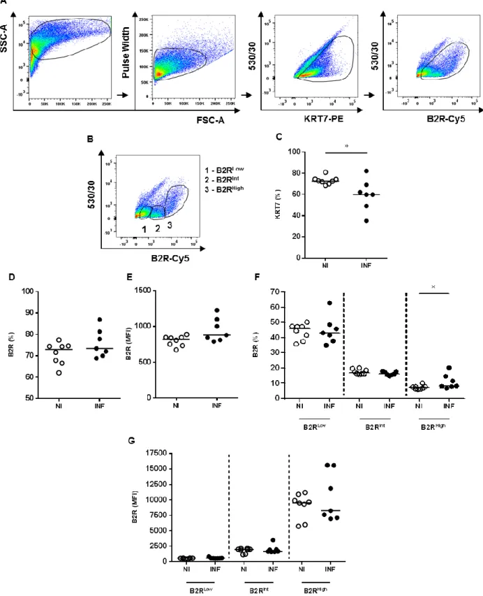
We observed an increase in parasitemia levels from G17 to G18, null variation on the maternal weight gain towards G18 and a reduction of foetal weight in infected pregnant mice (Fig. 5). This late time-point gathers particular characteristics of the model that may be an indicative of impaired placental homeostasis and function. This might be the best time window to look at the impact that infection has on the expression of vasoactive factors namely the bradykinin-receptor 2 (B2R). Modulation of B2R expression could suggest impaired signaling and function of the associated pathway. To confirm if the levels of the receptor are modulated by infection, we assessed the surface expression of the B2R in trophoblasts isolated at G18 from non-infected (NI) and infected (INF) mice. Freshly isolated trophoblasts were stained and analysed by flow cytometry. Depicted gating strategy (Fig. 6A) shows the selection of events with cellular morphology (FSC-A x SSC-A) from which very few doublets were excluded (FSC-A x Pulse Width). Trophoblasts were identified as the events positive for cytokeratin 7 (KRT7), a specific marker for this cell type. KRT7+ events were analysed for B2R expression by gating the events that were positive for Cy5 (Autofluor-FITC x B2R-Cy5). Parameter labelled as “530/530” was used as an empty channel to sort out autofluorescent events that were KRT7 negative (KRT7-). The percentage (%) of trophoblasts was significantly reduced (p=0.027) in infected samples (median of 72.7% in NI to 59.9% in INF) (Fig. 6C), suggesting that infection might be inducing death of the trophoblasts. No variation was observed neither in the overall percentage of trophoblasts expressing B2R (Fig. 6D) nor in the levels of expression of the receptor (median fluorescence intensity, MFI) (Fig. 6E). Additionally, it was possible to separate trophoblasts with different levels of B2R expression. Gating strategy illustration (Fig. 6B) shows the identification of the trophoblast populations with low (KRT+B2RLow), intermediate (KRT+B2RInt) and high (KRT+B2RHigh) expression of the receptor. Populations were distributed in an unequal frequency (Fig. 6F); KRT+B2RLow is the most abundant population, followed by KRT+B2RInt and then by KRT+B2RHigh. Population that expresses high levels of B2R (KRT+B2RHigh) was the only one showing a significant increase (p=0.014) in frequency during infection (Fig. 6F). In opposition, no variation was seen between these populations regarding B2R levels of expression (Fig. 6G). Taken together, the increase in percentage of the KRT7+B2RHigh population may be an indicative of receptor’s de novo synthesis and/or upregulation of the expression by mobilizing intracellular receptors to the membrane as it was previously shown to occur in some cell types (Takano and Matsuyama, 2014).18
Figure 6. Impact of Plasmodium infection on trophoblasts B2R expression in vivo. A) Schematic
representation of the gating strategy. Cells were gated A x SSC-A) and doublets were excluded (FSC-A x Pulse Width). Events positives for KRT7 (KRT7+) (KRT7-PE x 530/530) were identified as trophoblasts and B2R expression was evaluated (KRT7+B2R+) (B2R-Cy5 x 530/530). B) Identification of three populations of trophoblasts with differentially expression of B2R: 1 - B2RLow, 2 - B2RInt, 3 - B2RHigh. C) Frequency (%) of KRT7+ events (trophoblasts) and D) trophoblasts-expressing B2R (KRT7+B2R+). E) B2R expression by trophoblasts plotted as median fluorescence intensity (MFI). F) Frequency (%) and G) expression (MFI) of the different populations of trophoblasts-expressing B2R. Comparisons were done between trophoblasts from non-infected (NI) and infected (INF) placentas for the different parameters. * p<0.05 (Mann-Whitney test); line refer to median.
19
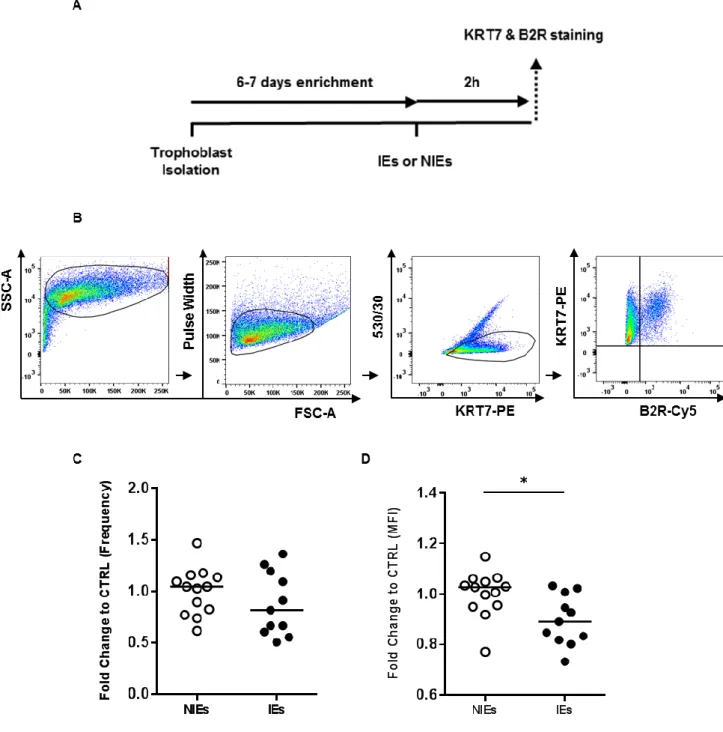
Plasmodium infection modulates B2R levels of expression in vitro
Evidences suggest that Plasmodium infection influences B2R expression by trophoblasts in our in vivo model (Fig. 6). Infection of trophoblasts in vitro allows to attribute any modulation on the B2R levels of expression to the presence of P. berghei-IEs since there is no other systemic factor that could interfere with the B2R expression. To evaluate if B2R modulation could be a consequence of the interaction with infected erythrocytes (IEs), we generated an
in vitro model (Fig. 7A) where primary trophoblasts were cultured with IEs or non-infected
erythrocytes (NIEs) for 2 hours. The choice for a 2-hour culture was based on our previous experimental set-up. We cultured trophoblasts with IEs or NIEs for 1 hour and 2 hours. Both conditions induced reduction of B2R expression on infected trophoblasts (compared to NIEs) but fold change was higher at 2-hour incubation compared to the 1-hour time-point (data not shown). Percentages of B2R+ trophoblasts were not altered in none of the two conditions. After incubation period, non-phagocytosed IEs or NIEs were removed with ACK, trophoblasts were detached from plates, stained with anti-KRT7 and anti-B2R fluorescent antibodies and analysed by flow cytometry. Figure 7B illustrates the gating strategy used to evaluate B2R expression on cultured trophoblasts. Cells were identified by morphology (FSC-A x SSC-A) followed by doublets exclusion (FSC-A x Pulse Width). Trophoblasts were identified as positive events for the KRT7 marker (KRT7+) while removing autofluorescent events using the 530/30 filter (KRT7-PE x 530/530). Finally, trophoblasts expressing B2R were identified inside the double positive quadrant (KRT7+B2R+) were frequency and median fluorescence intensity were measured. Our data showed that IEs do not significantly alter the frequency of trophoblasts expressing B2R after 2 hours of infection (Fig. 7C). On the other hand, it was observed an impact on B2R expression levels (Fig. 7D); IEs lead to a significant decrease of the MFI (p=0.0128). These results support the idea that IEs contact with trophoblasts might have direct consequences on the expression of B2R.
20
Figure 7. Plasmodium infection alters B2R levels of expression in vitro. A) Experimental design.
Isolated trophoblasts were plated and enriched for 6-7 days for infection with synchronized schizonts (IEs). Non-infected erythrocytes (NIEs) were added as control. After 2 (h)ours, cells were detached from plate and stained for KRT7 and B2R. B2R expression was measured by flow cytometry. B) Schematic representation of the gating strategy. Cells were gated (FSC-A x SSC-A) and doublets were excluded (FSC-A x Pulse Width). Events positives for KRT7 (KRT7+) (KRT7-PE x 530/530) were identified as trophoblasts and B2R expression was evaluated (KRT7+B2R+) (B2R-Cy5 x KRT7-PE). C) Frequency and D) MFI of B2R expression in trophoblasts co-cultured with IEs or NIEs for 2h. Comparisons were done by normalizing data from IEs to NIEs. * p<0.05 (Mann-Whitney Test); line refer to median.
21
Acute Plasmodium infection lead to a prolonged modulation of B2R
expression
We have observed that 2-hour incubation with P. berghei-IEs leads to reduction of expression levels of B2R on trophoblasts (Fig. 7D). To understand if the constant presence of IEs is needed to induce a modulatory phenotype we incubated primary trophoblasts with IEs or NIEs for 2 hours, removed non-phagocytosed IEs with ACK and then incubated trophoblast with complete medium (Ø) for an additional 2 hour period (Fig. 8A). Trophoblasts incubated with NIEs were submitted to the same conditions. IEs lead to a significant increase of the percentage of trophoblasts expressing B2R in the subsequent incubation period (p=0.0207) (Fig. 8B), yet the expression levels were not significantly altered (p=0.2786) (Fig. 8C) when comparing to non-infected control. This apparent reduction of the MFI levels after the additional incubation period suggests that the constant presence of the parasite is not needed to induce a prolonged downregulation of B2R. Moreover, by comparing the levels of expression between cells incubated for the additional 2-hour period after IEs exposure to conditions without the extra period (only submitted to IEs), we observed that the MFI levels are maintained (Fig. 8E). These results suggest that the downregulatory effect induced by infection might be prolonged throughout time independently of the presence of the parasite. We also observed an increase frequency of trophoblasts expressing B2R induced during the additional 2-hour period of culture without IEs (p=0.0003) (Fig 8D). Regarding non-infected cultures, no differences were observed neither in the frequency of trophoblasts-expressing B2R (Fig. 8D) nor in the expression levels (Fig. 8E). Together these results suggest that B2R modulation in infected cultures occurs due to interaction between parasite and trophoblasts. Additionally, the percentage of trophoblasts expressing B2R increases, which is in line with our in vivo studies.
22
Figure 8. Plasmodium infection leads to a prolonged modulation of B2R expression in vitro. A)
Experimental design. Isolated trophoblasts were plated and enriched for 6-7 days for infection with synchronized schizonts (IEs). Non-infected erythrocytes (NIEs) were added as control. After 2 (h)ours, cells were washed with ACK to remove non-phagocytosed erythrocytes and incubated with medium (Ø) for an additional period of 2h (+). Cells were detached from plate and stained for KRT7 and B2R. B2R expression was measured by flow cytometry. B) Frequency and C) MFI of B2R expression in trophoblasts co-cultured with IEs or NIEs for 2h with further incubation with medium (Ø) for 2h (+). Data from IEs were normalized to NIEs. D) Frequency and E) MFI of B2R expression in trophoblasts co-cultured with IEs or NIEs for 2h, additionally incubated with medium (Ø) for another 2h (+). Data from IEs and NIEs (+) were normalized to the controls that went only through a 2h incubation with IEs or NIEs (-), respectively. Comparisons were done by normalizing test conditions to their internal controls. * p<0.05, *** p<0.001 (Mann-Whitney test); line refer to median.
23
Discussion
In the present study, we show that P. berghei infection can modulate the expression of bradykinin-receptor 2 (B2R) on trophoblasts. Using a well-established PM mouse model, we looked at B2R expression on late gestation (G18). We observed three distinct populations regarding the levels of B2R expression and that the percentage of B2R+ cells were increased in the B2RHigh subpopulation compared to non-infected mice. We also show that in
vitro P. berghei-IEs induced modulation of B2R expression levels after 2-hour incubation
with IEs. Yet, no changes were observed in the percentage of cells expressing B2R on the first 2-hour incubation with IEs. However, frequency of trophoblasts expressing B2R increased after the additional 2-hour incubation period without IEs. Altogether, these results suggest that modulation of the B2R expression might occur due to direct interaction between IEs and trophoblasts.
The bradykinin-signaling pathway exerts its functional effects mainly through the activation of B2R upon bradykinin binding inducing vasomodulation and vascular permeability (Leeb-Lundberg et al., 2005). We hypothesized that impaired signaling through B2R may lead to impaired vasodilation and control of placental perfusion, which can have an impact on foetal weight gain and survival. As such, we assessed B2R surface expression on trophoblasts isolated from non-infected and infected pregnant mice euthanized at G18. Expression was evaluated at this late time-point for different reasons: 1) high values of parasitemia were reached at this time-point; 2) a significant decrease in foetal weight was observed specifically at G18 and 3) downregulation of the B2R gene was only observed at G18. Using flow cytometry, we identified trophoblasts as positive events for the cytokeratin 7 marker (KRT7+) (Maldonado-Estrada et al., 2004). Placentas infected with Plasmodium had a significant reduction in the percentage of trophoblasts suggesting cell death caused by infection. After identification of three subpopulations of trophoblasts with different levels of B2R expression, we observed that the subpopulation with high levels of expression (KRT7+B2RHigh) was increased during infection. It is possible that increase in percentage of B2R+ trophoblast during infection is reflecting the upregulation of the Bdkrb2 gene at G17 supporting de novo synthesis. On the other hand, it is possible that the exposure of the B2R at the surface does not depend on de novo synthesis (since the mRNA levels are decreased at G18) but on the intracellular distribution of the receptor, which can be recycled to the membrane (Takano and Matsuyama, 2014). Stainings were performed on trophoblasts from pooled placentas due to technical restrictions: it is not possible to isolate a sufficient amount of cells from individual placentas to perform proper stainings. Taking this into account, it is possible that these conditions masked a possible differential expression of the receptor between infected placentas that could have different degrees of infection.
24
Our data on evaluation of B2R surface expression on G18, together with the unpublished previous work that shows downregulation of the mRNA levels of Bdkrb2 gene at the same time-point, support the idea that the parasite is capable of modulating B2R expression. To clarify this, primary trophoblasts isolated from non-infected placentas were co-cultured with
P. berghei-IEs. This in vitro system does not include systemic factors that may be influencing
B2R expression in vivo. Trophoblasts incubated with parasite for 2 hours showed a decrease on B2R levels of expression but no significant changes were observed regarding the percentage of cells expressing the receptor. Yet, a significant increase in the frequency of trophoblasts expressing B2R was observed after an additional 2-hour incubation period with medium. The impact on the levels of expression seemed to be maintained after this period since the MFI levels were 1) decreased when comparing with the non-infected control that had the additional incubation period, although not significantly and 2) maintained when comparing to the infected control only stimulated for 2 hours. No changes were observed in uninfected conditions. Since IEs and NIEs were lysed with ACK before the second incubation, it is possible that trophoblasts suffered a prolonged modulation of the receptor without being in the constant presence of the parasite. B2R activation upon BK binding is known to cause downregulation, redistribution and internalization of the receptor (Takano and Matsuyama, 2014). These dynamics are known to be dependent of phosphorylation and dephosphorylation events (Blaukat et al., 1996) on the serine and threonine residues of the C-terminal tail of the receptor (Pizard et al., 1999), reducing the levels of available B2R at the cell surface. In our in vitro experiments, we observed downregulation of B2R expression levels after IEs infection. It is possible that contact between parasite and trophoblasts may trigger cytosolic kinases’ activity promoting phosphorylation of intracellular proteins leading to internalization of B2R as previously described (Takano and Matsuyama, 2014). Similar events were shown to occur in syncytiotrophoblast and in BeWo cell line where P.
falciparum-IEs were capable of inducing phosphorylation of tyrosine residues of cytosolic
proteins (Lucchi et al., 2006).
We observed that downregulation of B2R was followed by an increase in the percentage of trophoblasts expressing the receptor after 4 hours in culture in which the first 2 hours were incubated with IEs. Although we have not assessed underlying mechanisms to explain the increase of B2R+ cells, we speculate that this response could be consequence of proinflammatory cytokine secretion by trophoblasts after IE contact. It has already been shown that Plasmodium induces the production of proinflammatory cytokines namely IFN-γ (interferon- γ) and TNF-α (tumor necrosis factor α) by the placenta during pregnancy (Fried et al., 1998). Poovassery and co-workers showed that TNF-α was secreted by cultured cells isolated from the ectoplacental cone (murine trophoblasts) after exposure to IEs