https://doi.org/10.1590/0004-282X20180099
ARTICLE
The effect of CA1 administration of orexin-A
on hippocampal expression of COX-2 and
BDNF in a rat model of orofacial pain
O efeito da administração de CA1 de orexina-A na expressão hipocampal de COX-2 e BDNF
em um modelo de dor orofacial em ratos
Razieh Kooshki1, Mehdi Abbasnejad1, Saeed Esmaeili-Mahani1, Maryam Raoof2,3
1Shahid Bahonar University of Kerman, Faculty of Sciences, Department of Biology, Kerman, Iran;
2Kerman University of Medical Sciences, Institute of Neuropharmacology, Neuroscience Research Center, Laboratory of Molecular Neuroscience, Kerman, Iran; 3Kerman University of Medical Sciences, Endodontology Research Center, Kerman, Iran.
Correspondence: Mehdi Abbasnejad; Department of Biology, Faculty of Sciences, Shahid Bahonar University of Kerman, Kerman P.O. Box: 76135-133, Iran. E-mail: [email protected]
Conflict of interest: There is no conflict of interest to declare.
Received 25 March 2018; Received in final form 01 June 2018; Accepted 12 June 2018.
ABSTRACT
The neuropeptide orexin-A and its receptors are widely distributed in both hippocampal circuitry and pain transmission pathways. Objective:
Involvement of the CA1 orexin 1 receptor (OX1R) on the modulation of orofacial pain and pain-induced changes in hippocampal expression of cyclooxygenase-2 (COX-2) and brain-derived neurotrophic factor (BDNF) was investigated. Methods: Orofacial pain was induced by an intra-lip injection of capsaicin (100 μg). Reverse transcription polymerase chain reaction and immunoblot analysis were used to indicate changes in hippocampal BDNF and COX-2 expression, respectively. Results: Capsaicin induces a significant pain response, which is not affected by either orexin-A or SB-334867-A, an OX1R antagonist. However, an increased expression of COX-2 and decreased expression of BDNF was observed in the hippocampus of animals that received capsaicin or SB-334867-A (80 nM) plus capsaicin. Meanwhile, orexin-A (40 pM) attenuated the effects of capsaicin on the expression of COX-2 and BDNF. Conclusions: CA1 OX1R activation moderates capsaicin-induced neuronal inflammation and neurotrophic deficiency.
Keywords: Orofacial pain; orexins; brain-derived neurotrophic factor; cyclooxygenase 2, rats.
RESUMO
O neuropeptídeo orexina-A e seus receptores estão amplamente distribuídos nos circuitos do hipocampo e nas vias de transmissão da dor.
Objetivo: O envolvimento do receptor de orexina 1 CA1 (OX1R) na modulação da dor orofacial e alterações induzidas pela dor na expressão do hipocampo de ciclooxigenase-2 (COX-2) e fator neurotrófico derivado do cérebro (BDNF) foi investigado. Métodos: A dor orofacial foi induzida por injeção intra-labial de capsaicina (100 μg). A reação em cadeia da polimerase de transcrição reversa e a análise de imunotransferência foram utilizadas para indicar alterações na expressão de BDNF e COX-2 no hipocampo, respectivamente. Resultados: A capsaicina induz uma resposta significativa à dor, que não é afetada pela orexina-A ou pelo SB-334867-A, um antagonista do OX1R. No entanto, uma expressão aumentada de COX-2 e uma expressão diminuída de BDNF foi observada no hipocampo de animais que receberam capsaicina ou SB-334867-A (80 nM) mais capsaicina. Enquanto isso, a orexina A (40 pM) atenuou os efeitos da capsaicina na expressão de COX-2 e BDNF. Conclusões: A ativação de CA1 OX1R modera a inflamação neuronal induzida por capsaicina e a deficiência neurotrófica.
Palavras-chave: Dor facial; orexinas; fator neurotrófico derivado do encéfalo; ciclo-oxigenase 2; ratos.
Orofacial pain is one of the most prevalent and debilitat-ing pain conditions that arise from oral and facial structures. It is also characterized by significant mood distresses and
neuronal deficiencies. It is widely accepted that the trigemi
-nal subnucleus caudalis serves as the critical brainstem relay for orofacial nociception1,2.
Transient receptor potential vanilloid 1 channels are expressed in a subpopulation of trigeminal sensory neurons.
Activation of transient receptor potential vanilloid 1 chan-nels by chemical agents such as capsaicin, the active
com-pound of chili pepper, promotes calcium influx and sub
-sequent release of pro-inflammatory mediators and pain
molecules in trigeminal pathways3,4. In particular,
in initiation and progression of inflammatory responses5. Spinal COX-2 up-regulation has been associated with pain hypersensitivity following peripheral inflammation. It is thought to be mediated through the activation of the
NF-kB-associated pathways6.
Brain-derived neurotrophic factor (BDNF) is an agonist of tropomyosin receptor kinase B, a member of the tyrosine kinases family7. The binding of BDNF to tropomyosin recep
-tor kinase B activates the downstream molecules and initi
-ates the signaling events that are essential for neuronal sur-vival and synaptic plasticity8. Brain-derived neurotrophic factor depletion in definite brain regions has been reported in neurodegenerative and psychiatric disorders9. In addition,
BDNF is well known as a pain mediator in spinal and tri
-geminal nociceptive synapses. It has been shown that BDNF expression and induction is altered during either nociceptive
or inflammatory processes10.
Orexin-A and orexin-B are hypothalamic peptides that
stimulate target cells via two G-protein-coupled recep
-tors, named orexin 1 receptor (OX1R) and orexin 2 recep-tor (OX2R). Orexin-A equally binds to both OX1R and OX2R, whereas orexin-B shows a much higher affinity for OX2R than that for OX1R11. Orexinergic neurons distrib-ute through the central nervous system and regulate vari-ous physiological functions12,13. Specifically, orexin-A and OX1R are densely expressed in hippocampal formation14. It has been indicated that hippocampal orexin-A
micro-injection increases synaptic plasticity and memory effi
-ciency in rats15. Orexin-A also acts as a potent analgesic16,17. Specifically, the orexin-A modulatory effect on trigeminal
nociception has been demonstrated using different behav
-ioral and electrophysiological studies18,19. Amazingly,
orexin-A treatment was also able to diminish learning and mem
-ory loss in capsaicin-treated rats19. However, the underlying
mechanism(s) of orexin-A effects on the modulation of oro
-facial pain and pain-induced cognitive deficiency are poorly understood. In the present study, we investigated the role of CA1 OX1R in the modulation of capsaicin-induced
neu-ronal inflammation and neurotrophic deficiency in the hip
-pocampus, with emphasis on alterations in hippocampal expression of COX-2 and BDNF.
METHODS
Animals
Adult male Wistar rats, with a weight of 230-255 g were
selected for the experiment. Animals were kept in a tem
-perature-controlled room at 22°C ± 1°C, with a standard
12-hour light/dark cycle. The animals were given ad libitum
access to food and water. The protocol was approved by the ethical committee of the Kerman University of Medical Sciences, Kerman, Iran (EC: 93.26). All efforts were made to minimize suffering.
Surgery
The rats were deeply anesthetized by intraperitoneal injection of ketamine (100 mg/kg) combined with xylazine
(10 mg/kg). The animals were placed in a stereotaxic appara
-tus and stainless steel guide cannulas (22-gauge) were bilat
-erally implanted in the CA1 region according to the Paxinos and Watson atlas (3.8 mm posterior to the bregma, 2.2 mm lateral from the midline and 3.2 mm depth to the cortical sur-face). Two screws were inserted into the skull. Dental cement mixed with acrylic liquid was used to firmly hold the guide cannula. The cannulas were then closed by tightly-fitting screws. All animals were allowed at least one week to recover from surgery, before the microinjection of drugs. At the end of the experiment, methylene blue was injected through the guide cannula to confirm the correct cannula placement. If the cannula was not fixed in the exact place, that data was discarded from the analysis.
Drugs
Capsaicin (Sigma-Aldrich, USA) was dissolved in a combi
-nation of Tween 80/ethanol/distilled water (1:1:8). Orexin-A
and SB-334867-A (both Tocris, USA) were dissolved in nor
-mal saline and dimethyl sulfoxide, respectively.
Microinjection
Orexin-A and SB-334867-A were microinjected into the CA1 using an injection needle (27-gauge) that was attached by polyethylene tubing to a 5 μl Hamilton microsyringe. The injection needle was inserted 1 mm beyond the tip of the guide cannula. Infusions were delivered in a total injection volume of 2 μl (1 μl each side). The needle was left in place for another 30 seconds to avoid back flush.
Experimental groups
Four experimental groups of rats (n = 6) were used as
follows: sham group, which received no injection; capsa
-icin group, which received subcutaneous intra-lip injection of capsaicin (100 μg/10 µg); and OX1R agonist and antago -nist pretreated groups, which received orexin-A (40 pM) or SB-334867-A (80 nM) into the CA1, prior to capsaicin injection. The future use of sham surgery as a control may
be necessary to validate the results. However, previous stud
-ies have shown that strictness of capsaicin-induced orofacial
pain was not affected by surgical procedures19,20. So, in the
present study, a sham surgical group was not included.
Pain induction
area with its fore or hind paws, for a period of 40 minutes19. Twenty-four hours after nociceptive assessment, the animals were sacrificed under deep anesthesia by exposure to high concentrations of carbon dioxide and the hippocampal tis-sues were separated and stored at -80°C.
Western blot analysis
Rat hippocampal tissues were lysed in RIPA buffer con
-taining 10 mM Tris–HCl, pH 7.4, 150 mM NaCl, 1 Mm eth-ylenediaminetetraacetic acid, 0.1% sodium dodecyl sulfate, 0.1% Na-deoxycholate, 1% NP-401% NP-40 and protease
inhibitors (1 mM phenylmethylsulfonyl fluoride, 2.5 μg/ ml of
leupeptin, 10 μg/ml of aprotinin) and 1 mM sodium orthovan-adate. Equal amounts of protein from each sample (40 μg) were separated using sodium dodecyl sulfate polyacrylamide gel electrophoresis and transferred to a polyvinyl difluoride membrane. Blots were then blocked with 3% nonfat milk in
0.1% Tween-Tris-buffered saline for two hours at room tem
-perature, followed by overnight (4°C) incubation with COX-2 primary antibody (1:15,000, Santa Cruz Biotechnology, USA). The primary antibody was detected with goat mouse horseradish peroxidase-conjugated secondary
anti-body (1:15,000, Santa Cruz Biotechnology, USA). The anti
-body-antigen complexes were identified using the ECL
system and exposed to Lumi-Film chemiluminescent detec
-tion film (Roche, Germany). Lab Works analyzing software (UVP, UK) was used to evaluate the intensity of the blotting bands. β-actin (1:10,000) was used as the loading control. The
expression values were presented as tested proteins / β-actin
ratio for each rat.
Reverse transcription polymerase chain reaction (RT-PCR)
Total RNA was isolated from the hippocampus by a modi
-fied version of the guanidine isothiocyanate-phenol-chloro
-form method using RNX+ reagent. The RT-PCR reaction was
performed using Oligo-dT primer and M-MuLV reverse tran
-scriptase, based on the manufacturer’s protocol (Fermentas GMBH, Germany). The reactions were incubated at 42°C for 60 minutes and then inactivated at 70°C for 10 minutes. Three separate PCR reactions were used for studying gene expression in the samples obtained from each rat. Each PCR reaction was carried out using selective forward and reverse primers for β-actin (as an internal standard) and BDNF genes. The sequence of the primers used was: BDNF forward:
5′TCC ATT CAG CAC AAG CC-3′; BDNF reverse: 5′GAG CCC
AGT CAG GTA ACC AC-3′; β-actin forward: 5′-CCC AGA
GCA AGA GAG GCA TC-3′; β-actin reverse: 5′-CTC AGG
AGG AGC AAT GAT CT-3′. Taq DNA polymerase (Cinaclon,
Iran) was used for DNA amplification and reactions were set
according to the manufacturers’ instructions. The PCR reac
-tions were incubated at 94°C for five minutes, followed by 35 cycles of thermal cycling (45 seconds at 94°C, 45 seconds at 60°C and 45 seconds at 72°C). The final cycle was followed
by a five-minute extension step at 72°C. For analysis of PCR products, prestained (EtBr) 1.5% agarose LMMP (Roche, Germany) gel was used.
Statistical analysis
Data are presented as means ± standard error of the mean. Statistical analysis comprised one-way analysis of variance followed by post-hoc Tukey’s test. The p-value < 0.05 was considered statistically significant.
RESULTS
Nociceptive behavior assessment
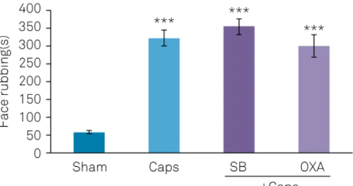
Capsaicin-treated rats showed significant increases in nociceptive responses (p < 0.001). Pretreatment with either orexin-A (40 pM) or SB-334867-A (80 nM), into the CA1, had no significant effect on capsaicin-induced nociceptive behavior (Figure 1).
The effects of CA1 microinjection of OX1Rs agonist and antagonist on hippocampal COX-2 induction in capsaicin-treated rats
Immunoblot analysis showed significant differences in
hippocampal COX-2 protein levels among the different experi
-mental groups. As shown in Figure 2, COX-2 induction in cap
-saicin (p < 0.01) and SB-334867-A-pretreated (p < 0.001) groups were significantly increased compared with the sham group; while pretreatment with orexin-A (40 pM) diminished the effects of capsaicin on COX-2 expression (p < 0.001) (Figure 2).
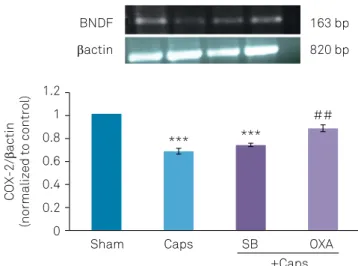
The effects of CA1 microinjection of OX1Rs agonist and antagonist on hippocampal BDNF expression in capsaicin-treated rats
In capsaicin-treated rats, the BDNF mRNA expression was significantly decreased compared with the sham group (p < 0.01). However, in rats microinjected with orexin-A (40 pM) prior to capsaicin application, BDNF expression was up-regulated compared with the capsaicin group (p < 0.001) (Figure 3).
Sham Caps
*** *** ***
SB
+Caps OXA 400
350 300 250 200
Face rubbing(s)
150 100 50 0
SEM: standard error of the mean; Caps: capsaicin; OXA: orexin-A; SB: SB-334867-A; ***p < 0.001 versus sham group.
DISCUSSION
In the present study, high levels of COX-2 protein in the
hippocampus were detected following capsaicin-induced oro
-facial pain. Interestingly, CA1 administration of orexin-A was able to attenuate the effects of capsaicin on COX-2 induction. COX-2 is a potent neuro-inflammatory molecule that has a critical role in nociceptive modulation and neurological chal-lenges. In the central nervous system, COX-2 is highly distrib-uted in hippocampal neurons and its expression is correlated with the induction of pro-inflammatory cytokines and pain molecules21. In the study by Gao and Duan, increased COX-2
in the trigeminal subnucleus caudalis of rats was observed fol
-lowing tooth movement pain5. Capsaicin activates transient
receptor potential vanilloid 1 channels on a subpopulation of primary afferent sensory neurons, specifically C fibers, involved in nociception. It causes prolonged and burning pain responses via induction of pain neuropeptides and mediators including neurokinin-A, substance P and CGPR in trigeminal nerves3. Furthermore, capsaicin can induce central sensitiza-tion and pain hypersensitivity by increased sensitivity within the peripheral trigeminal nerves. Increasing central
transmis-sion of nociceptive signals results in enhancement of inflam
-matory pathways in brain circuitry22.
Here, orexin-A showed an anti-inflammatory effect
through down-regulation of COX-2. Consistent with the cur
-rent data, in the study by Xiong et al., tumor necrosis factor alpha, an important pro-inflammatory cytokine, production
by lipopolysaccharide-stimulated microglial BV2 cells was sig
-nificantly reduced by orexin-A pretreatment23. It has been also
reported that the orexin system has a protective effect against focal ischemia by modulation of inflammatory responses in rats24. Rodent studies have shown that administration of orexin-A results in significant suppression of formalin and
capsaicin-induced inflammation17,19. There are strong correla
-tions between the up-regulation of pro-inflammatory media
-tors and the risk of neuronal damage and neurodegeneration. Particularly, it has been reported that hippocampal-dependent functions such as memory performance, synaptic plasticity and neurogenesis are disturbed during pain and inflammatory processes25. Orexin-A and OX1R receptors are widely expressed in the hippocampus and improve cognitive performances and synaptic plasticity in hippocampal formation13,26. Moreover,
there are various well-documented reports on orexin-A’s neu
-roprotective and anti-apoptotic properties, which may be beneficial in mediating the suppression of capsaicin-induced
neuronal inflammation27,28. Interestingly, Raoof et al. reported
that rat hippocampal OX1R expression is down-regulated
in responses to inflammatory tooth pain20. So, the inhibitory
effect of orexin-A on capsaicin-induced COX-2 expression may be due to orexin-A’s neuroprotective potentials.
In addition, our data showed that intra-lip capsaicin administration reduces BDNF mRNA expression in the hip-pocampus. This result is supported by the findings of Duric et al. that showed down-expression of hippocampal BDNF following formalin or complete Freund’s adjuvant-induced
inflammatory pain29. It has also been reported that
neuro-pathic pain induces spatial learning and memory impair-ments as well as BDNF down-regulation in the hippocampus of rats30. A recent study suggested that trigeminal neuralgia
induces cognitive deficits in rats. Moreover, it was associ
-ated with decreased activities of the c-AMP-responsive ele
-ment binding protein (CREB)/BDNF pathway, as a critical
pathway involved in synaptic remodeling and memory con
-solidation31,32. Other studies, however, indicated that the BDNF/TrkB-mediated signaling pathway is involved after tissue injury and in the development of nerve injury-induced neuropathic pain33,34.
Sham Caps
** ***
###
SB +Caps
OXA 1.8
1.6 1.4 1.2 1
COX-2/
actin
(normalized to control)
COX-2
actin
74 KDa
42 kDa
0.8 0.6 0.2 0
SEM: standard error of the mean; Caps: capsaicin; SB: SB-334867-A; OXA: orexin-A; **p < 0.01 versus sham group; ***p < 0.001; ###p < 0.001 versus
Caps and Caps+SB-334867-A treated groups.
Figure 2. Effects of CA1 administration of orexin-A (40 pM) and SB-334867-A (80 nM) on capsaicin-induced COX-2 expression in the hippocampus. Values represent mean ± SEM.
Sham Caps
*** ***
##
SB
+Caps OXA 1.2
1 0.8 0.6
COX-2/
actin
(normalized to control)
BNDF
actin
163 bp
820 bp
0.4 0.2 0
SEM: standard error of the mean; Caps: capsaicin; SB: SB-334867-A; OXA: orexin-A; ***p < 0.001 versus sham group; ##p < 0.01 versus Caps and Caps+
SB-334867-A treated groups.
References
1. Sessle BJ. Neural mechanisms and pathways in craniofacial pain. Can J Neurol Sci. 1999 Nov;26(1 Suppl 3):S7-11. https://doi.org/10.1017/S0317167100000135
2. Delwel S, Binnekade TT, Perez RS, Hertogh CM, Scherder EJ, Lobbezoo F. Oral health and orofacial pain in older people with dementia: a systematic review with focus on dental hard tissues. Clin Oral Investig. 2017 Jan;21(1):17-32. https://doi.org/10.1007/s00784-016-1934-9 3. Bae YC, Oh JM, Hwang SJ, Shigenaga Y, Valtschanoff JG.
Expression of vanilloid receptor TRPV1 in the rat trigeminal sensory nuclei. J Comp Neurol. 2004 Oct;478(1):62-71. https://doi.org/10.1002/cne.20272
4. Eid SR, Crown ED, Moore EL, Liang HA, Choong KC, Dima S et al. HC-030031, a TRPA1 selective antagonist, attenuates inflammatory- and neuropathy-induced mechanical hypersensitivity. Mol Pain. 2008 Oct;4:48. https://doi.org/10.1186/1744-8069-4-48
5. Gao Y, Duan YZ. Increased COX2 in the trigeminal nucleus caudalis is involved in orofacial pain induced by experimental tooth movement. Anat Rec (Hoboken). 2010 Mar;293(3):485-91. https://doi.org/10.1002/ar.21078
6. Lee KM, Kang BS, Lee HL, Son SJ, Hwang SH, Kim DS et al. Spinal NF-kB activation induces COX-2 upregulation and contributes to inflammatory pain hypersensitivity. Eur J Neurosci. 2004 Jun;19(12):3375-81. https://doi.org/10.1111/j.0953-816X.2004.03441.x
7. Gottmann K, Mittmann T, Lessmann V. BDNF signaling in the formation, maturation and plasticity of glutamatergic and GABAergic synapses. Exp Brain Res. 2009 Dec;199(3-4):203-34. https://doi.org/10.1007/s00221-009-1994-z
8. Lu Y, Christian K, Lu B. BDNF: a key regulator for protein synthesis-dependent LTP and long-term memory? Neurobiol Learn Mem. 2008 Mar;89(3):312-23. https://doi.org/10.1016/j.nlm.2007.08.018
9. Nagahara AH, Tuszynski MH. Potential therapeutic uses of BDNF in neurological and psychiatric disorders. Nat Rev Drug Discov. 2011 Mar;10(3):209-19. https://doi.org/10.1038/nrd3366
10. Merighi A, Salio C, Ghirri A, Lossi L, Ferrini F, Betelli C, et al. BDNF as a pain modulator. Prog Neurobiol. 2008 Jul;85(3):297-317. https://doi.org/10.1016/j.pneurobio.2008.04.004
11. Sakurai T, Amemiya A, Ishii M, Matsuzaki I, Chemelli RM, Tanaka H et al. Orexins and orexin receptors: a family of hypothalamic neuropeptides and G protein-coupled receptors that regulate feeding behavior. Cell. 1998;92(4):573-85 https://doi.org/10.1016/S0092-8674(00)80949-6
12. Li J, Hu Z, de Lecea L. The hypocretins/orexins: integrators of multiple physiological functions. Br J Pharmacol. 2014 Jan;171(2):332-50. https://doi.org/10.1111/bph.12415
13. Nambu T, Sakurai T, Mizukami K, Hosoya Y, Yanagisawa M, Goto K. Distribution of orexin neurons in the adult rat brain. Brain Res. 1999 May;827(1-2):243-60. https://doi.org/10.1016/S0006-8993(99)01336-0 14. Marcus JN, Aschkenasi CJ, Lee CE, Chemelli RM, Saper CB,
Yanagisawa M et al. Differential expression of orexin receptors 1 and 2 in the rat brain. J Comp Neurol. 2001 Jun;435(1):6-25. https://doi.org/10.1002/cne.1190
15. Aou S, Li XL, Li AJ, Oomura Y, Shiraishi T, Sasaki K et al. Orexin-A (hypocretin-1) impairs Morris water maze performance and CA1-Schaffer collateral long-term potentiation in rats. Neuroscience. 2003;119(4):1221-8. https://doi.org/10.1016/S0306-4522(02)00745-5 16. 16. Azhdari Zarmehri H, Semnanian S, Fathollahi Y, Erami E,
Khakpay R, Azizi H et al. Intra-periaqueductal gray matter microinjection of orexin-A decreases formalin-induced nociceptive behaviors in adult male rats. J Pain. 2011 Feb;12(2):280-7. https://doi.org/10.1016/j.jpain.2010.09.006
The present data also indicated that capsaicin-induced
BDNF down-regulation is blocked following CA1 admin
-istration of orexin-A. In line with this finding, Harada et al. indicated that suppression of postischemic glucose intoler-ance by orexin-A leads to the prevention of cerebral ischemic neuronal damage. They suggested that hypothalamic BDNF
plays a noticeable role in this effect of orexin-A35. Activation
of orexin receptors leads to the mobilization of excitatory intracellular cascades including cAMP-dependent protein kinase and the mitogen-activated protein kinase pathways
as well as transducing Ca2+-dependent signals. These events
have critical effects on regulation of transcriptional factors
involved in the CREB pathway activity and BDNF expres
-sion36. Moreover, BDNF signaling can be also activated
fol-lowing neuronal depolarization37. So, orexin-A-induced
neu-ronal excitability might be an important mechanism for BDNF induction.
It has been already reported that orexin-A is a strong analgesic. However, in this study, the effects of orexin-A on the expression of BDNF and COX-2 in the hippocampus were not associated with the alterations in nociceptive responses,
suggesting a site-dependent effect of orexin-A. The hippo
-campal formation is connected to a number of pain centers in the brain; however, it is a secondary rather that a primary
site for controlling pain signals such as the periaqueductal gray matter and some of the brainstem nuclei38,39. Decreased levels and activities of neurotrophic factors, such as BDNF, have been associated with impaired synaptic plasticity and cognitive performances in a number of pathological
condi-tions including inflammatory diseases40. So, it is likely that
administration of orexin-A into the CA1 region is able to
decrease pain-related neuronal deficiency via increased hip
-pocampal BDNF; however, it could not block the transmis
-sion of trigeminal nociceptive signals. Specifically, it has been reported that orexin-A can suppress nociceptive signals
fol-lowing the administration into a number of spinal and supra
-spinal sites that are directly involved in pain-signal propaga-tion and modulapropaga-tion17,19.
In conclusion, the results provided evidence that CA1 OX1R activation mitigates capsaicin-induced neuronal
inflammation and neurotrophic deficiency in the hippocam
-pus, mainly through attenuation of capsaicin-induced COX-2 and BDNF expressions. However, further studies are needed to explain the detailed role(s) and exact mechanism(s) of orexin-A in this regard.Acknowledgments: This work
would not have been possible without the financial sup
17. Yamamoto T, Nozaki-Taguchi N, Chiba T. Analgesic effect of intrathecally administered orexin-A in the rat formalin test and in the rat hot plate test. Br J Pharmacol. 2002 Sep;137(2):170-6. https://doi.org/10.1038/sj.bjp.0704851
18. Holland PR, Akerman S, Goadsby PJ. Modulation of nociceptive dural input to the trigeminal nucleus caudalis via activation of the orexin 1 receptor in the rat. Eur J Neurosci. 2006 Nov;24(10):2825-33. https://doi.org/10.1111/j.1460-9568.2006.05168.x
19. Kooshki R, Abbasnejad M, Esmaeili-Mahani S, Raoof M. The role of trigeminal nucleus caudalis orexin 1 receptors in orofacial pain transmission and in orofacial pain-induced learning and memory impairment in rats. Physiol Behav. 2016 Apr;157:20-7. https://doi. org/10.1016/j.physbeh.2016.01.031
20. Raoof R, Esmaeili-Mahani S, Abbasnejad M, Raoof M, Sheibani V, Kooshki R et al. Changes in hippocampal orexin 1 receptor expression involved in tooth pain-induced learning and memory impairment in rats. Neuropeptides. 2015 Apr;50:9-16. https://doi.org/10.1016/j.npep.2015.03.002 21. Minghetti L. Cyclooxygenase-2 (COX-2) in inflammatory and
degenerative brain diseases. J Neuropathol Exp Neurol. 2004 Sep;63(9):901-10. https://doi.org/10.1093/jnen/63.9.901
22. Iwaoka E, Wang S, Matsuyoshi N, Kogure Y, Aoki S, Yamamoto S et al. Evodiamine suppresses capsaicin-induced thermal hyperalgesia through activation and subsequent desensitization of the transient receptor potential V1 channels. J Nat Med. 2016 Jan;70(1):1-7. https://doi.org/10.1007/s11418-015-0929-1
23. Xiong X, White RE, Xu L, Yang L, Sun X, Zou B et al. Mitigation of murine focal cerebral ischemia by the hypocretin/orexin system is associated with reduced inflammation. Stroke. 2013 Mar;44(3):764-70. https://doi.org/10.1161/STROKEAHA.112.681700
24. Kitamura E, Hamada J, Kanazawa N, Yonekura J, Masuda R, Sakai F et al. The effect of orexin-A on the pathological mechanism in the rat focal cerebral ischemia. Neurosci Res. 2010 Oct;68(2):154-7. https://doi.org/10.1016/j.neures.2010.06.010
25. Leszek J, Barreto GE, Gąsiorowski K, Koutsouraki E, Ávila-Rodrigues M, Aliev G. Inflammatory mechanisms and oxidative stress as key factors responsible for progression of neurodegeneration: role of brain innate immune system. CNS Neurol Disord Drug Targets. 2016;15(3):329-36. https://doi.org/10.2174/1871527315666160202125914
26. Akbari E, Motamedi F, Davoodi FG, Noorbakhshnia M, Ghanbarian E. Orexin-1 receptor mediates long-term potentiation in the dentate gyrus area of freely moving rats. Behav Brain Res. 2011 Jan;216(1):375-80. https://doi.org/10.1016/j.bbr.2010.08.017 27. Esmaeili-Mahani S, Vazifekhah S, Pasban-Aliabadi H, Abbasnejad
M, Sheibani V. Protective effect of orexin-A on 6-hydroxydopamine-induced neurotoxicity in SH-SY5Y human dopaminergic
neuroblastoma cells. Neurochem Int. 2013 Dec;63(8):719-25. https://doi.org/10.1016/j.neuint.2013.09.022
28. Pasban-Aliabadi H, Esmaeili-Mahani S, Abbasnejad M. Orexin-A protects human neuroblastoma SH-SY5Y cells against
6-OHDA-induced neurotoxicity: involvement of PKC and PI3K signaling pathways. Rejuvenation Res. 2017 Apr;20(2):125-33. https://doi.org/10.1089/rej.2016.1836
29. Duric V, McCarson KE. Persistent pain produces stress-like alterations in hippocampal neurogenesis and gene expression. J Pain. 2006 Aug;7(8):544-55. https://doi.org/10.1016/j.jpain.2006.01.458 30. Hu Y, Yang J, Hu Y, Wang Y, Li W. Amitriptyline rather than lornoxicam
ameliorates neuropathic pain-induced deficits in abilities of spatial learning and memory. Eur J Anaesthesiol. 2010 Feb;27(2):162-8. https://doi.org/10.1097/EJA.0b013e328331a3d5
31. Zhang L, Ding X, Wu Z, Qian X, An J, Tian M. Trigeminal neuralgia induced by cobra venom leads to cognitive deficits associated with downregulation of CREB/BDNF Pathway. Pain Physician. 2017 Feb;20(2):53-68.
32. Mizuno M, Yamada K, Maekawa N, Saito K, Seishima M, Nabeshima T. CREB phosphorylation as a molecular marker of memory processing in the hippocampus for spatial learning. Behav Brain Res. 2002 Jul;133(2):135-41. https://doi.org/10.1016/S0166-4328(01)00470-3 33. Geng SJ, Liao FF, Dang WH, Ding X, Liu XD, Cai J et al. Contribution
of the spinal cord BDNF to the development of neuropathic pain by activation of the NR2B-containing NMDA receptors in rats with spinal nerve ligation. Exp Neurol. 2010 Apr;222(2):256-66. https://doi.org/10.1016/j.expneurol.2010.01.003
34. Ren K, Dubner R. Pain facilitation and activity-dependent plasticity in pain modulatory circuitry: role of BDNF-TrkB signaling and NMDA receptors. Mol Neurobiol. 2007 Jun;35(3):224-35. https://doi.org/10.1007/s12035-007-0028-8
35. Harada S, Yamazaki Y, Tokuyama S. Orexin-A suppresses postischemic glucose intolerance and neuronal damage through hypothalamic brain-derived neurotrophic factor. J Pharmacol Exp Ther. 2013 Jan;344(1):276-85. https://doi.org/10.1124/jpet.112.199604 36. Guo Y, Feng P. OX2R activation induces PKC-mediated ERK and
CREB phosphorylation. Exp Cell Res. 2012 Oct;318(16):2004-13. https://doi.org/10.1016/j.yexcr.2012.04.015
37. Park H, Poo MM. Neurotrophin regulation of neural circuit development and function. Nat Rev Neurosci. 2013 Jan;14(1):7-23. https://doi.org/10.1038/nrn3379
38. Liu MG, Chen J. Roles of the hippocampal formation in pain information processing. Neurosci Bull. 2009 Oct;25(5):237-66. https://doi.org/10.1007/s12264-009-0905-4