Research Article
Gastroenteropancreatic Neuroendocrine Neoplasia
Characterization in Portugal: Results from the NETs Study
Group of the Portuguese Society of Endocrinology, Diabetes
and Metabolism
A. P. Santos
,
1J. Vinagre
,
2,3,4P. Soares
,
2,3,5I. Claro,
6A. C. Sanches,
1L. Gomes,
7I. Fernandes,
8,9A. L. Catarino,
10J. Preto,
4,5B. D. Pereira,
11A. P. Marques,
12F. Rodrigues,
13C. Amaral,
14G. Rocha,
15J. C. Mellidez,
16H. Simões,
6J. M. Lopes
,
2,3,4,5M. J. Bugalho,
8,9and On behalf of the NETs Study Group of the Portuguese Society of Endocrinology, Diabetes
and Metabolism
171Instituto Português de Oncologia do Porto, Francisco Gentil (IPOPFG), 4200-162 Porto, Portugal 2Instituto de Investigação e Inovação em Saúde (i3S), 4200-135 Porto, Portugal
3Instituto de Patologia e Imunologia Molecular da Universidade do Porto (IPATIMUP), 4200-465 Porto, Portugal 4Faculdade de Medicina da Universidade do Porto (FMUP), 4200-319 Porto, Portugal
5Centro Hospitalar de São João (CHSJ), 4200-319 Porto, Portugal
6Centro Hospitalar de Lisboa Ocidental (CHLO), 1349-019 Lisboa, Portugal
7Centro Hospitalar e Universitário de Coimbra (CHUC), 3000-075 Coimbra, Portugal 8Centro Hospitalar Lisboa Norte, EPE (CHLN), 1649-035 Lisboa, Portugal
9Centro Académico de Medicina de Lisboa (CAML), 1649-035 Lisboa, Portugal 10Hospital da Luz, 1500-650 Lisboa, Portugal
11Hospital Garcia de Orta, EPE, 2801-951 Almada, Portugal
12Unidade Local de Saúde de Matosinhos, 4464-513 Senhora da Hora, Portugal
13Instituto Português de Oncologia de Coimbra, Francisco Gentil (IPOCFG), 3000-075 Coimbra, Portugal 14Centro Hospitalar do Porto-Hospital Santo António, 4099-001 Porto, Portugal
15Centro Hospitalar Gaia/Espinho (CHGE), 4434-502 Vila Nova de Gaia, Portugal 16Centro Hospitalar do Baixo Vouga (CHBV), 3810-501 Aveiro, Portugal
17Portuguese Society of Endocrinology, Diabetes and Metabolism, Rua Fernando Vicente Mendes, 1B1600-892 Lisboa, Portugal Correspondence should be addressed to A. P. Santos; anapaulasantos@ipoporto.min-saude.pt
Received 16 October 2018; Accepted 21 January 2019; Published 1 August 2019 Academic Editor: Giuseppe Reimondo
Copyright © 2019 A. P. Santos et al. This is an open access article distributed under the Creative Commons Attribution License, which permits unrestricted use, distribution, and reproduction in any medium, provided the original work is properly cited. Background. The incidence of gastroenteropancreatic neuroendocrine neoplasms (GEP-NENs) has been increasing in the lastfive decades, but there is no large-scale data regarding these tumours in Portugal. We conducted a cross-sectional, multicentric study in main Portuguese centers to evaluate the clinical, pathological, and therapeutic profile of GEP-NENs. Methods. From November, 2012, to July, 2014, data from 293 patients diagnosed with GEP-NENs from 15 centers in Portugal was collected and registered in an online electronic platform. Results. Median age at diagnosis was 56.5 (range: 15-87) years with a preponderance of females (54.6%). The most frequent primary sites were the pancreas (31.1%), jejunum-ileum (24.2%), stomach (13.7%), and rectum (8.5%). Data regarding hormonal status was not available in most patients (82.3%). Stratified by the tumour grade (WHO 2010 classification), we observed 64.0% of NET G1, 24.7% of NET G2, and 11.3% of NEC. Poorly differentiated tumours occurred mainly in older patients (p = 0 017), were larger (p < 0 001), and presented more vascular (p = 0 004) and lymphatic (p = 0 001) invasion. At the time of diagnosis, 44.4% of GEP-NENs presented metastatic disease. Surgery (79.6%) and somatostatin Volume 2019, Article ID 4518742, 10 pages
analogues (30.7%) were the most frequently used therapies of GEP-NENs with reported grading. Conclusion. In general, Portuguese patients with GEP-NENs presented similar characteristics to other populations described in the literature. This cross-sectional study represents thefirst step to establish a national database of GEP-NENs that may aid in understanding the clinical and epidemiological features of these tumours in Portugal.
1. Introduction
Neuroendocrine neoplasms (NENs) are a heterogeneous group of rare malignancies originating from endodermal cells with secretory capacity within the neuroendocrine sys-tem. Gastroenteropancreatic- (GEP-) NENs represent a sub-type of these tumours, located either in the pancreas or in the gastrointestinal tract [1]. Although the incidence is low, it has
been increasing significantly in the recent years; the
age-adjusted incidence rate increased 6.4-fold from 1973 (1.09 per 100,000 persons) to 2012 (6.98 per 100,000 persons) [2]. Due to the long survival rate of patients with these tumours, the estimated 20-year limited-duration prevalence of NENs in the USA on January 1, 2014, was 171,321 [2]. The long survival reflects, besides the intrinsic biologic char-acteristics of neuroendocrine cells, the advances in diagnostic techniques and the awareness among clinicians [3].
NENs can be classified into functional and nonfunctional
tumours according to the presence or absence of clinical symp-toms associated with hormone overproduction [4]. Nonspe-cific symptoms are evident in the majority of nonfunctional cases resulting in a delay in diagnosis. NENs have been a sub-ject of long debate regarding nomenclature, grading, and clas-sification. The 2010 World Health Organization (WHO)
classi
fication,developedtogetherwiththeEuropeanNeuroen-docrine Tumour Society (ENETS), presented a significant progress by using two separate and complementary
classifica-tion tools: histologic grading and site-specific staging system,
classifying NENs according to the proliferation index (fraction of Ki-67 staining or number of mitotic counts) into grade 1 (G1), grade 2 (G2), and neuroendocrine carcinoma (NEC) [5]. In 2017, this WHO classification was updated, and the NENs are now divided into 3 main categories: mixed neuroendocrine-nonneuroendocrine neoplasms (MiNEN),
NEN G1/G2/G3 (well-differentiated NEN), and NEC G3
(poorly differentiated NEN, large or small cell subtypes). The main differences in comparison with the 2010 classification are the Ki-67 index of NEN G1 tumours that was altered to less
than 3% (instead of≤2%) and an additional NEN G3
subcate-gory that was added to the well-differentiated NENs, with a
labelling index of more than 20% for Ki-67 or more than 20 mitotic counts per 10 HPF. NEC G3 (poorly differentiated car-cinomas) also require a Ki-67 proliferative index higher than 20%, as well as more than 20 mitotic counts per 10 HPF [6].
The aims of the available treatment options are to pro-mote symptom relief, improve life quality, and ideally, a disease-free setting in patients which is largely dependent on primary tumour size and localization. These therapies vary from conservative procedures to pharmacologic and surgical management, and patterns of care differ between hospitals and countries depending on medical teams, experi-ence and available resources.
Due to paucity of data on GEP-NENs in Portugal, the Neuroendocrine Tumours Study Group (GE-TNE) of the Portuguese Society of Endocrinology, Diabetes and Metabo-lism (SPEDM) sought to perform an observational study to present an outline of GEP-NEN patients followed up at the main Portuguese hospitals regarding their sociodemographic
and clinical profiles (spectrum of symptoms at presentation,
methods used in the diagnosis, and treatment modalities
applied). These data will contribute towards the effort of
developing a National Registry for effective monitoring of NENs and emphasize its importance as well as the need for multidisciplinary involvement for a comprehensive manage-ment of GEP-NENs in Portugal.
2. Materials and Methods
We designed a cross-sectional multicenter evaluation of patients diagnosed with GEP-NENs in 15 Portuguese centers that agreed to participate in the study. Inclusion criteria were
patients with more than 18 years of age, a confirmed
diagno-sis of GEP-NEN based on histopathological, cytological,
and/or biochemical/nuclear imaging findings, and a signed
informed consent for study inclusion. Patients were consecu-tively enrolled in the study as they attended their medical appointment during a continuous 18-month period of the study. At the time of enrollment, data were collected directly
from patients and from clinicalfiles and submitted to an
elec-tronic platform. Variables included age, gender, GEP-NEN subtype, site of the primary tumour, WHO 2010 grading
classification, tumour stage at diagnosis, symptoms at
pre-sentation, diagnostic procedures, hormonal and biochemical evaluations, treatment procedures, and duration of follow-up. Carcinoid syndrome was defined as values of 5-hydroxyindoleacetic acid (5-HIAA) equal or greater than
twice the upper limit of the normal range plus flushing
and/or diarrhea. Insulinoma diagnosis was based on hypo-glycemic symptoms, Whipple triad, and/or a positive 72-hour prolonged fasting test. Gastrinoma diagnosis was based on clinical picture and gastrin levels greater than ten times the upper limit of the normal range, after excluding chronic atrophic gastritis and PPI (proton pump inhibitors) use. Ima-giological procedures were evaluated according to primary tumour location. The tumour stage was classified as localized (confined to the organ of origin), regional (invasion of the surrounding organs or tissues or regional lymph nodes), or distant (spread to distant organs).
Ethical principles concerning ESP-GPP (Expanded Scope of Practice-Good Pharmacy Practicing), Helsinki
Declara-tion, and National Legislation requirements were fulfilled.
Statistical analysis was performed with SPSS® statistics (software version 15.0). Categorical and continuous variables were summarized using descriptive statistics (frequencies for
categorical variables and mean/standard deviation or media-n/interquartile range for continuous variables, as appropri-ate). Proportions were compared by the Chi-squared test or
Fisher’s exact test, as appropriate. Means were compared
using thet-test or ANOVA.
3. Results
3.1. General Characteristics of the Population. A total of 314 cases were collected, whereas only 293 patients were included in the present study; the remaining 21 patients were excluded as they did not meet the inclusion criteria, such as lack of clinical information or the absence of informed consent. Data are summarized in Table 1.
Briefly, the cohort presented a 1 : 1.2 male to female ratio
(133 males and 160 females), with a median age at diagnosis
of 56.5 years (range: 15–87). The primary tumour site was
predominantly the pancreas (31.1%), followed by the jejunum-ileum (24.2%), the stomach (13.7%), and the rec-tum (8.5%).
Clinically/hormonal functional syndrome was identified in 16.5% of patients: 17 presented criteria for carcinoid syn-drome, 11 for insulinoma, and 4 for gastrinoma. No other hypersecreting tumours were detected in this series.
The majority of cases were diagnosed by histopathology or cytopathology, 86.7% and 5.8%, respectively, and less fre-quently (1.7%) by biochemistry, namely, in insulinomas.
According to the WHO 2010 classification, cases where
graded as NET G1 (n = 158, 64.0%), NET G2 (n = 61,
24.7%), and NEC (n = 28, 11.3%); in 46 cases, data was not
available. Information regarding extension of the disease was available in 214 cases and revealed localized disease in 35.5% of cases (including gastric, duodenum, and colorectal polyps) and distant disease in 44.4%. Regional spread was present in 20.1% of the cases.
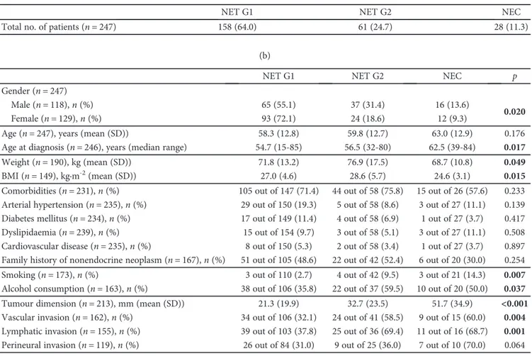
The sociodemographic and clinical features of GEP-NEN patients, according to the tumour grade, are summarized in Table 2. NET G1 were more frequently detected in females (72.1%), whereas NET G2 and NEC were more common in
males, 31.4% and 13.6%, respectively, (p = 0 020). There
was a significant association between the WHO 2010 tumour
grading and age at diagnosis (p = 0 017), with NEC being
diagnosed at a median age of 62.5 years (range: 39–84) vs. 56.5 years (range: 32–80) for NET G2 and 54.7 years (range:
15–85) for NET G1. Patients with well-differentiated NENs
presented a significantly higher mean body mass index
(BMI) (p = 0 015) in comparison with NEC patients. There
was a significant association of smoking and alcohol
con-sumption with NET G2 (p = 0 007) and NEC (p = 0 037).
NEC patients had less comorbidities than patients of the other two groups of NENs (57.6% vs. 71.4% in NET G1 and 75.8% in NET G2); these results were not statistically signif-icant. There was a significant association between WHO 2010 tumour grading groups and primary tumour size at
diagnosis, higher in NEC (p < 0 001). Vascular and
lym-phatic invasions were significantly more frequent in NEC
(p = 0 004 and p = 0 001, respectively), whereas perineural invasion presented the same trend without statistical
signifi-cance (p = 0 064).
Table 1: Patient general characteristics. Gender (n = 293)
Male,n (%) 133 (45.4)
Female,n (%) 160 (54.6)
Age (years,n = 293)
Median (range) 59.9 (22-89)
Age at diagnosis (years,n = 291)
Median (range) 56.5 (15‡-87)
Race (n = 293)
Caucasian,n (%) 285 (97.3)
African,n (%) 1 (0.3)
Other or not specified, n (%) 7 (2.4)
Type of diagnosis (n = 293)
Histopathological,n (%) 254 (86.7)
Cytological,n (%) 17 (5.8)
Biochemical,n (%) 5 (1.7)
Other or not specified, n (%) 17 (5.8)
Primary tumour by localization (n = 293)
Pancreas,n (%) 91 (31.1)
Head,n (%) 28 (30.7)
Body,n (%) 29 (31.9)
Tail,n (%) 32 (35.2)
Not specified, n (%) 2 (2.2)
Jejunum-ileum,n (%) 71 (24.2)
Stomach,n (%) 40 (13.7)
Type 1,n (%) 23 (57.5)
Type 2,n (%) 9 (22.5)
Type 3,n (%) 7 (17.5)
Not specified, n (%) 1 (2.5)
Rectum,n (%) 25 (8.5)
Duodenum,n (%) 20 (6.8)
Appendix,n (%) 20 (6.8)
Colon,n (%) 16 (5.5)
Oesophagus,n (%) 3 (1.0)
Unknown primary tumour 7 (2.4)
Tumour group by secretion
Carcinoid syndrome∗,n positive/total
studied∗∗(%) 17/115 (14.8)
Gastrinoma$,n positive/total studied$$(%) 4/55 (7.3) Insulinoma&,n positive/total studied&&(%) 11/24 (45.8) Tumour group by grade (n = 247); WHO, 2010
NET G1,n (%) 158 (64.0)
NET G2,n (%) 61 (24.7)
NEC,n (%) 28 (11.3)
Tumour group by stage (n = 214); TNM (ENETS)
Localized,n (%) 76 (35.5)
Locoregional,n (%) 43 (20.1)
Disseminated,n (%) 95 (44.4)
‡
Patient was 15 y old at diagnosis, currently 22 y old at the time of the study;
∗carcinoid syndrome criteria: 5− HIAA > 2 times the normal value and
flushing and/or diarrhea;∗∗cases with 5-HIAA quantification;$gastrinoma
criteria: gastrin≥ 10 times the normal value and exclusion of types I and II gastric tumours; $$cases with gastrin quantification; &insulinoma criteria: hypoglycemic symptoms, Whipple triad, and/or positive 72-hour prolonged fasting test;$$cases with insulin quantification.
Multiple endocrine neoplasia type 1 (MEN-1) syndrome was diagnosed in 4 patients; two patients had pancreatic tumours and two patients with gastric tumours. All patients
with MEN-1 syndrome had primary hyperparathyroidism, and two patients had a pituitary adenoma and an adrenal adenoma, respectively.
Table 2: Sociodemographic and clinical features of patients and tumour characteristics according to WHO tumour classification (grading). (a)
NET G1 NET G2 NEC
Total no. of patients (n = 247) 158 (64.0) 61 (24.7) 28 (11.3)
(b)
NET G1 NET G2 NEC p
Gender (n = 247)
Male (n = 118), n (%) 65 (55.1) 37 (31.4) 16 (13.6)
0.020
Female (n = 129), n (%) 93 (72.1) 24 (18.6) 12 (9.3)
Age (n = 247), years (mean (SD)) 58.3 (12.8) 59.8 (12.7) 63.0 (12.9) 0.176
Age at diagnosis (n = 246), years (median range) 54.7 (15-85) 56.5 (32-80) 62.5 (39-84) 0.017
Weight (n = 190), kg (mean (SD)) 71.8 (13.2) 76.9 (17.5) 68.7 (10.8) 0.049
BMI (n = 149), kg·m-2(mean (SD)) 27.0 (4.6) 28.6 (5.7) 24.6 (3.1) 0.015
Comorbidities (n = 231), n (%) 105 out of 147 (71.4) 44 out of 58 (75.8) 15 out of 26 (57.6) 0.233 Arterial hypertension (n = 235), n (%) 29 out of 150 (19.3) 5 out of 58 (8.6) 3 out of 27 (11.1) 0.139 Diabetes mellitus (n = 234), n (%) 17 out of 149 (11.4) 4 out of 58 (6.9) 1 out of 27 (3.7) 0.417 Dyslipidaemia (n = 239), n (%) 15 out of 154 (9.7) 3 out of 58 (5.1) 3 out of 27 (11.1) 0.508 Cardiovascular disease (n = 235), n (%) 8 out of 150 (5.3) 2 out of 58 (3.4) 1 out of 27 (3.7) 0.897 Family history of nonendocrine neoplasm (n = 167), n (%) 51 out of 105 (48.6) 22 out of 42 (52.4) 6 out of 20 (30.0) 0.254 Smoking (n = 173), n (%) 3 out of 110 (2.7) 4 out of 42 (9.5) 3 out of 21 (14.3) 0.007 Alcohol consumption (n = 163), n (%) 38 out of 106 (35.8) 22 out of 37 (59.5) 10 out of 20 (50.0) 0.037 Tumour dimension (n = 213), mm (mean (SD)) 21.3 (19.9) 32.7 (23.5) 51.7 (34.9) <0.001 Vascular invasion (n = 162), n (%) 34 out of 106 (32.1) 24 out of 41 (58.5) 9 out of 15 (60.0) 0.004 Lymphatic invasion (n = 155), n (%) 39 out of 103 (37.8) 25 out of 36 (69.4) 11 out of 16 (68.7) 0.001 Perineural invasion (n = 119), n (%) 26 out of 84 (31.0) 9 out of 25 (36.0) 7 out of 10 (70.0) 0.064
(c)
NET G1 NET G2
Hormonal status
Functioning (n = 32)a 17 out of 32 (53.1) 6 out of 32 (18.6)
Carcinoid (n = 17)b 8 out of 17 (47.0) 5 out of 17 (29.4)
Gastrinoma (n = 4)c 2 out of 4 (50.0) 1 out of 4 (25.0)
Insulinoma (n = 11)d 7 out of 11 (63.6) 0
Nonfunctioning, (n = 20)e 12 out of 20 (60.0) 5 out of 20 (25.0)
(d)
NET G1 NET G2 NEC p
MEN-1 syndrome (n = 213)§ 2 out of 137 (1.5) 2 out of 51 (3.9) 0 out of 25 (0.0) 0.575 Stage (n = 186)
Localized,n (%) 51 out of 114 (44.7) 11 out of 48 (22.9) 4 out of 24 (16.7)
0.001
Locoregional,n (%) 26 out of 114 (22.8) 10 out of 48 (20.8) 2 out of 24 (8.3) Disseminated,n (%) 37 out of 114 (32.5) 27 out of 48 (56.3) 18 out of 24 (75.0)
Cases missing WHO tumour classification grading:an = 9,bn = 4,cn = 1,dn = 4, anden = 3.§Cases reported as not presenting MEN-1 syndrome clinical features
3.2. Biochemical Tests. Biochemical data analysis concerning hormonal hypersecretion was informative in 32 patients (10.9%). Chromogranin A (CgA) equal or greater than twice the normal value was detected in 86 (51.2%) of the 165 patients evaluated (Table 3). Concerning specific markers, urinary 5-HIAA was evaluated in 115 patients and was positive in 47 (40.9%); of these, 17 patients presented car-cinoid syndrome criteria. Insulinoma was identified in 11
patients (3.6%) either by Whipple’s triad criteria and/or
positive prolonged fasting test. Four sporadic gastrinomas
were identified (Table 1).
3.3. Imaging Studies. The imaging modalities used as a diagnostic procedure—either for primary tumours or for metastases—are presented in Table 4. A computerized tomography (CT) scan was performed in 233 (79.5%) of the 293 patients and identified primary and/or metastatic tumour location in 79.5% of the evaluated cases. Octreos-can® was performed in 121 (41.3%) of the 293 patients and was informative in 63.6% of the evaluated cases. A
68Ga-positron emission tomography- (PET-) SSTR scan
was used in 99 (33.8%) of the 293 patients and was
infor-mative in 75.8% of the evaluated cases. 111In-pentetreotide
(111In-octreoscan®) (Octreoscan®) and 68Ga-PET-SSTR
scan were mainly used in NET G1 and NET G2 patients, 89.8% and 93.1%, respectively. Fluorodeoxyglucose- (FDG-) PET was evaluated in 36 (12.3%) of 293 patients. Upper gastrointestinal endoscopy presented the highest efficiency in localizing oesophageal (3 out of 3, 100%), gastric (27 out of 30, 90%), and duodenal (17 out of 19, 89.5%) tumours. Echoendoscopy was valuable in the detection of duodenal (6 out of 6, 100%), pancreatic (25 out of 28, 89.3%), and gastric (7 out of 13, 53.8%) tumours. A colonoscopy was the main diagnostic procedure in colonic NEN detection (12 out of 12, 100%), as well as in rectal NENs (21/22, 95.5%). For midgut tumours, magnetic resonance imaging (MRI), CT, and video capsule were the mostly used imag-ing procedures; PET for somatostatin receptors (SSTR),
68Ga-PET-SSTR, demonstrated to be the most sensitive
(94.1%) imaging tool.
3.4. Extension of the Disease. Extension of the disease was evaluated in 186 patients (Figure 1 and Table 2). Localized disease was more frequent in NET G1 (44.7%). Regional
dis-ease was detected in 20.1% of the patients: 22.8% with NET G1, 20.8% with NET G2, and 8.3% with NEC. Metastases were present in 32.5% of patients with NET G1, in 56.3% with NET G2, and in 75.0% with NEC. Among cases with
distant metastases at presentation (n = 82), 30.5% presented
liver metastases. Bone metastases were detected in one patient with a NET G2 and two patients with NEC. Only one patient with NEC had lung metastases. Other sites of
dis-tant metastases included the peritoneum (five patients: one
NET G1, one NET G2, and three NEC), adrenal glands (one patient with NEC), ovary (one patient with NET G1), and inferior vena cava (one patient with NET G1).
3.5. Treatment Procedures. Endoscopic removal of the tumours was possible in 40 patients with localized gastric, duodenal, and colorectal NENs. According to the WHO 2010 classification, either curative or cytoreductive surgery was performed in 125 out of 155 cases (80.6%) of NET G1, 48 out of 60 cases (80.0%) of NET G2, and 18 out of 25 cases (72.0%) of NEC (Table 5); overall, 191 of 240 patients (79.6%) were treated with surgery. Concerning patients with disseminated disease, 22 patients (18.2%) with NET G1, 9 patients (20.5%) with NET G2, and 8 patients (44.4%) with NEC were submitted to debulking surgery, mainly liver metastasectomy.
Although 95 patients presented liver metastases at diagnosis, locoregional ablative therapy, such as transarter-ial embolization (TAE), transartertransarter-ial chemoembolization (TACE), radioembolization, or radiofrequency (RF)/ther-moablation (TA), was only performed in 14 patients with well-differentiated NETs; 70.0% of the cases submitted to TAE and 75.0% submitted to RF/TA were NET G1. Only four patients were submitted to radioembolization, being three NET G1 and one NET G2.
Systemic therapy included somatostatin analogues
(SSAs), interferon-α2b, target therapies with tyrosine kinase
inhibitors and mTOR inhibitors, peptide receptor radiother-apy (PRRT), and chemotherradiother-apy (Table 5, Figure 2).
SSAs were mostly used in well-differentiated NETs (p < 0 001), comprising 20.4% of NET G1, 59.3% of NET G2, and 32.0% of NEC. Only 4 patients received combined
treatment with SSAs and interferon-α2b. Target therapies
as sunitinib and everolimus were used in seven (3.0%) patients: two with NET G1, two with NET G2, and three with NEC. Peptide receptor radiotherapy (PRRT) was used in nine (3.9%) of the patients, mainly well-differentiated NETs (33.3% NET G1 and 66.7% NET G2). Chemotherapy treat-ment was performed in 20 patients, mostly in NEC of the
colon and the pancreas (11 patients;p < 0 001).
4. Discussion
GEP-NENs have been historically considered a rare and heterogeneous group of neoplasms. They comprise approx-imately 0.5% of all human cancers and 2% of gastrointesti-nal tumours [1]. New data from SEER 18 [2] reported a 6.5-fold increase in the annual incidence from 1973 to 2012 in NENs [2], reinforcing the need for research in this field. GEP-NENs often exhibit relatively indolent clinical
Table 3: Biochemical tests.
Biochemical tests Positive results,n (%)
Chromogranin A (n = 168) 86 (51.2) 5-HIAA (n = 115) 47 (40.9) Insulin (n = 25) 11 (44.0) Gastrin (n = 55) 25 (45.5) Glucagon (n = 8) 0 VIP (n = 9) 0 ACTH (n = 17) 0 GH (n = 12) 0
VIP: vasoactive intestinal peptide; ACTH: adrenal corticotrophin; GH: growth hormone.
Table 4: Imaging mod alities used for the diagnostic procedure either for primary sites and metastasis. Oesophagus Gastric Pancreas Appendiceal Duodenum Jejunum-ileum Colon Rectum UP T ∗ Positi ve/total exams Upp er endoscopy 3/3 (100.0) 27/30 (90.0) —— 17/19 (89.5) —— — 1/4 (25.0) 48/56 (85.7) Echo endoscopy — 7/13 (53.8) 25/28 (89.3) — 6/6 (100.0) —— 10/10 (100.0) — 48/57 (84.2) Video capsule — — ——— 8/9 (88.9) —— — 8/9 (88.9) Do uble balloon — — ——— 1/1 (100.0) —— — 1/1 (100.0) Colonoscopy — — ——— 12/33 (36.4) 12/12 (100.0) 21/22 (95.5) — 45/67 (67.2) Enter o-CT — — ——— 4/4 (100.0) —— — 4/4 (100.0) Enter o-MRI — — ——— 11/11 (100.0) 1/1 (100.0) —— 12/12 (100.0) US scan — 5/12 (41.7) 33/41 (80.5) 1/4 (25.0) 2/2 (100.0) 23/27 (85.2) 1/2 (50.0) 0/3 (0.0) 1/1 (100.0) 66/92 (71.7) CT scan 3/3 (100) 10/22 (45.5) 71/77 (92.2) 4/11 (36.4) 13/17 (76.5) 52/62 (83.9) 13/15 (86.7) 9/20 (45.0) 6/6 (100.0) 181/233 (77.7) MR I — 0/3 (0.0) 38/44 (86.4) 1/1 (100.0) 5/5 (100.0) 11/13 (84.6) 2/4 (50.0) 4/9 (44.4) 2/2 (100.0) 63/81 (77.8) 111 In-pentetreotide ‡ — 6/17 (35.3) 26/36 (72.2) 2/6 (33.3) 8/12 (66.7) 25/30 (83.3) 5/8 (62.5) 2/9 (22.2) 3/3 (100.0) 77/121 (63.6) 68 Ga-PET-SRP — 5/12 (41.7) 26/31 (83.9) 2/5 (40%) 1/2 (50.0) 32/34 (94.1) 2/2 (100.0) 5/11 (45.5) 2/2 (100.0) 75/99 (75.8) PET -FDG 2/2 (100.0) 0/5 (0.0) 10/17 (58.8) 0/1 (0.0) 1/1 (100.0) 2/4 (50.0) 3/5 (60.0) — 1/1 (100) 19/36 (52.8) ‡ Octr eoscan®; ∗UP T: unk nown primary tumo ur; CT: compute d tomo graph y; MRI: ma gnetic resonance imaging ; PET-FDG : positr on emission tomog raphy-( 18 F) fluo rodeoxy glucose.
courses and a delay in the diagnosis and tend to present metastases at the time of diagnosis, preserving the potential for lethal progression.
The present study was designed to characterize the over-all scenario of GEP-NENs in Portugal, namely, the incidence and epidemiology of these tumours, sociodemographic and
clinical profiles of the patients, and the patterns of care in a
multicenter audit. Our results provide a comprehensive and relevant information on a group of neoplasm still poorly characterized, particularly, in Southern Europe. Published data from GEP-NEN in European countries is available in a French registration study [7], in a Spanish study of the Neu-roendocrine Tumours Study Group Registry of Spain (RGETNE) [8], in an Italian epidemiological study [9], in a prospective Greek registry [10], and in the United Kingdom
and Northern European countries [11–13]. Worldwide, the
most characterized cohorts are from the United States of America (USA) [2, 14], and there is data available from Asian countries, such as China [15] and Japan [16].
Overall, ourfindings are in accordance with reports of
NENs from other countries and corroborate that they are a heterogeneous group of tumours with a wide range of clinical presentation. We observed a similar gender ratio with a slight preponderance for females, as observed in a USA series [13, 14], Canadian series [17], and Italian study [9]. In our series, the pancreas was the most frequent primary tumour site, followed by the jejunum-ileum and the stomach.
These findings are in agreement with data from Southern
European countries, as the Italian and Greek cohorts [9, 10] as well as in China [18], but in contrast with other pub-lished studies [2, 7, 8, 17], where the gastrointestinal tract was reported as the most frequent primary site. These inconsistencies may be due to a referral bias and may suggest geographic and ethnic variation in the carcino-genesis of GEP-NENs. A recent publication stresses the differences in geographic and ethnic distribution, other
than NEN fortuitous location and identification related
to the current accuracy of the diagnostic methods [19], and points to the possibility of involved environmental
risk factors. Prospective and larger studies will be useful
to further clarify these findings.
The present study provides a comprehensive report on diagnostic and therapeutic procedures used in the current clinical practice in Portugal. Like the Spanish results reported by the RGETNE, in Portugal, there is a limited overall use of biochemical tests at diagnosis, namely, the general marker
serum chromogranin A or urinary 5-HIAA quantification
for midgut tumours.
In our cohort, as in another series [8], the most frequent functioning tumour was NEN with carcinoid syndrome, followed by insulinoma and apparently sporadic gastrinoma. No glucagonoma, VIPoma, somatostatinoma, or other rare syndromes were identified. It should also be taken into con-sideration that in 71.7% of the cases, the hormonal secretion by the tumour was not evaluated. This seems to reflect a low referral rate of patients to specialized centers, low participa-tion of endocrinologists in the oncology team, and/or a lim-ited laboratory support in some of the institutions that participated in this study. Our results highlight the ongoing demand for an adequate management of diagnostic, treat-ment, and follow-up work-out for patients with GEP-NENs. Most of the international epidemiological studies report data about localization, histological classification, and staging of GEP-NENs, but information about their hormonal secretion is sparse. Biochemical evaluation is important, not only for diagnostic purposes but also for therapeutic decision and monitoring of treatment responses, and an adequate assessment of tumour secretion is strongly encouraged. Genetic testing is also important when clinically indicated, as it allows for (1) a personalized life-long screen-ing for prototypic tumours and their timely treatment, (2) the
identification of affected family members that may benefit
from this screening, and (3) appropriate genetic counseling. In our series, the majority of the cases lacked genetic evalua-tion for clinical suspicion of hereditary syndromes.
Histological classification of NENs is evolving as the WHO revised the nomenclature and classification of GEP-NENs in 2010 [5] and updated it in 2017 [6]. Histopatholog-ical characterization with immunohistochemistry markers such as chromogranin and synaptophysin is essential to make the diagnosis. The mitotic index and/or immunohisto-chemistry for Ki-67 labelling index is mandatory to generate the tumour grading [4]; these are minimum requirements for an accurate pathological classification. At the time of the inclusion of the patients in the present study, the histological classification was performed according to the 2010 WHO cri-teria, the up-to-date guidelines used for this study. Overall, in
this study, the frequency of NET G1, NET G2, and NECfits
with other reports.
Tumour metastases at diagnosis represent an important prognostic marker [2]. In this series, distant metastases were detected in 44.4% of patient (NET G1: 32.5%; NET G2: 56.3%; and NEC: 75.0). This is consistent with other studies, as the Spanish and Italian studies [8, 9], where distant metas-tases were observed in 44% and 42% of patients, respectively, and contrasts with a lower rate of distant metastases at diag-nosis in the Greek [10], Chinese [15], and Canadian [17] studies (25.0%, 6.0%, and 20.8%, respectively), as well as the
60 50 40 30 N um b er o f cas es 20 10 0
Localized Locoregional Disseminated NET G1
NET G2 NEC
Figure 1: Extension of disease according to WHO 2010 classification.
SEER Registry (21.0%) [14]. An explanation for these di ffer-ences may be due to the inclusion of cases from oncological institutions, where the proportion of metastatic disease is considerably higher. In this study, the oncological institu-tions, from Lisbon and Porto, contributed with 46% of the patients included.
Endoscopic therapy is the mainstay for types 1 and 2 gastric endocrine tumours and for localized duodenal and colorectal NENs. In this cohort, endoscopic therapy was performed mainly in those cases.
Surgery remains the treatment of choice for GEP-NENs, with curative intent whenever feasible. If the tumour is unre-sectable, several approaches are available to induce tumour debulking as a manner to control life-threatening symptoms
due to hormone secretion and to increase patient survival and quality of life [20, 21]. In experienced centers, ablative therapies are a good option to treat liver metastatic disease [22]. Our results show that either primary or cytoreductive surgery was performed in the majority of the hospitals included and mainly in well-differentiated NENs. Ablative therapies were used in less than 5% of the patients proba-bly due to the fact that few centers have this treatment
available. This finding indicates the need of a referral of
the patients to centers where they can benefit from these therapeutic options.
Currently, the standard of care for systemic treatment in
advanced NET treatment is SSA, which proved to be effective
in controlling excessive hormonal secretion [23, 24] and allowing long-term improvement in secretory symptoms in 30–70% of patients. Recent studies report an additional anti-proliferative role of SSA in nonfunctioning midgut [25], pan-creatic, and lung NENs [26], which reflected in the significant progression-free survival in the treated patients when compared with placebo. Other therapeutic options include biologic agents interfering with specific molecules of cell sig-naling pathways, e.g., the mammalian target of rapamycin (mTOR) and vascular endothelial growth factor (VEGF), with everolimus and sunitinib, respectively, both approved for pancreatic NENs [27, 28]. Everolimus was also approved for the treatment of advanced nonfunctioning lung and gas-trointestinal NENs [29]. Studies using oral chemotherapy with temozolomide and capecitabine are demonstrating promising results in well-differentiated pancreatic NENs [30]. However, classic cytotoxic drugs still continue to be
the first-line therapy for poorly differentiated GEP-NENS
and are effective (up to 60% response rates) in
well-Table 5: Treatments administered to patients with GEP-NETs. (a)
Gastric,n (%) Duodenum,n (%) Rectum,n (%)
Endoscopic therapy (n = 40) 21 (52.5) 4 (10.0) 15 (37.5)
(b)
NET G1 NET G2 NEC p
Surgical therapy (n = 240)$ 125 out of 155 (80.6) 48 out of 60 (80.0) 18 out of 25 (72.0) 0.607 Surgery of metastases (n = 183)$ 22 out of 121 (18.2) 9 out of 44 (20.5) 8 out of 18 (44.4) 0.055 Liver ablative therapy
TAE (n = 199)$ 7 out of 131 (5.3) 3 out of 49 (6.1) 0 out of 19 (0.0) 0.781
RFA (n = 101)$ 3 out of 61 (4.9) 1 out of 27 (3.7) 0 out of 13 (0.0) >0.999
Systemic therapies
Somatostatin analogues (n = 231)$ 31 out of 152 (20.4) 32 out of 54 (59.3) 8 out of 25 (32.0) <0.001 Interferon (n = 231)$+SSAs 3 out of 152 (2.0) 1 out of 55 (1.8) 0 out of 24 (0.0) >0.999 Target therapies∗(n = 231)$ 2 out of 153 (1.3) 2 out of 53 (3.8) 3 out of 25 (12.0) 0.020
PRRNT∗∗(n = 230)$ 3 out of 150 (2.0) 6 out of 55 (10.9) 0 out of 25 (0.0) 0.021
Chemotherapy (n = 244)$ 3 out of 157 (1.9) 6 out of 60 (10.0) 11 out of 27 (40.7) <0.001 $
Number of cases with information. TAE = transhepatic arterial embolization; RFA = radiofrequency ablation; PRRNT: peptide receptor radionuclide therapy.
∗Sunitinib;∗∗177Lu-THERA. 100 80 60 P er cen ta ge o f cas es tr ea te d (%) 40 20 0 SSAs Interferon+ SSAs Target therapies PRNNT Chemotherapy NET G1 NET G2 NEC
Figure 2: Cases submitted to different systemic therapies according to WHO 2010 classification.
differentiated pancreatic NETs; however, early relapses often occur [31]. Concerning the therapeutic options in the present study, endoscopic therapies, either curative or cytoreductive surgery, and SSA treatment were the preferred options for the majority of patients. Somatostatin analogues (SSAs) were the most frequently used drugs in our study. Locoregional ablative therapy, PRRT, and target therapies were rarely used. Remarkably, PRRNT was more frequently chosen than target therapies. This fact was remarkable, as in the Portuguese National Health System, only one center offered this thera-peutic modality at the time of the present study. As in other series and according to the guidelines, chemotherapy was the treatment of choice in NEC and was also an option in well-differentiated nonpancreatic NETs, which may reflect the inclusion of older cases and/or the absence of a referral to specialized centers.
The results obtained in this study represent the first
comprehensive registry of GEP-NENs in Portugal per-formed by the Neuroendocrine Study Group of the Portu-guese Society of Endocrinology, Diabetes and Metabolism. These provide a valuable insight into the epidemiology, current clinical practice, and therapy strategies of this het-erogeneous disease and will set the ground for the devel-opment of a National Registry of NENs. These reinforce the need for a national clinical framework for GEP-NENs, in order to ensure a systematic surveillance of the disease and ultimately improve the diagnosis, clinical manage-ment, and outcome of NEN patients.
Data Availability
The clinical data used to support thefindings of this study are
included within the article.
Disclosure
The study sponsor did not participate in the study design, data collection, analysis, preparation of the manuscript, and/or decision to publish.
Conflicts of Interest
The authors declare no conflict of interest.
Authors’ Contributions
Santos AP and Vinagre J contributed equally. Lopes JM and Bugalho MJ supervised this work equally.
Acknowledgments
This study was industry-sponsored by the pharmaceutical
company Ipsen Portugal. João Vinagre
(CEE-CIND/00201/2017) and Paula Soares receive funding from the Operational Program for Competitiveness and Interna-tionalization (POCI), Portugal 2020; Portuguese funds through FCT (Fundação para a Ciência e a Tecnologia)/-Ministério da Ciência, Tecnologia e Inovação in the
frame-work of the project“Institute for Research and Innovation
in Health Sciences” (POCI-01-0145-FEDER-007274) and
the project “Advancing Cancer Research: From Basic
Knowledge to Application”
(NORTE-01-0145-FEDER-000029); and “Projetos Estruturados de I&D&I,” funded
by Norte 2020—Programa Operacional Regional do Norte. Further funding was from the European Regional Develop-ment Fund (ERDF) through the Operational Program for
Competitiveness and Internationalization (COMPETE
2020) and Portuguese national funds via FCT (Fundação
para a Ciência e a Tecnologia), under the project
“POCI-01-0145-FEDER-016390: CANCEL STEM.” Maria João Bugalho and João Vinagre were funded by the Sociedade Portuguesa de Endocrinologia, Diabetes e Metabolismo through the Study Group for Neuroendocrine Tumours.
References
[1] K. Oberg and B. Eriksson,“Endocrine tumours of the pan-creas,” Best Practice & Research Clinical Gastroenterology, vol. 19, no. 5, pp. 753–781, 2005.
[2] A. Dasari, C. Shen, D. Halperin et al.,“Trends in the incidence, prevalence, and survival outcomes in patients with neuroendo-crine tumors in the United States,” JAMA Oncology, vol. 3, no. 10, pp. 1335–1342, 2017.
[3] M. Fraenkel, M. Kim, A. Faggiano, W. W. de Herder, G. D. Valk, and Knowledge NETwork,“Incidence of gastroentero-pancreatic neuroendocrine tumours: a systematic review of the literature,” Endocrine-Related Cancer, vol. 21, no. 3, pp. R153–R163, 2014.
[4] D. S. Klimstra, I. R. Modlin, N. V. Adsay et al.,“Pathology reporting of neuroendocrine tumors: application of the Del-phic consensus process to the development of a minimum pathology data set,” The American Journal of Surgical Pathol-ogy, vol. 34, no. 3, pp. 300–313, 2010.
[5] F. T. Bosman, F. Carneiro, R. H. Hruban, and N. D. Theise, WHO Classification of Tumours of the Digestive System, World Health Organization, 2010.
[6] R. V. Lloyd, R. Y. Osamura, G. Klöppel, J. Rosai, F. T. Bosman, E. S. Jaffe, S. R. Lakhani, and H. Ohgaki, Eds., WHO Classifica-tion of Tumours of Endocrine Organs, World Health Organiza-tion, 2017.
[7] C. Lombard-Bohas, E. Mitry, D. O’Toole et al., “Thirteen-month registration of patients with gastroenteropancreatic endocrine tumours in France,” Neuroendocrinology, vol. 89, no. 2, pp. 217–222, 2009.
[8] R. Garcia-Carbonero, J. Capdevila, G. Crespo-Herrero et al., “Incidence, patterns of care and prognostic factors for out-come of gastroenteropancreatic neuroendocrine tumors (GEP-NETs): results from the National Cancer Registry of Spain (RGETNE),” Annals of Oncology, vol. 21, no. 9, pp. 1794–1803, 2010.
[9] A. Faggiano, P. Ferolla, F. Grimaldi et al.,“Natural history of gastro-entero-pancreatic and thoracic neuroendocrine tumors. Data from a large prospective and retrospective Italian epidemiological study: the NET management study,” Journal of Endocrinological Investigation, vol. 35, no. 9, pp. 817–823, 2012.
[10] G. C. Nikou, K. Pazaitou-Panayiotou, D. Dimitroulopoulos et al., “Results of a prospective multicenter neuroendocrine tumor registry reporting on clinicopathologic characteristics of Greek patients,” BMC Endocrine Disorders, vol. 16, no. 1, p. 8, 2016.
[11] C. Lepage, B. Rachet, and M. P. Coleman, “Survival from malignant digestive endocrine tumors in England and Wales: a population-based study,” Gastroenterology, vol. 132, no. 3, pp. 899–904, 2007.
[12] K. Hemminki and X. Li,“Incidence trends and risk factors of carcinoid tumors: a nationwide epidemiologic study from Sweden,” Cancer, vol. 92, no. 8, pp. 2204–2210, 2001. [13] O. Hauso, B. I. Gustafsson, M. Kidd et al.,“Neuroendocrine
tumor epidemiology: contrasting Norway and North Amer-ica,” Cancer, vol. 113, no. 10, pp. 2655–2664, 2008.
[14] J. C. Yao, M. Hassan, A. Phan et al.,“One hundred years after “carcinoid”: epidemiology of and prognostic factors for neuro-endocrine tumors in 35,825 cases in the United States,” Journal of Clinical Oncology, vol. 26, no. 18, pp. 3063–3072, 2008. [15] X. Zhang, L. Ma, H. Bao, J. Zhang, Z. Wang, and P. Gong,
“Clin-ical, pathological and prognostic characteristics of gastroentero-pancreatic neuroendocrine neoplasms in China: a retrospective study,” BMC Endocrine Disorders, vol. 14, no. 1, p. 54, 2014. [16] T. Ito, H. Sasano, M. Tanaka et al.,“Epidemiological study of
gastroenteropancreatic neuroendocrine tumors in Japan,” Journal of Gastroenterology, vol. 45, no. 2, pp. 234–243, 2010. [17] J. Hallet, C. H. L. Law, M. Cukier, R. Saskin, N. Liu, and S. Singh,“Exploring the rising incidence of neuroendocrine tumors: a population-based analysis of epidemiology, metasta-tic presentation, and outcomes,” Cancer, vol. 121, no. 4, pp. 589–597, 2015.
[18] J. H. Fan, Y. Q. Zhang, S. S. Shi et al.,“A nation-wide retro-spective epidemiological study of gastroenteropancreatic neu-roendocrine neoplasms in China,” Oncotarget, vol. 8, no. 42, pp. 71699–71708, 2017.
[19] I. Huguet, A. B. Grossman, and D. O'Toole,“Changes in the epidemiology of neuroendocrine tumours,” Neuroendocrinol-ogy, vol. 104, no. 2, pp. 105–111, 2016.
[20] X. M. Keutgen, N. Nilubol, J. Glanville et al.,“Resection of pri-mary tumor site is associated with prolonged survival in met-astatic nonfunctioning pancreatic neuroendocrine tumors,” Surgery, vol. 159, no. 1, pp. 311–319, 2016.
[21] J. Guo, Q. Zhang, X. Bi et al.,“Systematic review of resecting primary tumor in MNETs patients with unresectable liver metastases,” Oncotarget, vol. 8, no. 10, pp. 17396–17405, 2017. [22] F. Cavalcoli, E. Rausa, D. Conte, A. F. Nicolini, and S. Massironi,“Is there still a role for the hepatic locoregional treatment of metastatic neuroendocrine tumors in the era of systemic targeted therapies?,” World Journal of Gastroenterol-ogy, vol. 23, no. 15, pp. 2640–2650, 2017.
[23] C. G. Moertel,“Karnofsky memorial lecture. An odyssey in the land of small tumors,” Journal of Clinical Oncology, vol. 5, no. 10, pp. 1502–1522, 1987.
[24] K. Öberg, L. Kvols, M. Caplin et al.,“Consensus report on the use of somatostatin analogs for the management of neuroen-docrine tumours of the gastroenteropancreatic system,” Annals of Oncology, vol. 15, no. 6, pp. 966–973, 2004. [25] A. Rinke, H. H. Müller, C. Schade-Brittinger et al.,
“Placebo-controlled, double-blind, prospective, randomized study on the effect of octreotide LAR in the control of tumor growth in patients with metastatic neuroendocrine midgut tumors: a report from the PROMID Study Group,” Journal of Clinical Oncology, vol. 27, no. 28, pp. 4656–4663, 2009.
[26] M. E. Caplin, M. Pavel, J. B.Ćwikłaetal., “Lanreotideinmetasta-tic enteropancrea“Lanreotideinmetasta-tic neuroendocrine tumors,”TheNewEngland Journal of Medicine, vol. 371, no. 3, pp. 224–233, 2014.
[27] J. C. Yao, M. H. Shah, T. Ito et al.,“Everolimus for advanced pancreatic neuroendocrine tumors,” The New England Journal of Medicine, vol. 364, no. 6, pp. 514–523, 2011.
[28] E. Raymond, L. Dahan, J. L. Raoul et al.,“Sunitinib malate for the treatment of pancreatic neuroendocrine tumors,” The New England Journal of Medicine, vol. 364, no. 6, pp. 501–513, 2011.
[29] J. C. Yao, N. Fazio, S. Singh et al.,“Everolimus for the treat-ment of advanced, non-functional neuroendocrine tumours of the lung or gastrointestinal tract (RADIANT-4): a rando-mised, placebo-controlled, phase 3 study,” The Lancet, vol. 387, no. 10022, pp. 968–977, 2016.
[30] J. R. Strosberg, R. L. Fine, J. Choi et al.,“First-line chemother-apy with capecitabine and temozolomide in patients with met-astatic pancreatic endocrine carcinomas,” Cancer, vol. 117, no. 2, pp. 268–275, 2011.
[31] R. Garcia-Carbonero, A. Rinke, J. W. Valle et al.,“ENETS con-sensus guidelines for the standards of care in neuroendocrine neoplasms: systemic therapy - chemotherapy,” Neuroendocri-nology, vol. 105, no. 3, pp. 281–294, 2017.
Stem Cells
International
Hindawi www.hindawi.com Volume 2018 Hindawi www.hindawi.com Volume 2018 INFLAMMATIONEndocrinology
International Journal of Hindawi www.hindawi.com Volume 2018 Hindawi www.hindawi.com Volume 2018Disease Markers
Hindawi www.hindawi.com Volume 2018 BioMed Research InternationalOncology
Journal of Hindawi www.hindawi.com Volume 2013 Hindawi www.hindawi.com Volume 2018 Oxidative Medicine and Cellular Longevity Hindawiwww.hindawi.com Volume 2018
PPAR Research
Hindawi Publishing Corporation
http://www.hindawi.com Volume 2013 Hindawi www.hindawi.com
The Scientific
World Journal
Volume 2018 Immunology Research Hindawi www.hindawi.com Volume 2018 Journal ofObesity
Journal of Hindawi www.hindawi.com Volume 2018 Hindawi www.hindawi.com Volume 2018 Computational and Mathematical Methods in Medicine Hindawi www.hindawi.com Volume 2018Behavioural
Neurology
Ophthalmology
Journal of Hindawi www.hindawi.com Volume 2018Diabetes Research
Journal ofHindawi
www.hindawi.com Volume 2018
Hindawi
www.hindawi.com Volume 2018
Research and Treatment
AIDS
Hindawi
www.hindawi.com Volume 2018
Gastroenterology Research and Practice
Hindawi www.hindawi.com Volume 2018