ARTIGO DE REVISÃO
Magnetic Resonance Imaging of Small Bowel
Crohn’s Disease
A Ressonância Magnética na Doença de Crohn do Intestino Delgado
M.R., A.P.M., J.P.: Department of Radiology. Hospital Garcia de Orta. Almada. Portugal. V.H.: Department of Radiology. Hospital do Espírito Santo. Évora. Portugal. C.C.: Department of Gastroenterology. Hospital São Bernardo. Setubal. Portugal.
R.C.S.: Department of Radiology. University of North Carolina at Chapel Hill. Chapel Hill, North Carolina. United States of America. J.D.F.: Department of Gastroenterology. Hospital Garcia de Orta. Almada. Portugal.
Recebido: 15 de Dezembro de 2011 - Aceite: 27 de Fevereiro de 2012 | Copyright © Ordem dos Médicos 2012
Miguel RAMALHO, Vasco HERÉDIA, Cláudia CARDOSO, António P. MATOS, João PALAS, João DE FREITAS, Richard C. SEMELKA
Acta Med Port 2012 Jul-Aug;25(4):231-240
RESUMO
A doença de Crohn (DC) é uma doença inflamatória intestinal de carácter crónico e recidivante, que atinge em muitos casos doentes jovens. Os métodos de imagem são indispensáveis no seu diagnóstico, na monitorização da progressão da doença e na resposta ao tratamento. Na prática clínica actual, a avaliação imagiológica da DC é efectuada de forma crescente por técnicas de imagem sec-cional, em particular a tomografia computorizada e a ressonância magnética (RM), permitindo a visualização simultânea do lumen e da parede intestinal, bem como a extensão extra-entérica. A enterografia por RM permite uma avaliação segura e não-invasiva destes doentes, sem necessidade de exposição a radiação ionizante. O novo paradigma de imagem deve contemplar a segurança do doente como um aspecto essencial na escolha do método de imagem. Por esta razão, a ressonância magnética poderá ser o método de avaliação preferido para a avaliação do intestino delgado, especialmente em doentes jovens com DC, considerando que a maioria irá realizar estudos de repetição. Além disso, a informação sobre a actividade da doença não é equiparável por qualquer outro método de imagem. Neste artigo de revisão os autores discutem os aspectos essenciais da utilização da RM na DC, incluindo o protocolo e os principais achados de imagem, fazendo ainda referência às suas vantagens comparativamente a outros métodos, nomeadamente no que diz respeito à segurança, à acuidade diagnóstica e à potencial importância na abordagem terapêutica desta doença.
ABSTRACT
Crohn’s disease (CD) is a chronic relapsing inflammatory disease of the gastrointestinal tract, which mostly affects young patients. Imaging techniques form a very important part for the evaluation of CD and for monitoring disease progression or response to ther-apy. Currently, imaging of CD is increasingly being performed by cross-sectional modalities, i.e. multidetector computed tomography (MDCT) and magnetic resonance imaging (MRI), since these techniques allow for simultaneous visualization of luminal, mural and extraintestinal disease extension. MR enterography has the potential to safely and noninvasively accomplish the imaging needs of patients with Crohn disease without exposing them to ionizing radiation. The new imaging paradigm should contemplate patient safety as a very important aspect when assessing the role of an imaging modality in comparison with others. For this reason, MRI may be the preferred modality for evaluation of small bowel disease, especially in young patients in the setting of CD, considering that the major-ity will undergo frequent repeat studies. Also, the information on disease activmajor-ity is not matched by any other imaging method. In this review article, the authors discuss the essential aspects of MR evaluation of CD, including protocol and imaging findings, also referring the advantages over other radiological studies, concerning safety, accuracy and potential importance for therapeutic approach.
INTRODUCTION
Crohn’s disease (CD) is a chronic relapsing inflamma-tory disease of the gastrointestinal tract involving all the lay-ers of the bowel wall that eventually may progress to fistu-las, abscesses, and strictures formation. The incidence of CD seems to be bimodal; the first peak occurs in the second and third decades of life, while a second, smaller increase in incidence can be seen between the fifth and seventh de-cades of life.1,2
Although any segment of the gastrointestinal tract may become involved with CD, it most commonly involves the terminal ileum, and frequently in association with disease in the right colon. Involvement of the terminal ileum occurs in approximately 70% of patients, with combined terminal ileal and cecal disease present in 40% of the total and isolated terminal ileal involvement in the remaining 30%. Although endoscopy and histologic examination have served as the
gold standard for the diagnosis of CD, diagnosing lesions in the small bowel from the distal duodenum to the terminal ileum has been a challenge.
The course of the disease is prolonged and unpredict-able with alternating exacerbations and remissions and vari-able response to medical or surgical therapy. The Crohn’s Disease Activity Index (CDAI) is currently the gold standard for clinical evaluation of disease activity and for monitoring response to therapy,3 despite the fact that the CDAI is in part based on subjective criteria (‘general well being’ and ‘intensity of abdominal pain’). Assessment of the disease activity is a major clinical problem and has important conse-quences for patient management.4–6
In the last decade, many new therapeutic strategies have been developed that have allowed the gastroenterolo-gist and surgeon to treat virtually all forms of CD effectively.
ARTIGO DE REVISÃO The success of these treatments depends on accurate
di-agnosis of the nature and extent of disease. Therefore it is no longer sufficient to detect the presence of CD, it is mandatory to accurately assess its subtype, location, and severity.
Imaging in Crohn’s disease
In the last 15 years, imaging of the small bowel has im-proved significantly. Traditionally, imaging of the small bow-el has rbow-elied on barium examinations. Conventional imaging techniques for evaluation of the small bowel have included small bowel follow-through and fluoroscopic enteroclysis. These studies allow direct evaluation of the mucosal layer, but fail to depict extraluminal complications, as much of this information on these studies is derived from indirect and non-specific secondary signs. Also, the presence of over-lapping loops can mask disease.
Wireless capsule endoscopy (WCE) has been recently introduced and provides direct mucosal visualization in the elective investigation of small bowel diseases.7,8 Segments of the small intestine that could not be reached by push en-teroscopy and retrograde ileoscopy are visualized by WCE. While there is little doubt that WCE provides a level of in-traluminal image detail comparable to standard endoscopy there are still certain limitations to capsule studies such as safety and performance issues.9,10 Localization of the dis-ease process can also be challenging.
All these methods are able to demonstrate mucosal dis-ease; however the major disadvantage they all suffer from is their inability to provide deep mural and extra-luminal in-formation. This is especially important for patients with CD, as the disease affects the full thickness of the bowel wall and also the adjacent fat and mesentery. These features have substantial clinical implications also because of the mucosa’s high regenerative capacity; its evaluation alone might under characterize the true extent and activity of dis-ease.
Currently, there is considerable consensus about re-placing the conventional fluoroscopic examinations with Computerized Tomography (CT) or Magnetic Resonance Imaging (MR) of the small bowel. Cross sectional tech-niques have several advantages, including their ability to display the entire thickness of the bowel wall, to visualize deep ileal loops in the pelvis without superimposition, and to evaluate the surrounding mesentery and perienteric fat. Another intrinsic advantage is the possibility to assess solid organs and provide a global overview of the abdomen. Ad-vances in CT and MR imaging technology have led to im-proved spatial and temporal resolution allowing nowadays high-resolution imaging and also enterography and entero-clysis techniques.
Traditionally, multidetector computed tomography (MDCT) has been the predominant cross-sectional tech-nique in patients with CD because of its proven efficacy in the evaluation of intestinal diseases and extraintestinal complications. However, MDCT has the disadvantages of exposure to a substantial burden of ionizing radiation and
iodinated contrast material and does not lend itself to dy-namic imaging due to radiation dose issues (as multiple post contrast acquisitions are needed), which are of particular importance especially in the follow-up evaluation of younger patients. The cumulative amount of lifetime radiation expo-sure for these patients may not be trivial.11,12 The estimated radiation effective dose for CT small bowel follow-trough is about 16 mSv. (Corresponding to 800 chest x-rays), which could be variably higher according to the implemented CT protocols and equipment used.13 At this point, the biologic impact of this type of radiation exposure is not yet known. The US Food and Drug Administration (FDA) estimates that a CT examination with an effective dose of 10 mSv may be associated with an increased chance of developing fatal malignancy for approximately one patient in 200014 and the National Academy of Science BEIR VII lifetime risk model predicts that approximately one individual in 1000 will de-velop cancer from an exposure to 10 mSv (0.01 Sv) of low-dose radiation.15 The limitations and risks associated with traditional advanced CT techniques, and the need for mul-tiple follow-up examinations should shift interest to an alter-native non-radiation imaging method.
Recent technological developments have dramatically improved the quality of abdominal MRI extending its role in the evaluation of the gastrointestinal tract. Hardware devel-opments with faster gradients, new coil technology, parallel imaging, and new software design with fast and ultrafast pulse-sequence developments have become essential req-uisites for MRI evaluation of the small bowel. MR imaging has emerged as a valuable tool in evaluation of small bowel Crohn’s disease. MRI provides several advantages to other imaging modalities, including the lack of ionizing radiation, multiplanar capability, and functional information.
The efficacy of CT and MRI have individually been ex-tensively studied in CD, however only a few studies have compared them, with most noting a similar diagnostic per-formance.16-19 As noted in earlier studies, the image qual-ity is usually considered superior for CT, because it is not degraded by motion artifact, especially in patients unable to cooperate with the required apneas, reflecting the fast acquisition time by new MDCT technology. With recent development and clinical application of motion-resistant T1-weighted sequences,20-22 capable of acquiring post-con-trast images in a free-breathing manner, we believe that this potential drawback will be substantially reduced. In addi-tion, MRE has the potential advantage of providing func-tional and quantitative information about bowel wall (e.g., diffusion, perfusion, motility) that cannot be obtained by CT. Technical considerations
Similar to other imaging techniques, homogeneous opacification and adequate luminal distention of the small bowel is desirable since poorly distended loops can simu-late disease23 or hide pathologic processes especially in less experienced hands.
Consistent distension of the small bowel is achieved by fluid administration after nasojejunal intubation
(entero-ARTIGO DE REVISÃO
clysis). However, the placement of the catheter is techni-cally challenging and invariably unpleasant and stressful. Additionally, placement of the tube still requires ionizing radiation. The improved distention achieved with enterocly-sis does not necessarily translate into an improvement in diagnostic effectiveness24,25 and peroral large volume fluid administration is an effective and most often satisfactory means of achieving small bowel distention.26,27 (Fig.1) A variety of intraluminal contrast agents are in use with oral administration. Apart from obvious safety and patient acceptance issues, required attributes of an oral contrast agent include lack of significant intestinal absorption, uni-form distribution, and a dilution-resistant effect on intralu-minal signal intensity. Although both positive (bright lumen) and negative (dark lumen) contrast agents have been pro-posed, biphasic contrast agents (water-based) are usually preferred because they are easy to implement and may provide excellent signal characteristics, resulting in bright lumen on T2-weighted, and dark lumen on T1-weighted se-quences.
In order to slow intestinal absorption of water, osmotic and viscosity agents may be added. We routinely have used the addition of 2% sorbitol, a nondigestable carbohydrate, and 2% of barium, to provide an osmotic load that slows water absorption and locust bean gum, which is commonly used in the food industry as a thickening agent.27,28 The lat-ter substance has the property of retaining a large volume of water up to 20 to 30 times its own volume, resulting in improved small bowel distention.
Between 1000 mL and 1500 mL of the intraluminal
con-trast is given for oral administration 45 to 55 minutes before the examination and 20 mg of metoclopramide may be add-ed directly to the oral contrast to promote gastric emptying. Adverse effects are rare, usually mild and transitory, and experienced mainly after the termination of the MR exami-nation.27
We regularly administer 1mg of glucagon intravenously immediately before the injection of intravenous contrast to produce small bowel paralysis.
MRE Technique
Placing the patient in the prone position may facilitate separation of small bowel loops while decreasing the vol-ume of peritoneal cavity to be image and, as a result, the number of coronal sections to be acquired.29 However many patients may not tolerate laying prone in the MR system, and as such the supine position is almost always adequate. A variety of pulse sequences are currently used for in-testinal MRI applications.
Despite considerable evolution, body MR imaging is still based on T1-weighted and T2-weighted sequences plus or minus fat suppression and postgadolinium T1-weighted se-quences.
A small bowel MR protocol should employ Half-Fourier single-shot echo-train spin echo T2-weighted sequences (SSETSE) in the coronal and axial plane with and without fat suppression, steady-state free-precession (SSFP) in the coronal and axial planes and gadolinium enhanced dynamic imaging (18 sec, 50 sec, 120 sec, 180 sec, 240 sec and 300 sec) with either two-dimensional (2D) or three-dimensional
Fig. 1 – Coronal T2-weighted SSETSE (a) and steady state free precession (b) images in a young patient with suspected Crohn’s disease. SSETSE sequence (a) demonstrates detailed structures and thickness of the bowel wall even without the use of spasmolytic agents. These sequences are susceptible to flow voids due to peristaltic motion, which are seen as multiple areas of low signal intensity within the bowel lumen. This drawback is compensated with steady state free precession (b) sequences which are robust to these artifacts.
(3D) spoiled gradient-echo T1 - weighted sequences. A set of coronal diffusion-weighted images (b = 0 - 50; b = 600 - 800 sec/mm2) was recently added to our protocol since it has been shown to aid in the assessment of disease acti-vity.
This comprehensive protocol is designed to overcome specific disadvantages of each of the sequences involved. Occasionally it may be useful to add repeated SSFP se-quences in the same slice position to create a cine-analysis of a bowel loop proximal to a potential stricture. To perform this properly, a stricture must be identified prospectively, usually requiring direct supervision by a radiologist. The high sensitivity for intraluminal fluid, lack of mag-netic susceptibility artifacts and insensitivity to motion ar-tifacts from bowel peristalsis make the SSETSE sequence the ideal T2-weighted sequence for imaging the bowel.29 These techniques are sequential single-section techniques, in which each slice takes approximately 1 second to ac-quire. The normal bowel wall usually has low signal intensity on these sequences, whereas fat and water possess high signal intensity. Single-shot echo-train spin echo sequences are susceptible to flow artifacts, and thus intraluminal flow voids can be seen as areas of low signal intensity within the bowel lumen.
SSFP technique offers T1 / T2-weighted image contrast. These sequences can be performed quickly and are com-plementary to SSETSE sequences and the preferred pulse sequence to evaluate the mesentery. The ratio of T1 / T2 contrast provides images that appear primarily T2-weight-ed, with very high signal for all types of fluid, as repetition time and echo time are so short that T1 is almost constant. This feature allows good evaluation of the bowel wall,
par-ticularly in the definition of edema and of bowel wall layering appearance.30 Similar to SSETSE sequences, these are re-sistant to motion artifact and also to intraluminal flow voids due to the balanced and symmetric gradient design (Fig. 2). Cine-analysis can also be performed with this technique allowing supplementary functional information. We gener-ally acquire 15 – 25 phases per section location during free breathing. These images may then be displayed as a cine loop to assess bowel motility, exclude or confirm fixed ste-noses and segmental dilatation, and detect adhesions.31-33 However, it should be noticed that if cine imaging of the bowel is performed, it should be completed prior to gluca-gon administration. Drawbacks are chemical shift artifacts, which can be resolved with the use of fat saturation,34 and high sensitivity to susceptibility artifacts that may hamper diagnosis in segments near to gas filled colon segments. Contrast-enhanced T1-weighted gradient echo se-quences with fat suppression are performed to assess for areas of increased mural enhancement. Information from dynamic post gadolinium three-dimensional T1-weighted gradient-echo fat suppressed sequences correlate well with the severity of inflammation.35-42 Fat suppression and gado-linium administration allow very good conspicuity between pathologically enhancing bowel wall and both the dark bow-el lumen and suppressed peri-enteric fat. These sequences are performed as either 2D or 3D techniques, and on new MR systems, the most commonly used is the 3D-gradient-echo with fat suppression. Pre and postcontrast coronal and axial series should be performed in the same plane to permit the measurement of signal enhancement.
Although 3D gradient echo sequences provide better spatial resolution, there is increased image blurring and a
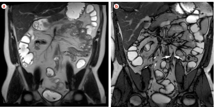
Fig. 2 – Coronal T2-weighted SSETSE (a) and steady state free precession (b) images in a 32-year old woman with inactive Crohn’s disease. Frozen mesentery appearance on SSETSE sequence (a) is due to k-space filtering effects. Steady state free precession (b) sequences are the ideal for the display of the vascular structures and the display of the mesentery. Small lymph nodes are well demon-strated on these images (small arrows). Note also the chemical-shift artifact that occurs at the interface of mesenteric fat and mesenteric structures (arrows).
decreased signal-to-noise ratio in comparison with the 2D sequences, principally noticeable in old systems, which are partly compensated after antiperistaltic drugs and contrast administration, respectively.
Disease activity and clinical use
Several findings suggest active inflammation in Crohn’s disease and correlate well with acute-phase laboratory pa-rameters.43-45 It must be emphasized that patients with CD can have multiple lesions at different stages of inflammation or even different types of the disease.46
Increased bowel wall enhancement is an important and well-known finding indicative of active inflammation in pa-tients with CD.38,47,48 Many study groups have focused on gadolinium enhanced T1-weighted images for assessment of disease activity,35-42 which may approach 100% of sensi-tivity.
A relatively simple and accurate approach for evalua-tion of CD activity may be based on the associaevalua-tion of T2-weighted and T1-T2-weighted post gadolinium sequences. This combination allows comprehensive evaluation and discrimi-nation between quiescent disease and active inflammation and for evaluation of complications including abscesses or fistulas.49 Several studies demonstrate that inflamed thick-ened bowel wall usually displays high signal intensity on T2-weighted images due to edema (Fig. 3), while fibrosis shows low signal intensity.35,50,51 Fat suppression may im-prove the conspicuity of the high signal changes in the wall as well as in the perienteric fat, which may also be related with active disease. It is challenging to discriminate be-tween edema and fibrosis on MDCT imaging, and it may be largely for this reason that MDCT findings have not corre-lated well with disease activity. In patients with long-lasting non-active CD, there may remain persistent low signal wall thickening on T2-weighted images with lack of increased enhancement. Acute on chronic involvement is suggested by marked enhancement of the mucosa with substantial low
T2-weighted signal intensity and minimal enhancement of the outer layer, and appreciation of this may have a role in the evaluation of acute exacerbations of CD.52 This dif-ferentiation is of utmost clinical importance, as obstructive fibrotic disease should be surgically treated while inflamma-tory disease may benefit from medical treatment.
The presence of intramural fat (high-signal intensity on T2-weighted and SSFP images), which is also related with past or chronic inflammation can be accurately identified when combining features from steady state free precession and fat suppressed T2-weighted images (Fig. 4).
Concomitant sensitive ancillary findings of active inflam-mation may be present. Engorgement of the affected bowel segment vasa recta -referred to as the Comb sign and en-hancement of local mesenteric lymph nodes, are the most consistent.53 These are easily identified on steady-state free-precession and post gadolinium T1-weighted images. Complications of CD are also well shown in MRI and include fistulas, phlegmons, abscesses and bowel obstruc-tion (Fig. 5). Fistulas and sinus tracts are demonstrated by the high signal intensity of their fluid content on steady-state free-precession and T2-weighted single-shot fast spin echo images, and enhancement of the linear tract on the post gadolinium T1-weighted sequences.54 Entero-enteric fistulas should be suspected when crowded retracted and angulated small bowel loops, known as star sign, are ap-preciated (Fig. 6). Abscesses can be recognized by their fluid content and increased contrast enhancement of the abscess wall (Fig. 7). The irregular morphology of an ab-scess cavity, and appreciation of its rounded configuration by studying the site on multiple planes, allows distinction from tubular-shaped bowel.
Assessment of severity
Assessment of inflammatory activity of CD is important to identify patients with active inflammation so that appropri-ate therapy may be prescribed.55 Given the advent of new
Fig. 3 – Coronal T2-weighted fat suppressed SSETSE (a), steady state free precession (b) and immediate post gadolinium T1-weighted
3D GRE fat suppressed (c, d) images in a 55-year old womman with severe non-fibrotic active Crohn’s disease. The SSETSE sequence (a) reveals abnormal bowel wall thickening involving the terminal ileum, displaying moderately high signal intensity in T2-weighted fat sup-pressed images consistent with submucosal edema (arrow). Mesenteric edema is also depicted (asterisk). Steady state free precession images (b) don’t demonstrate submucosal edema but clearly depicts mesenteric lymph nodes and comb sign, both indicators of disease activity. Post gadolinium images (c,d) show extensive mucosal enhancement in affected bowel segments (arrows), coomb sign and en-larged enhancing mesenteric lymph nodes reflecting active disease. All sequences display well fibro-fatty proliferation.
medications some with significant side effects (such as TNF alpha inhibitors), objective measures of activity are needed to justify their use and judge their effectiveness.56 Currently, there is no gold standard for determination of CD activity. Evaluation is multifactorial and based on a combination of clinical scoring, biologic indices, endoscopy, and radiologic imaging.57 The most widely used clinical scoring system is the CDAI. The limitations of this index are well documented and include inter-observer variability, inclusion of the sub-jective variables such as general well-being and intensity of abdominal pain, and the fact that it is based on a diary completed by the patient seven days before assessment, which prevents its use in everyday practice.58 Additionally, it underestimates disease in patients with fistulas or steno-ses. Similarly, correlation with laboratory biomarkers is use-ful but imperfect. As the disease affects the use-full thickness of the bowel wall, the adjacent fat and mesentery, due to mu-cosa’s high regenerative capacity, endoscopic evaluation might also under characterize the true extent and activity of disease.
Dynamic contrast-enhanced MRI and diffusion weight-ed imaging are two promising techniques for the detection
of active small bowel inflammation, providing quantitative measures of bowel perfusion and diffusion that can differ-entiate actively inflamed small bowel segments from normal small bowel in CD.
Dynamic contrast-enhanced semi-quantitative MR ap-proach allows the quantification of signal variations over time after intra-venous injection of paramagnetic contrast material. This has been shown to reflect the alterations of tissue microcirculation in inflammatory conditions, such as in rheumatoid arthritis.59 In inflammatory conditions, early, rapid, and marked contrast enhancement is related to in-creased vascularity, while late interstitial accumulation of contrast material is due to increased capillary permeability.60 In the specific case of CD, local vascularization is known to increase with the severity of the disease and has been found to be highly correlated with tissue enhancement.60-62 More recently, strong mucosal angiogenic acti-vity in CD has been detected histopathologically by Danese, et al.,63 confirming the role of inflammation-dependent neoangio-genesis on local microvasculature.
Based on these pathophysiological principles, a strong, early increase of contrast enhancement after intravenous
Fig. 4 – Coronal T2-weighted SSETSE non-fat suppressed (a) and fat suppressed (b), and coronal immediate (c) and axial 60 second (d)
post gadolinium T1-weighted 3D GRE fat suppressed images in a 47-year old woman. Skipped segmental wall thickening is appreciated involving the terminal ileum, while intramural fat deposition can be recognized by the high signal intensity depicted on the T2-weighted image (a) with signal suppression on the fat-suppressed image (b), indicative of long standing Crohn’s disease. Post gadolinium images (c and d) show marked mucosal enhancement consistent with acute on chronic Crohn’s disease (fatty, non fibrotic). Moderate fibro-fatty proliferation and mild comb sign are also depicted.
A liver hemangioma was also depicted in the right lobe.
Fig. 5 – Coronal T2-weighted SSETSE (a), steady state free precession (b, c) and immediate post gadolinium coronal T1-weighted 3D
GRE fat suppressed (c) images in a patient with active Crohn’s disease involving a long segment of the ileum. There is narrowing of the involved bowel segment with increased wall thickness (arrows, a, b) and prestenotic dilatation of unaffected bowel. Steady state free precession images (b, c) doesn’t demonstrate submucosal edema but clearly depicts comb sign suggestive of disease activity. Post gadolinium images (d, e) show extensive mucosal enhancement in affected bowel segments, with pseudo-polyps and comb sign, reflect-ing active disease. The presence of submucosal edema excludes late fibrotic changes of the bowel wall suggestreflect-ing potential response to medical therapy.
Fig. 6 – Coronal steady state free precession (a), coronal T2-weighted SSETSE (b) and 2,5-minute post gadolinium T1-weighted 3D GRE
fat suppressed (c) images in a 25-year old man with non-fibrotic active Crohn’s disease with entero-enteric fistulas involving ileal loops. Crowded retracted and angulated small bowel loops (star sign) are appreciated in all sequences. Post gadolinium images (c) shows extensive mucosal and mesenteric enhancement reflecting active disease. Fibro-fatty proliferation and the comb sign are appreciated in all sequences.
administration of a bolus of paramagnetic contrast material may be interpreted as a direct marker of inflammation of small bowel loops affected by active CD.61
Fast and marked contrast enhancement has been shown to occur only in actively inflamed bowel loops and this should be interpreted as a consequence of bowel loop inflammation. Giusti, et al.61 described two different types of time–signal intensity curves. More specifically, type I curves, found in active disease, are characterized by a steep upslope with a high peak followed by a plateau, on the other hand, type II curves may be considered represen-tative of inactive CD, with lower upslope, smaller peak, and progressive wash-out.61
Actively inflamed small bowel segments in CD patients demonstrate increased perfusion. Dynamic contrast-en-hanced MRI allows reliable differentiation between active and inactive CD of the small bowel by means of quantitative parameters.62
Diffusion-weighted imaging has been investigated re-cently in the assessment of bowel inflammation in CD. 63,64-66 Initial results from studies evaluating both small bowel and colon appear promising despite their limitations such as small sample size or an imperfect reference standard. The use of DWI with parallel imaging for detection of bowel wall inflammation in CD has been recently addressed in two small series.65,66 A recent study states that diffusion-weighted imaging (DWI) provides quantitative measures of small bowel inflammation that can differentiate actively inflamed small bowel segments from normal small bowel in CD.67 Increased cell density and viscosity, dilated lym-phatic channels and granuloma development can narrow the extracellular space and contribute to restricted diffusion of water molecules in the inflamed bowel wall, neverthe-less, the exact mechanism for restricted diffusion in actively inflamed bowel wall remains unclear. ADC parameter alone can provide high sensitivity for detection of active
inflamma-tion but the combinainflamma-tion of DWI and DCE-MRI parameters can potentially improve specificity.67
In perspective, these findings may reveal a potential role for MRI as an alternative technique to endoscopy for non-invasive evaluation of CD, with particular reference to the evaluation and grading of disease activity.
CONClUSION
The new imaging paradigm should contemplate patient safety as an important aspect of assessing the role of an im-aging modality. State of the art MRE has rapidly emerged as successful small bowel imaging modality, offering detailed morphologic information, and also permitting evaluation of extra-intestinal manifestation of disease. The main draw-backs are still related with present availability and economic constrains.
Intrinsic advantages and the lack of ionizing radiation may make MRE the preferred modality for evaluation of small bowel disease, especially in young patients in the setting of CD, considering that the majority will undergo frequent repeat studies. Pregnant patients and those with iodine contrast agents allergy or decreased renal clearance may also benefit from shifting to this technique.
The information on disease activity is not matched by any other imaging method, with recent studies describing also a potential role of MRI for noninvasive evaluation of disease activity by means of quantitative measurements. These features have now been shown to alter physician level of confidence and management plans, including medi-cal or surgimedi-cal approaches in patients with bowel disease. CONFlICT OF INTERESTS
None stated. FUNDING SOURCES None stated.
Fig. 7 – Coronal T2-weighted SSETSE non-fat suppressed (a), coronal steady state free precession (b) and 2-minute and 2,5-minute
post gadolinium T1-weighted 3D (c) and 2D GRE fat suppressed (d) images in a 24-year old man with long-standing Crohn’s disease. Segmental wall thickening is appreciated involving the proximal jejunum; there is two mesenteric fluid collections adjacent to the diseased bowel segment with high signal intensity in T2-weighted images (arrow, a). The irregular and thick enhancing rim (c, d) is characteristic of the reactive inflammatory capsule associated to the abscess. Also the round configuration in multiple planes allows confident diagnosis of fluid collections. Coronal post gadolinium images (d) show marked mucosal and serosal (arrows, d) enhancement consistent with acute Crohn’s disease. Notice that 2D acquisition enable less blurring and higher image quality in comparison with 3D acquisition.
Moderate fibro-fatty proliferation and mild comb sign are also depicted.
REFERENCES
1. Kim SC, Ferry GD. Inflammatory bowel diseases in pediatric and ado-lescent patients: clinical, therapeutic, and psychosocial considerations. Gastroenterology 2004;126:1550–1560.
2. Loftus EV Jr. Clinical epidemiology of inflammatory bowel disease: in-cidence, prevalence, and environmental influences. Gastroenterology 2004;126:1504–1517.
3. Sandborn WJ, Feagan BG, Hanauer SB, Lochs H, Löfberg R, Mod-igliani R, et al. A review of activity indices and efficacy endpoints for clinical trials of medical therapy in adults with Crohn’s disease. Gastro-enterology 2002;122:512–530.
4. Best W, Becktel J, Singleton J, Kern F. Development of a Crohn_s disease activity index: National Cooperative Crohn_s Disease Study. Gastroenterology 1976; 70:439–444.
5. Jørgensen LG, Fredholm L, Hyltoft Petersen P, Hey H, Munkholm P, Brandslund I. How accurate are clinical activity indices for scoring of disease activity in inflammatory bowel disease (IBD)?. Clin Chem Lab Med 2005;43:403–411.
6. Best WR. Predicting the Crohn_s disease activity index from the Har-vey-Bradshaw Index. Inflamm Bowel Dis 2006;12:304–310.
7. Swain CP, Gong F, Mills TN. Wireless transmission of a colour televi-sion moving image from the stomach using a miniature CCD camera, light source and microwave transmitter. GUT 1996;39:A26.
8. Iddan GI, Swain CP. History and development of capsule endoscopy. Gastrointest Endosc Clin N Am 2004;14:1–9.
9. Rondonotti E, Herrerias JM, Pennazio M, Caunedo A, Mascarenhas-Saraiva M, de Franchis R. Complications, limitations, and failures of capsule endoscopy: a review of 733 cases. Gastrointest Endosc 2005; 62(5):712-716.
10. Barkin J, Friedman S. Wireless capsule endoscopy requiring surgical intervention: the words experience. Am J Gastroenterol 2002;97:5298. 11. Donnelly LF. Reducing radiation dose associated with pediatric CT by decreasing unnecessary examinations. AJR Am J Roentgenol 2005;184:655–657.
12. Semelka RC, Armao D, Júnior JE, Huda W. Imaging Strategies to Re-duce the Risk of Radiation in CT Studies, Including Selective Substitu-tion With MRI. J Magn Reson Imaging 2007;25:900-909.
13. Picano E, Vano E, Semelka R, Regulla D. The American College of Ra-diology white paper on radiation dose in medicine:deep impact on the practice of cardiovascular imaging. Cardiovasc Ultrasound 2007;5:37. 14. Jaffe TA, Gaca AM, Delaney S, Yoshizumi TT, Toncheva G, Nguyen
G, et al. Radiation Doses from Small-Bowel Follow-Through and Abdominopelvic MDCT in Crohn’s Disease. AJR Am J Roentgenol 2007;189:1015–1022.
15. What are the radiation risks from CT? U.S. Food and Drug Administra-tion, http://www.fda.gov/cdrh/ct/risks.html; 2005.
16. Executive Summary. Board on Radiation Effects Research – Division on Earth and Life Studies editor Health Risks from Exposure to Low Levels of Ionizing Radiation: BEIR VII – Phase 2. Washington, DC: National Academy Press; 2005.
17. Lee SS, Kim AY, Yang SK, Chung JW, Kim SY, Park SH, et al. Crohn disease of the small bowel: comparison of CT enterography, MR en-terography, and small-bowel follow-through as diagnostic techniques. Radiology 2009;251:751-761.
18. Siddiki HA, Fidler JL, Fletcher JG, Burton SS, Huprich JE, Hough DM,
et al. Prospective comparison of state-of-the-art MR enterography and
CT enterography in small-bowel Crohn’s disease. AJR Am J Roent-genol 2009;193(1):113-121.
19. Fiorino G, Bonifacio C, Peyrin-Biroulet L, Minuti F, Repici A, Spinelli A,
et al. Prospective comparison of computed tomography enterography
and magnetic resonance enterography for assessment of disease ac-tivity and complications in ileocolonic Crohn’s disease. Inflamm Bowel Dis 2011;17:1073-1080.
20. Jensen MD, Ormstrup T, Vagn-Hansen C, Østergaard L, Rafaelsen SR. Interobserver and intermodality agreement for detection of small bowel Crohn’s disease with MR enterography and CT enterography. Inflamm Bowel Dis 2011;17:1081-1088.
21. Herédia V, Ramalho M, de Campos RO, Lee CH, Dale B, Vaidean GD,
et al. Comparison of a single shot T1-weighted in- and out-of-phase
magnetization prepared gradient recalled echo with a standard two-di-mensional gradient recalled echo: Preliminary findings. J Magn Reson Imaging 2011;33:1482-1490.
22. Azevedo RM, de Campos RO, Ramalho M, Herédia V, Dale BM, Se-melka RC. Free Breathing Three-Dimensional T1-Weighted Gradient-Echo Sequence Using Radial Data Sampling In Abdominal Imaging: Preliminary Observations. AJR Am J Roentgenol 2011;197:650-657. 23. Ferreira A, Ramalho M, de Campos RO, Heredia V, Azevedo RM, Dale
B, et al. Comparison of t1-weighted in- and out-of-phase single shot magnetization-prepared gradient-recalled-echo with three-dimensional gradient-recalled-echo at 3.0t: preliminary observations. J Magn Re-son Imaging. 2011 Nov 29. [completar referência com volume e pági-nas de inicio e fim].
24. Gore RM, Balthazar EJ, Ghahremani GG, Miller FH. CT features of ulcerative colitis and Crohn’s disease. AJR Am J Roentgenol 1996;167:3–15.
25. Negaard A, Paulsen V, Sandvik L, Berstad AE, Borthne A, Try K, et
al. A prospective randomized comparison between two MRI studies of
the small bowel in Crohn’s disease, the oral contrast method and MR enteroclysis. Eur Radiol 2007;17:2294-2301.
26. Schreyer AG, Geissler A, Albrich H, Schölmerich J, Feuerbach S, Ro-gler G, et al. Abdominal MRI after enteroclysis or with oral contrast in patients with suspected or proven Crohn’s disease. Clin Gastroenterol Hepatol 2004;2:491–497.
27. Mazzeo S, Caramella D, Battolla L, Melai L, Masolino P, Bertoni M,
et al. Crohn disease of the small bowel: spiral CT evaluation after
oral hyperhydration with isotonic solution. J Comput Assist Tomogr 2001;25:612–616.
28. Ajaj W, Goehde SC, Schneemann H, Ruehm SG, Debatin JF, Lauen-stein TC. Oral contrast agents for small bowel MRI: comparison of dif-ferent additives to optimize bowel distension. Eur Radiol 2004;14:458– 464.
29. Ajaj W, Goyen M, Schneemann H, Kuehle C, Nuefer M, Ruehm SG,
et al. Oral contrast agents for small bowel distension in MRI: influence
of the osmolarity for small bowel distention. Eur Radiol 2005;15:1400– 1406.
30. Gourtsoyiannis N, Papanikolaou N, Grammatikakis J, Maris T, Prasso-poulos P. MR enteroclysis protocol optimization: comparison between 3D FLASH with fat saturation after intravenous gadolinium injection and true FISP sequences. Eur Radiol 2001;11:908–913.
31. Gourtsoyiannis NC, Grammatikakis J, Papamastorakis G, Koutroum-bakis J, Prassopoulos P, Rousomoustakaki M, et al. Imaging of small intestinal Crohn’s disease: comparison between MR enteroclysis and conventional enteroclysis. Eur Radiol 2006;16:1915–1925.
32. Torkzad MR, Vargas R, Tanaka C, Blomqvist L. Value of cine MRI for better visualization of the proximal small bowel in normal individuals. Eur Radiol 2007;17:2964–2968.
33. Buhmann-Kirchhoff S, Lang R, Kirchhoff C, Steitz HO, Jauch KW, Re-iser M, et al. Functional cine MR imaging for the detection and map-ping of intraabdominal adhesions: method and surgical correlation. Eur Radiol 2008;18:1215–1223.
34. Girometti R, Zuiani C, Toso F, Brondani G, Sorrentino D, Avellini C, et
al. MRI scoring system including dynamic motility evaluation in
assess-ing the activity of Crohn’s disease of the terminal ileum. Acad Radiol 2008;15:153–164.
35. Kim SC, Ferry GD. Inflammatory bowel diseases in pediatric and ado-lescent patients: clinical, therapeutic, and psychosocial considerations. Gastroenterology 2004;126:1550–1560.
36. Semelka RC, Shoenut JP, Silverman R, Kroeker MA, Yaffe CS, Micf-likier AB. Bowel disease: prospective comparison of CT and 1.5-T pre- and postcontrast MR imaging with T1-weighted fatsuppressed and breath-hold FLASH sequences. J Magn Reson Imaging 1991;1:625– 632.
37. Marcos HB, Semelka RC. Evaluation of Crohn’s disease using halffou-rier RARE and gadolinium-enhanced SGE sequences: initial results. Magn Reson Imaging 2000;18:263–268.
38. Low RN, Francis IR, Politoske D, Bennett M. Crohn’s disease evalua-tion: comparison of contrast-enhanced MR imaging and single phase helical CT scanning. J Magn Reson Imaging 2000;11:127– 135. 39. Low RN, Sebrechts CP, Politoske DA, Bennett MT, Flores S, Snyder
RJ, et al. Crohn disease with endoscopic correlation: single-shot fast spin-echo and gadoliniumenhanced fat-suppressed spoiled gradient-echo MR imaging. Radiology 2002;222:652–660.
40. Shoenut JP, Semelka RC, Silverman R, Yaffe CS, Micflikier AB. Mag-netic resonance imaging in inflammatory bowel disease. J Clin Gastro-enterol 1993;17:73–78.
41. Shoenut JP, Semelka RC, Magro CM, Silverman R, Yaffe CS, Micflikier AB. Comparison of magnetic resonance imaging and endoscopy in dis-tinguishing the type and severity of inflammatory bowel disease. J Clin Gastroenterol 1994;19:31–35.
42. Albert JG, Martiny F, Krummenerl A, Stock K, Lesske J, Göbel CM,
et al. Diagnosis of small bowel Crohn’s disease: a prospective
com-parison of capsule endoscopy with magnetic resonance imaging and fluoroscopic enteroclysis. Gut 2005;54:1721–1727.
43. Florie J, Wasser MN, Arts-Cieslik K, Akkerman EM, Siersema PD, Stoker J. Dynamic contrastenhanced MRI of the bowel wall for as-sessment of disease activity in Crohn’s disease. AJR Am J Roentgenol 2006;186:1384–1392.
44. Maccioni F, Viscido A, Broglia L, Marrollo M, Masciangelo R, Caprilli R, et al. Evaluation of Crohn disease activity with magnetic resonance imaging. Abdom Imaging 2000;25:219–228.
45. Kettritz U, Isaacs K, Warshauer DM, Semelka RC. Crohn’s disease. Pilot study comparing MRI of the abdomen with clinical evaluation. J Clin Gastroenterol 1995;21:249–253.
46. Sempere GA, Martinez Sanjuan V, Medina Chulia E, et al. MRI evalua-tion of inflammatory activity in Crohn’s disease. AJR Am J Roentgenol 2005;184:1829–1835.
47. Maglinte DD, Gourtsoyiannis N, Rex D, Howard TJ, Kelvin FM. Classi-fication of small bowel Crohn’s subtypes based on multimodality imag-ing. Radiol Clin North Am 2003;41:285–303.
48. Maccioni F, Bruni A, Viscido A, Colaiacomo MC, Cocco A, Montesani C,
et al. MR imaging in patients with Crohn disease: value of T2- versus
T1-weighted gadolinium- enhanced MR sequences with use of an oral superparamagnetic contrast agent. Radiology 2006;238:517–530. 49. Oommen J, Oto A. Contrast-enhanced MRI of the small bowel in
Crohn’s disease. Abdom Imaging 2011;36:134–141.
50. Martin DR, Lauenstein T, Sitaraman SV. Utility of Magnetic Reso-nance Imaging in Small Bowel Crohn’s Disease. Gastroenterology 2007;133:385–390.
51. Madsen SM, Thomsen HS, Schlichting P, Dorph S, Munkholm P. Evalu-ation of treatment response in active Crohn’s disease by lowfield mag-netic resonance imaging. Abdom Imaging 1999;24:232–239. 52. Schunk K, Kern A, Oberholzer K, Kalden P, Mayer I, Orth T, et al.
Hy-dro-MRI in Crohn’s disease: appraisal of disease activity. Invest Radiol 2000;35:431–437.
53. Martin D, Altun E, Elias JR J, Elazzazi M, Ramalho M, Lee C-H, et al. Gastrointestinal tract. In: R.C. Semelka, editor. Abdominal-Pelvic MRI. 23rd edition. Wiley. 2006;784–796.
54. Gourtsoyiannis N, Papanikolaou N, Grammatikakis J, Papamastorakis G, Prassopoulos P, Roussomoustakaki M. Assessment of Crohn’s dis-ease activity in the small bowel with MR and conventional enteroclysis: preliminary results. Eur Radiol 2004;14:1017–1024.
55. Prassopoulos P, Papanikolaou N, Grammatikakis J, Rousomoustakaki M, Maris T, Gourtsoyiannis N. MR Enteroclysis Imaging of Crohn Dis-ease. Radiographics 2001;21:S161–S172.
56. Malagò R, Manfredi R, Benini L, D’Alpaos G, Mucelli RP. Assessment of Crohn’s disease activity in the small bowel with MR-enteroclysis: clinico-radiological correlations. Abdom Imaging 2008;33:669–675. 57. Bodily KD, Fletcher JG, Solem CA, Johnson CD, Fidler JL, Barlow JM,
et al. Mural attenuation and thickness at contrast-enhanced CT
enter-ography—correlation with endoscopic and histologic findings of inflam-mation. Radiology 2006; 238:505–516.
58. Martínez MJ, Ripollés T, Paredes JM, Blanc E, Martí-Bonmatí L. As-sessment of the extension and the inflammatory activity in Crohn’s disease: comparison of ultrasound and MRI. Abdom Imaging 2009;34:141–148.
59. Pupillo VA, Di Cesare E, Frieri G, Limbucci N, Tanga M, Masciocchi C. Assessment of inflammatory activity in Crohn’s disease by means of dynamic contrast-enhanced MRI. Radiol Med 2007;112:798–809. 60. Ostergaard M, Stoltenberg M, Løvgreen-Nielsen P, Volck B,
Sonne-Holm S, Lorenzen I. Quantification of synovitis by MRI: correlation be-tween dynamic and static gadolinium-enhanced magnetic resonance imaging and microscopic and macroscopic signs of synovial inflamma-tion. Magn Reson Imaging 1998;16:743–754.
61. Horsthuis K, Lavini C, Bipat S, Stokkers PC, Stoker J. Perianal Crohn disease: evaluation of dynamic contrast-enhanced MR imaging as an indicator of disease activity. Radiology 2009;251:380–387.
62. Giusti S, Faggioni L, Neri E, Fruzzetti E, Nardini L, Marchi S, et al. Dynamic MRI of the small bowel: usefulness of quantitative contrast-enhancement parameters and time–signal intensity curves for differen-tiating between active and inactive Crohn’s disease. Abdom Imaging 2010;35:646–653.
63. Oommen J, Oto A. Contrast-enhanced MRI of the small bowel in Crohn’s disease. Abdom Imaging 2011;36:134–141.
64. Taylor SA, Punwani S, Rodriguez-Justo M, Bainbridge A, Greenhalgh R, De Vita E, et al. Mural Crohn disease: correlation of dynamic con-trast-enhanced MR imaging findings with angiogenesis and inflamma-tion at histologic examinainflamma-tion–pilot study. Radiology 2009;251:369– 379.
65. Danese S, Sans M, de la Motte C, Graziani C, West G, Phillips MH, et
al. Angiogenesis as a novel component of inflammatory bowel disease
pathogenesis. Gastroenterology 2006;130:2060–2073.
66. Oto A, Fan X, Mustafi D, Jansen SA, Karczmar GS, Rubin DT, et al. Quantitative analysis of dynamic contrast enhanced MRI for assess-ment of bowel inflammation in Crohn’s disease pilot study. Acad Radiol 2009;16:1223–1230.
67. Oto A, Zhu F, Kulkarni K, Karczmar GS, Turner JR, Rubin D. Evaluation of diffusion-weighted MR imaging for detection of bowel inflammation in patients with Crohn’s disease. Acad Radiol 2009;16:597–603. 68. Kiryu S, Dodanuki K, Takao H, Watanabe M, Inoue Y, Takazoe M, et
al. Free-breathing diffusion- weighted imaging for the assessment
of inflammatory activity in Crohn’s disease. J Magn Reson Imaging 2009;29:880–886.
69. Oto A, Kayhan A, Williams JT, Fan X, Yun L, Arkani S, et al. Active Crohn’s Disease in the Small Bowel: Evaluation by Diffusion Weighted Imaging and Quantitative Dynamic Contrast Enhanced MR Imaging. J Magn Reson Imaging 2011;33:615–624.