Dement Neuropsychol 2017 December;11(4):343-355
343
Alves et al. Cerebrovascular disease: novel neuroimaging methods
Views
&
Reviews
A panel of clinical and neuropathological
features of cerebrovascular disease through
the novel neuroimaging methods
Gilberto Sousa Alves1, Luiza de Amorim de Carvalho1, Felipe Kenji Sudo2,3,
Lucas Briand1, Jerson Laks4,5, Eliasz Engelhardt6
ABSTRACT. The last decade has witnessed substantial progress in acquiring diagnostic biomarkers for the diagnostic workup of cerebrovascular disease (CVD). Advanced neuroimaging methods not only provide a strategic contribution for the differential diagnosis of vascular dementia (VaD) and vascular cognitive impairment (VCI), but also help elucidate the pathophysiological mechanisms ultimately leading to small vessel disease (SVD) throughout its course. Objective: In this review, the novel imaging methods, both structural and metabolic, were summarized and their impact on the diagnostic workup of age-related CVD was analysed. Methods: An electronic search between January 2010 and 2017 was carried out on PubMed/MEDLINE, Institute for Scientific Information Web of Knowledge and EMBASE. Results: The use of full functional multimodality in simultaneous Magnetic Resonance (MR)/Positron emission tomography (PET) may potentially improve the clinical characterization of VCI-VaD; for structural imaging, MRI at 3.0 T enables higher-resolution scanning with greater imaging matrices, thinner slices and more detail on the anatomical structure of vascular lesions. Conclusion: Although the importance of most of these techniques in the clinical setting has yet to be recognized, there is great expectancy in achieving earlier and more refined therapeutic interventions for the effective management of VCI-VaD. Key words: neuroimaging, vascular, PET, MRI, diffusion tensor imaging, DTI, novel methods.
UM PAINEL DE CARACTERÍSTICAS CLÍNICAS E NEUROPATOLÓGICAS DA DOENÇA CEREBROVASCULAR ATRAVÉS DOS NOVOS MÉTODOS DE NEUROIMAGEM
RESUMO. A última década vem testemunhando avanços substanciais na aquisição de marcadores biológicos para o diagnóstico da doença cerebrovascular (DCV). Os métodos de neuroimagem avançados não apenas fornecem uma contribuição estratégica para o diagnóstico diferencial do comprometimento cognitivo vascular (VCI) e demência vascular (VaD), mas contribuem substancialmente na elucidação dos mecanismos fisiopatológicos relacionados à doença de vasos pequenos (SVD) e sua progressão clínica. Objetivo: Nesta revisão, métodos de imagem estruturais e metabólicos foram descritos e sua importância diagnóstica analisada, particularmente na investigação da CVD relacionada ao envelhecimento. Métodos: uma pesquisa eletrônica de janeiro de 2010 a 2017 foi realizada através do PubMed/ MEDLINE, do Instituto de Informação Científica Web of Knowledge e da EMBASE. Resultados: O emprego de estudos de multimodalidade plenamente funcional com Ressonância Magnética (MR)/ Tomografia por Emissão de Pósitrons (PET) representa uma janela para a caracterização clínica mais detalhada da VCI-VaD; com relação à neuroimagem estrutural, a ressonância magnética em 3,0 T vem permitindo varreduras com maior resolução e matrizes de imagem mais elevadas, cortes mais delgados e maior detalhamento anatômico das lesões vasculares. Conclusão: Embora a importância da maior parte dessas técnicas no cenário clínico aguarde reconhecimento, há uma grande expectativa de que o seu uso favoreça intervenções terapêuticas progressivamente mais precoces e refinadas para o gerenciamento efetivo do VCI-VaD.
Palavras-chave: neuroimagem, vascular, PET, ressonância magnética, imagem de tensor de difusão, DTI, métodos inovadores.
This study was conducted at the Departamento de Medicina Interna, Universidade Federal do Ceará, CE, Brazil.
1Departamento de Medicina Interna, Universidade Federal do Ceará, CE, Brazil. 2Departamento de Psicologia, Pontifícia Universidade Católica do Rio de Janeiro,
RJ, Brazil. 3Instituto D’Or de Ensino e Pesquisa, Rio de Janeiro, RJ, Brazil. 4Instituto de Psiquiatria, Universidade Federal do Rio de Janeiro, RJ, Brazil. 5Programa de
Pós-Graduação em Biomedicina Translacional (BIOTRANS), Unigranrio, Duque de Caxias, RJ, Brazil. 6Setor de Neurologia Cognitiva e do Comportamento, Instituto
de Neurologia Deolindo Couto (INDC-CDA/IPUB), Rio de Janeiro, RJ, Brazil.
Gilberto Sousa Alves. Rua Prof. Costa Mendes 1608 / 4o andar – Fortaleza CE – Brazil. E-mail: [email protected]
Disclosure: The authors report no conflicts of interest.
Received: October 10, 2017. Accepted in final form November 14, 2017.
Dement Neuropsychol 2017 December;11(4):343-355
344 Cerebrovascular disease: novel neuroimaging methods Alves et al.
INTRODUCTION
V
ascular cognitive impairment (VCI) is an umbrella term denoting a continuum of behavioral and cog-nitive deicits associated with cerebrovascular disease (CVD).1-3 CVD is estimated to occur in one third of thepopulation, often being recognized as a pathological inding on conventional Magnetic Resonance Imaging (MRI).1,3,4 Depending on the site, intensity, and severity,
CVD may either cause or contribute to further cognitive impairment.2,5
Over the last decade, there has been substantial progress in acquiring diagnostic biomarkers for the diagnostic workup of neurodegenerative and vascular disorders.2,6In vivo brain imaging has been applied for
several decades to identify brain structural (disease-speciic atrophy) and functional (disease-(disease-speciic hypo-metabolism) abnormalities. Advanced neuroimaging methods not only provide a strategic contribution for the diferential diagnosis of vascular dementia (VaD), but also help elucidate the pathophysiological mecha-nisms ultimately leading to small vessel disease (SVD) throughout aging.6 One example of the growing
impor-tance of structural and functional imaging markers on the diagnostic work up of dementia is that the ifth edi-tion of the DSM (2013) has changed to include a broader deinition of cognitive impairment, the neurocognitive disorder. hese criteria stress the need to support the etiological diagnosis with neuroimaging markers.6
In a relatively short period, particularly in the last 15 years, structural neuroimaging has evolved from a quite artisanal approach – focused on the delimitation of pre-deined Region of interest (ROIs) – to powerful volumetric-based morphometry (VBM) analysis,7,8 a
measure based on a voxel-wise comparison of highly localized gray matter (GM) regions between two clini-cal groups. VBM tests for residual tissue concentration diferences that remain after spatial normalization into the same standardized stereotaxic space and method calculations rate the within-voxel concentrations of GM (i.e., diferences in the proportion of GM contained within a given voxel).9,10 Accordingly, the speciic
con-tribution of molecular imaging provided by nuclear medicine techniques such as Single Photon Emission Computed Tomography (SPECT) and Positron Emis-sion Tomography (PET) has been profound, with major improvements regarding speciicity, imaging resolution, and more recently, functional multimodality.
Early reports from structural studies have identi-ied subcortical hyperintensities as macroscopic white matter (WM) changes which have been cited by sev-eral reports as associated with CVD-VCI, mood
disor-ders, executive dysfunction and higher conversion to dementia. In functional terms, it is hypothesized that cognitive deicits observed in subcortical VCI arise when infarcts in the WM lead to the disruption of neuronal circuits connecting cortical and subcortical structures. Despite the substantial progress on the characteriza-tion of cognitive deicits and the early identiicacharacteriza-tion of minor vascular lesions, other important issues related to CVD, such as the neuropathological etiology of these lesions, remain a subject of intensive research in the last decade, with studies evolving to address the relationship between normal-appearing WM and amyloid angiopathy or Wallerian Degeneration.11-13 On the other hand,
cere-bral microbleeds (CMB) or cerecere-bral microhemorrhages (CMH) are small hypointense lesions with variable cut-of size – typically between 5 and 10 mm – that have been attracting growing interest in recent years. Previ-ous literature has shown an increased number of CMBs in MCI (around 11%) and there is an extensive debate on the signiicance of these lesions in terms of higher conversion to dementia.14
In this brief review, we aim to summarize some of these novel implementations, both in the macro and microanatomy and radiotracer aspects, and discuss their impact on the diagnostic workup of age-related cogni-tive disorders, focusing on the ield of VCI-VaD.
METHODS
A review of the literature (Table 1) published between January 2010 and 2017 was performed through searches on the electronic databases PubMed/MEDLINE (http://www.ncbi.nlm.nih.gov/pubmed/), Institute for Scientiic Information Web of Knowledge (http://www. isiknowledge.com) and EMBASE (http://www.embase. com), using the following terms: “structural neuroim-aging”, “cerebrovascular”, “vascular dementia”, “vascular cognitive impairment” “aging”, “difusion tensor imaging”, “DTI”, “MRI”, “VBM”, “molecular neuroim-aging”, “SPECT” and “PET” search. Firstly, the complete abstract was read, with the irst paper selection. A second selection included the full reading of the papers. Articles were included if they focused on clinical and therapeutic applications of novel neuroimaging techniques in the assessment of cognitive symptoms of VCI-CVD-VaD. Although we designed a non-systematic review, article retrieval and selection were performed following the main recommendations of the Moose guidelines.15
RESULTS
Dement Neuropsyc
hol 2017 December;11(4):343-355
345
Alves et al.
Cer
ebro
vascular disease:
no
vel neuroimaging methods
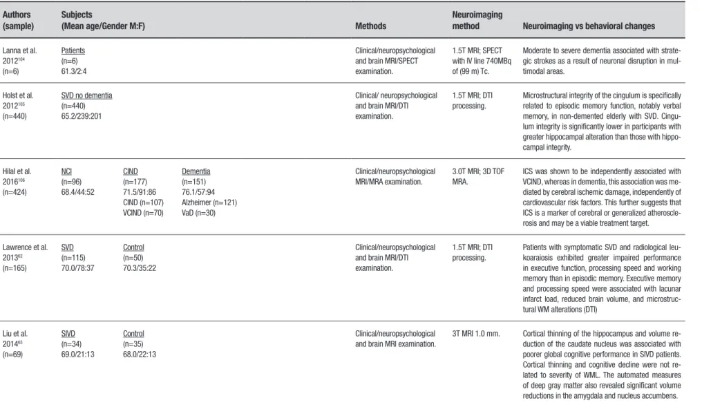
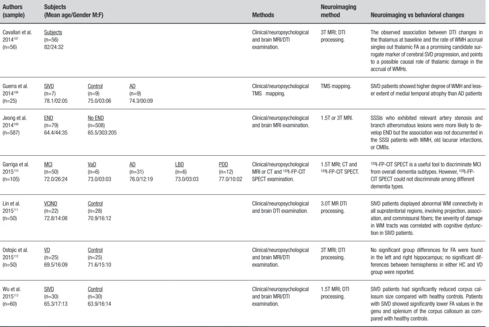
Table 1. Structural and metabolic imaging studies with VCI and VaD.
Authors (sample)
Subjects
(Mean age/Gender M:F) Methods
Neuroimaging
method Neuroimaging vs behavioral changes
Lanna et al. 2012104
(n=6)
Patients (n=6) 61.3/2:4
Clinical/neuropsychological and brain MRI/SPECT examination.
1.5T MRI; SPECT with IV line 740MBq of (99 m) Tc.
Moderate to severe dementia associated with strate-gic strokes as a result of neuronal disruption in mul-timodal areas.
Holst et al. 2012105
(n=440)
SVD no dementia (n=440) 65.2/239:201
Clinical/ neuropsychological and brain MRI/DTI examination.
1.5T MRI; DTI processing.
Microstructural integrity of the cingulum is specifically related to episodic memory function, notably verbal memory, in non-demented elderly with SVD. Cingu-lum integrity is significantly lower in participants with greater hippocampal alteration than those with hippo-campal integrity.
Hilal et al. 2016106
(n=424)
NCI (n=96) 68.4/44:52
CIND (n=177) 71.5/91:86 CIND (n=107) VCIND (n=70)
Dementia (n=151) 76.1/57:94 Alzheimer (n=121) VaD (n=30)
Clinical/neuropsychological MRI/MRA examination.
3.0T MRI; 3D TOF MRA.
ICS was shown to be independently associated with VCIND, whereas in dementia, this association was me-diated by cerebral ischemic damage, independently of cardiovascular risk factors. This further suggests that ICS is a marker of cerebral or generalized atheroscle-rosis and may be a viable treatment target.
Lawrence et al. 201362
(n=165)
SVD (n=115) 70.0/78:37
Control (n=50) 70.3/35:22
Clinical/neuropsychological and brain MRI/DTI examination.
1.5T MRI; DTI processing.
Patients with symptomatic SVD and radiological leu-koaraiosis exhibited greater impaired performance in executive function, processing speed and working memory than in episodic memory. Executive memory and processing speed were associated with lacunar infarct load, reduced brain volume, and microstruc-tural WM alterations (DTI)
Liu et al. 201465
(n=69)
SIVD (n=34) 69.0/21:13
Control (n=35) 68.0/22:13
Clinical/neuropsychological and brain MRI examination.
3T MRI 1.0 mm. Cortical thinning of the hippocampus and volume re-duction of the caudate nucleus was associated with poorer global cognitive performance in SIVD patients. Cortical thinning and cognitive decline were not re-lated to severity of WML. The automated measures of deep gray matter also revealed significant volume reductions in the amygdala and nucleus accumbens.
Dement Neuropsyc
hol 2017 December;11(4):343-355
346
Cer
ebro
vascular disease:
no
vel neuroimaging methods
Alves et al.
Table 1. Continuation.
Authors (sample)
Subjects
(Mean age/Gender M:F) Methods
Neuroimaging
method Neuroimaging vs behavioral changes
Cavallari et al. 2014107
(n=56)
Subjects (n=56) 82/24:32
Clinical/neuropsychological and brain MRI/DTI examination.
3T MRI; DTI processing.
The observed association between DTI changes in the thalamus at baseline and the rate of WMH accrual singles out thalamic FA as a promising candidate sur-rogate marker of cerebral SVD progression, and points to a possible causal role of thalamic damage in the accrual of WMHs.
Guerra et al. 2014108
(n=25)
SIVD (n=7) 78.1/02:05
Control (n=9) 75.0/03:06
AD (n=9) 74.3/00:09
Clinical/neuropsychological TMS mapping.
TMS mapping. SIVD patients showed higher degree of WMH and less-er extent of medial temporal atrophy than AD patients
Jeong et al. 2014109
(n=587)
END (n=79) 64.4/44:35
No END (n=508) 65.5/303:205
Clinical/neuropsychological and brain MRI examination.
1.5T or 3T MRI. SSSIs who exhibited relevant artery stenosis and branch atheromatous lesions were more likely to de-velop END but the association was not documented in the SSSI patients with WMH, old lacunar infarctions, or CMBs.
Garriga et al. 2015110
(n=105)
MCI (n=50) 72.0/26:24
VaD (n=6) 73.0/03:03
AD (n=31) 76.0/12:19
LBD (n=6) 73.0/03:03
PDD (n=12) 77.0/10:02
Clinical/neuropsychological MRI or CT and ¹²³I-FP-CIT SPECT examination.
1.5T MRI; CT and ¹²³I-FP-CIT SPECT.
¹²³I-FP-CIT SPECT is a useful tool to discriminate MCI from overall dementia subtypes. However, ¹²³I-FP-CIT SPECT could not discriminate among different dementia types.
Lin et al. 2015111
(n=50)
VCIND (n=22) 72.8/14:08
Control (n=28) 70.9/16:12
Clinical/neuropsychological and brain DTI examination.
3.0T MR DTI processing.
SIVD patients displayed abnormal WM connectivity in all supratentorial regions, involving projection, associ-ation, and commissural fibers; the severity of damage in WM tracts was correlated with cognitive dysfunc-tion in SIVD patients.
Ostojic et al. 2015112
(n=50)
VD (n=25) 69.5/16:09
Control (n=25) 71.6/15:10
Clinical/neuropsychological and brain MRI/DTI examination.
3T MRI; DTI processing.
No significant group differences for FA were found in the left and right hippocampus; no significant dif-ferences between hemispheres in either HC and VD group were reported.
Wu et al. 2015113
(n=60)
SIVD (n=30) 65.3/17:13
Control (n=30) 63.9/16:14
Clinical/neuropsychological and brain MRI/DTI examination.
1.5T MRI; DTI processing.
SIVD patients had significantly reduced corpus cal-losum size compared with healthy controls. Patients with SIVD showed significantly lower FA values in the genu and splenium of the corpus callosum as com-pared with healthy controls.
Dement Neuropsyc
hol 2017 December;11(4):343-355
347
Alves et al.
Cer
ebro
vascular disease:
no
vel neuroimaging methods
Table 1. Continuation.
Authors (sample)
Subjects
(Mean age/Gender M:F) Methods
Neuroimaging
method Neuroimaging vs behavioral changes
Baykara et al. 201689
(n=710)
CADASIL exploratory (n=113) 49.1/43:61
CADASIL validation (n=57) 53.4/38:19
Sporadic SVD (n=444) 65.3/243:201
Memory clinic patients with SVD (n=105) 74.9/54:51
Clinical/neuropsychological and brain MRI/DTI/PSMD examination.
MRI; DTI with PSMD processing.
The study established a novel-imaging marker (PSMD) for SVD. PSMD combined the analysis of DTI-WM tract skeletonization and MD histogram; PSMD was asso-ciated with SVD pathology but not with neurodegen-erative pathology. An important association between PSMD and processing speed deficits across all study samples was reported.
Veluw et al. 201659
(n=5)
Cases with CAA (n=5) 85.0/03:02
Clinical/neuropsychological and brain histopathological and in vivo and ex vivo MRI examination.
Ex vivo 7.0T MRI. CMBs and microinfarcts appeared to be the most fre-quent marker of focal bleeding and focal ischaemic injury in SVD (particularly CAA), and therefore are im-portant candidate biomarkers for clinical trials.
Thong et al. 201487
(n=100)
Control (n=25) 68.00/11:14
Mild VCIND (n=25) 66.20/19:06
MSVCI (n=30) 69.87/14:16
AD (n=20) 74.25/06:14
Clinical/neuropsychological and brain MRI/HARDI examination.
3T MRI 1 mm and HARDI 3 mm.
Compared to the Mild VCIND group, MSVCI subjects showed thinner cortex in the left superior frontal gy-rus, cingulate, temporal pole, and middle temporal gyrus; in contrast, no statistical differences between groups in cortical thickness in the right hemisphere were reported.
Mascalchi et al. 2016114
(n=36)
CADASIL (n=18) 42.9/07:11
Control (n=18) 41.2/10:8
Clinical/neuropsychological and brain MRI/DTI examination.
3T MRI ; 1mm ; DTI processing; TBSS.
CADASIL patients showed extensive almost symmet-ric areas of significantly increased radial and mean diffusivities and of significantly decreased axial dif-fusivity and FA that involved the cerebral WM, the thalami, and the corpus callosum.
Pasi et al. 2017115
(n=36)
CAA-ICH (n=191) 74.9/95:96
HTN-ICH (n=125) 66.1/73:52
Clinical/neuropsychological and brain MRI examination.
1.5T MRI. The topographic distribution of lacunes (lobar vs deep) helped distinguish the underlying SVD subtype (CAA vs HTN-SVD) in patients with primary ICH, regardless of age status, diagnosis of hypertension or other MRI markers of SVD severity. Lobar lacunes seemed to have a closer relationship with WMH, suggesting a possible common origin.
Dement Neuropsychol 2017 December;11(4):343-355
348 Cerebrovascular disease: novel neuroimaging methods Alves et al.
25 studies were subsequently considered eligible for inclusion and discussion.
PET and SPECT. Both PET and SPECT have been inten-sively applied in the last 20 years to quantify changes in regional brain function induced by age-related
disor-ders16,17 he most widely utilized PET tracer in
cogni-tive disorders is 2-[18F] luoro-2-Deoxy-D-glucose (FDG) PET for measurements of cerebral metabolic rate of glucose (CMRglc), an indicator of diferent parameters, e.g., neuronal activity, oxygen consump-tion, synaptic alterations and molecular changes.16,17
Studies have shown that CMRglc reductions occur in AD-risk states,14 in preclinical AD,18 and correlate with
disease progression19 with higher accuracy than the
Mini-Mental State Examination and ADAS-cog.20
Considerable technical improvements have been made in SPECT and PET methodology, propelling the introduction of modern hybrid technology,21 which
has improved structural – functional assignments that have paved the way for SPECT/CT and PET/CT towards accepted clinical imaging standards.16,22 he beneicial
efects of hybrid systems are clear for brain scans, since post-hoc software-based image fusion of independently acquired imaging data may be employed as a well-established and precise method.23 Advanced
technol-ogy implemented in modern hybrid devices ofers the opportunity to reduce image acquisition times,22 and to
use low-dose CT for accurate attenuation correction of brain scans on SPECT/CT and PET/CT. More recently, with the advent of simultaneous MR/PET,24 novel
solu-tions for adequate attenuation correction have been proposed,25-27 but also improved algorithms for head
motion correction utilizing MRI-based motion tracking in combination with PET list mode data motion correc-tion,28,29 having a major impact on the spatial resolution
of brain imaging studies.
True multimodality. he most fundamental advantage
of MR/PET is a major advancement in true multimo-dality,21,30 e.g. structural-functional and
functional-functional. Due to the distinctiveness of structural MRI sequences yielding quantitative MR applications,31,32
a more reined clinical structural depiction of brain lesions has become possible, along with the functional characterization provided by PET tracer measurements. Indeed, all the methodological improvements of recent years have also led to important enhancement in the diagnostic characterization of cognitive disorders, improving the speciicity and sensitivity of human imag-ing biomarker studies.19 For instance, the possibility of
full functional-functional multimodality in simultane-ous MR/PET; hybrid protocols ofer for instance parallel FDG PET and MRI-spectroscopic imaging,24 improving
the molecular imaging characterization of CVD and neurodegenerative disorders; or, parallel dynamic ligand acquisitions in combination with functional MRI tech-niques (e.g. BOLD fMRI, resting state fMRI, continu-ous arterial spin labeling, contrast-enhanced perfusion techniques, etc.) under pharmacological or non-phar-macological experimental challenges, are expected to advance molecular characterization of neurodegenera-tive disorders and CVD in clinical human neurosciences. Finally, improvements in PET imaging temporal resolu-tion have opened a ield for integrating time-of-light33,34
information into the reconstruction process, leading to the high resolution of today’s PET/CT systems, again, improving imaging capabilities, potentially reducing acquisition times and improving the speciicity of the imaging set-up.
While in AD the best-recognized biomarkers that can be detected in cerebrospinal luid and blood are amyloid-β, tau-protein and phosphorylated tau-protein (phospho-tau), for VaD no speciic biomarker is avail-able. Nevertheless, in VaD, FDG PET usually diferenti-ates widespread areas of focal cortical and subcortical hypometabolism from the pattern typically found in AD, with markedly lower metabolic rates in temporal-pari-etal lobes.17 Additionally, in VCI-VaD, the metabolic ratio
seems to be generally higher than in the AD group.17
Another study conducted by Kim et al.35 investigated
the proile of negative [(subcortical vascular dementia) (n=24)] and positive [(AD), (n=81)] amyloid-β patients using 11C PiB PET. When compared to AD, the negative amyloid-β (subcortical vascular patients) cases showed more pronounced cortical thinning in the bilateral infe-rior frontal, supeinfe-rior temporal gyri and orbitofrontal lobes. Findings also evidenced that, in these areas, SVD independently contributed to cortical atrophy through diferent mechanisms than those of AD.35
More recently, F-18 labeled amyloid PET tracers have been introduced, including compounds such as [18F]Flo-rbetaben,36-38 [18F]Flutemetamol39,40 and
[18F]Florbeta-pir.41,42 hese novel compounds ofer the major
advan-tages of a longer half-life of the radioactive label (110 min), allowing a much wider distribution of amyloid PET scans, even in institutions without an on-line cyclotron facility. Another innovative method to speciically char-acterize AD pathology by PET imaging is through the use of selective in-vivo tau PET tracers, which allow the quantifying of tau aggregation in the brain.43,44
Dement Neuropsychol 2017 December;11(4):343-355
349
Alves et al. Cerebrovascular disease: novel neuroimaging methods
between tau deposits, decreased cognitive function, and neurodegenerative changes, and selective tau imaging enables these associations to be explored in vivo.43
Although qualitative indings achieved with amyloid tracers are robust, quantitative measures of amyloid tracer retention show considerable variability across centers. herefore, standardization of acquisition pro-tocols, subject management, tracer administration, image quality control, and image processing and analy-sis methods has become an important issue for improv-ing the accuracy of quantitative amyloid PET measure-ments.45 his is of particular importance for longitudinal
multi-center studies, and for improving the sensitivity of intervention efects targeting amyloid clearance.46
Recently, a novel method for standardization denomi-nated ‘Centiloids’ was introduced, which attempts to standardize the quantitative amyloid data by relating “nonstandard” analysis methods to a ‘standard’ PIB PET data analysis and expressing the data after transforma-tion into the so-called ‘Centiloid scale’.45
Quantitative Susceptibility Mapping (QSM) of the Brain.
Many biological processes, including regulation of protein expression,47,48 oxygen transport and
neuro-transmission require the presence of iron. he accu-mulation of iron may be found throughout normal aging,49,50 for instance in the basal ganglia and
hippo-campus and also in subcortical regions.49 In a variety
of cognitive related disorders, including CVD, iron accu-mulation is thought to play an important role, possibly due to biochemical alterations related to neurodegener-ation (e.g., oxidative stress, abnormal neuronal connec-tivity), although the exact mechanisms are not fully understood. In rodent models, iron deposition was associated with WM disruption and atrophy by inducing endothelial cell damage.50,51 More recently, improved
quality and accuracy in the quantiication of iron by MRI has been achieved through quantitative suscepti-bility mapping (QSM). In the ield of vascular-related disorders, despite limited data, there is increasing evidence pointing to iron accumulation in putamen and caudate nucleus when compared to healthy controls.50,52 Similar indings were also reported with
CADASIL (cerebral autosomal-dominant arteriopathy with subcortical infarcts and leukoencephalopathy), a genetically deined form of early SVD.52,53 Although
most of the literature reported a predominant pattern of iron deposition in subcortical areas, accumulation in greater vessels has also been mentioned, in this case leading to more extensive vascular damage.50 Taken
together, these support the idea that iron
accumula-tion is a marker of neurodegeneraaccumula-tion and endothelial damage, regardless of the underlying process.
High field MRI of the Brain. he main advantage of 3.0 T over lower-ield MR scanners is a better sign-to-noise ratio (SNR), which increases roughly linearly with the strength of the magnetic ield.54 From 2004 on, a
number of studies have been conducted to assess white matter damage in diseases such as multiple sclerosis,32
Alzheimer’s disease (AD),55 and adrenoleukodystrophy19
with 3.0 or 4.0 T MR scanners, and evaluation of iron deposition in neurodegenerative and cerebro-vascular disease at 7 T.49,54 Consequently, imaging at
3.0 T enables higher-resolution scanning with larger imaging matrices, thinner slices and more detail on anatomical structure, without extending (or extending minimally) the scan acquisition time. hese advan-tages come with a trade-of of increased sensitivity to ield inhomogeneity (deviation of the local magnetic ield from its average value) and changes in relaxation times, in turn producing changes in image contrast.54
At comparable acquisition times, images obtained at 3.0 T have a higher quality with an improved resolu-tion over images obtained at 1.5 T. Alternatively, 3.0 T MRI can be used to obtain acceptable images, similar to those obtained at 1.5 T, but at a fraction of the time, thus reducing potential motion artifacts and providing greater comfort for patients.
Regarding the study of vascular-related pathology, a major development is underway, particularly with the advent of 7 T MRI; previous attempts have been made to distinguish between vascular occlusion and microinfarc-tion versus demyelinating disease, for instance in the diferential diagnosis at 7 T between Susac syndrome and Multiple Sclerosis.56,57 Accordingly, the anatomic
modiications involving dilated perivascular spaces have been investigated and quantiied with greater accuracy using 7 T MRI,58 showing diferent patterns and
quan-tiication, for instance, in stroke, migraine, CADASIL, dementia, AD, and mild cognitive impairment. Further-more, 7 T studies may be more successful in providing a more detailed picture of the neuropathology of closely related conditions, such as the case of cerebral micro-infarcts, whose regional distribution (intracortical and juxtacortical location)59 may indicate chronic or acute
lesions, with the latter being described as gliotic cere-bral microinfarcts with hemorrhagic components. hese indings can be conidently extended by in vivo MRI in
the context of aging and dementia.
Dement Neuropsychol 2017 December;11(4):343-355
350 Cerebrovascular disease: novel neuroimaging methods Alves et al.
(WHM). Some of the methods are based on the Expec-tation-Maximization (EM) algorithm,60 which
difer-entiates brain tissue into WM, GM and cerebrospinal luid (CSF). In addition, tissue segmentation from T1 and Flair images through the EM algorithm also enables the segmentation of lacunar infarcts.61 Finally, the
com-bination of high-ield MRI techniques and novel image sequences, particularly susceptibility-weighted imaging (SWI), has improved the detection of CMB.14
Cortical thinning and cortical surface analysis. Hippo-campal and GM reductions are often described in diferent forms of CVD and may indicate, with greater likelihood, conversion to dementia.7 In subjects with
subcortical ischaemic vascular dementia (SIVD), several studies have observed decreased gray matter both in the total volume62 or in regional territories,7,63
for instance the frontal and temporal lobes. In addi-tion, hippocampal volume atrophy, particularly in the CA1 subield, seems to be vulnerable to vascular-related events, for instance, hypoxia and ischemia, as suggested by rodent-model64 and structural
neuro-imaging65 investigations. More recently, it has been
shown that cortical volume, surface area and cortical thickness are closely related, and a reduction in cortical volume may afect either thickness or surface area (or both).65 Initial studies have attempted to investigate
cortical thinning and found reduction in the perisyl-vian, medial frontal area and posterior cingulate.35,66
More recently, surface area in gray matter has been found to be reduced in SIVD, particularly in the left temporal lobe and dorsolateral PFC.65 Possibly, cortical
thinning and atrophy play a greater role in cognitive decline than the occurrence of WML.67 Taken together,
these indings provide further support for the close relationship between vascular and neurodegeneration, highlighting the anatomical relevance of the perisyl-vian area as a highly sensitive territory to the efects of cortical thinning, possibly due to ischemic damage of lateral cholinergic pathways and disruption of ibers connecting the nucleus basalis of Meynert to frontopa-rietal and temporal areas.68
The use of support vector machines in CVD. he last decade has witnessed a great deal of efort in the devel-opment of methods to quantitatively assess speciic CVD markers and devise metrics allowing the quantii-cation of total CVD burden.69 Regarding data analysis,
machine learning-based algorithms for dementia clas-siication according to the expression of AD-typical metabolic patterns (but also other imaging parameters)
are recent developments and have been inluencing the ield of cerebrovascular disorders. he most employed algorithms include K-means clustering, artiicial neural network, random forest and support vector machine (SVM); the potential advantage of SVM is the classii-cation of more than one biomarker combined, further improving performance accuracy, as demonstrated by studies matching FDG-PET and structural MRI, which yielded higher accuracy rates compared to single modality classiication.70,71
Despite the enthusiasm and great potential of these methods, problems in deining threshold and cluster-ing approach and the statistical inference for SVM and the use of permutation tests that ignore SVM margin still limit the wide applicability of these methods. Fur-thermore, a number of drawbacks remain regarding the limited evaluation of indings, particularly the clinical interpretation of disease mechanisms according to the classiier’s decision.69 Possibly in the future, a better
quantiication of the total brain burden of CVD through machine learning-based algorithms will help promote both the stratiication of patients (rather than using individual features) and understanding of the cause-efect relationship between events that ultimately leads to CVD. Hence, the ambitious achievements of advanced methods of CVD open a large avenue to unravel the core neuropathological mechanisms (e.g., axonal degenera-tion, myelin breakdown) related to vascular disease.6
In the clinical scenario, there is great hope that, in the years to come, SVM can be employed to identify and reverse vascular tissue disease at earlier stages, before lesions become apparent.69
Diffusion tensor imaging (DTI) and tractography. Difu-sion tensor imaging (DTI) is a variant of MRI that non-invasively measures the difusion of water in vivo brain
tissues,72-74 that is highly sensitive for evaluating the
microstructure of WM, including the study of axonal organization, density of the ibers and even the integ-rity of the myelin sheath.72,75 One of the most common
proxies of DTI is fractional anisotropy or anisotropy fraction (FA), an indirect measure of the molecule direction in a given set of ibers and bundles of a brain structure.72,73,76 An increasing body of evidence
has demonstrated an association of WM microstruc-tural abnormalities and FA decreases in the deep WM and corpus callosum of patients with VCI compared to healthy controls.9 Although largely unknown and
struc-Dement Neuropsychol 2017 December;11(4):343-355
351
Alves et al. Cerebrovascular disease: novel neuroimaging methods
tural organization), and changes in membrane water permeability.77 Another promising approach of DTI in
a variety of neuropsychiatric disorders78-83 is the use of
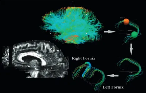
tractography, which allows non-invasive three-dimen-sional identiication of iber tracts74,84 and enables WM
bundle reconstruction typically found in post mortem analysis.85 Tractography is based on iber
connec-tivity probability and anisotropic water movement in a speciic group of ibers and their surroundings. he technique can be either global or local, probabilistic or deterministic86 (Figure 1).
he use of the isotropic HARDI technique, includ-ing sinclud-ingle shot sequences with no interslice gaps and use of the HARDI atlas, seems to provide more pow-erful computational analysis and higher precision ana-tomical examination of WM integrity.87,88 For instance,
moderate to severe VCI exhibited increased mean dif-fusivity (MD) in the temporal lobe and decreased FA in the corpus callosum, superior longitudinal fasciculus (SLF), internal capsule (IC), corona radiate (CR), thala-mus and uncinate fasciculus (UNC).87 Interestingly, no
diferences in MD or FA were found between AD and VCI and this may point to distinct trajectories of iber bundles compromised by these two conditions. In the neuroprogression of AD, for instance, while the poste-rior U-ibers of the supeposte-rior longitudinal fasciculus are often compromised, in VCI the most common pattern includes the involvement of neocortical anterior bun-dles, typically long association ibers, such as the UNC and IC/CR.87
Another interesting technique is the peak width of skeletonized mean difusivity (PSMD), which is based on the analysis of iber tracts and on the diference between the 95th and the 5th percentiles of the voxel-based MD
values within the WM skeleton.89 Increasing evidence
has shown that the PSMD may substantially reduce contamination from CSF and other spurious structures and enhance the sensitivity of SVD total burden mea-sures.69 Recent evidence also indicates greater sensitivity
of the PSMD for rating the progression of injury from SVD than the individual volumetric measures of WMH, lacunes and brain total volume. In a cross-sectional investigation of 69 patients with CADASIL, processing speed emerged as the most prominent cognitive domain afected.89 he use of STRIVE89,90 criteria on T2-weighted
gradient echo images has been employed for the identi-ication of CMB. Conversely, lacunar volumes could be rated by placing a seed-growing algorithm where a seed voxel is placed into a lacune on the 3D-T1 image.89
Magnetization transfer (MT). Magnetization transfer (MT) imaging, or magnetization transfer contrast (MTC) magnetic resonance imaging, is a modality of MRI technique based upon the exchange between bonded water in the brain tissue and proton magneti-zation in free water to characterize brain tissue proper-ties quantitatively.91,92 he magnetization transfer ratio
(MTR), derived from MT imaging, has been explored and used to evaluate brain injury in diferent brain diseases, e.g., multiple sclerosis, Alzheimer’s disease, stroke, and epilepsy. In the ield of vascular disease, although early reports have been available since 1999, only a small number of studies have employed this approach. One of these investigations included 56 subjects with WML and showed that periventricular WM had lower MTR than deep WM.93 Such indings were replicated by later
studies showing an association of cognitive impairment with either larger periventricular WM94-96 or reduced
Dement Neuropsychol 2017 December;11(4):343-355
352 Cerebrovascular disease: novel neuroimaging methods Alves et al.
MTR in normal appearing white matter (NAWM).97
Overall, these studies also support previous evidence showing reduced periventricular MTR in subjects with Binswanger disease (whose cognitive impairment is associated with a proportion of subcortical WML over 25%) compared to non-demented subjects with similar severity of WML, and seem to conirm the sensitivity of MTR for detecting clinically relevant CVD.
CONCLUSIONS
his review briely summarizes some of the most prom-ising neuroimaging techniques addressing brain struc-tural and metabolic changes in CVD. Indeed, much has been achieved in terms of unraveling the neuropatho-logical underpinnings and clinical correlates of VCI and VaD; a number of controversial issues, however, still remain. While on the one hand, neuroimaging has evolved quickly in the development of powerful and sensitive methods for studying in vivo brain architecture
in CVD and related cognitive disorders, most structural techniques are limited by a number of pitfalls. he source of criticism centers on multiple aspects, including the large variability in imaging modalities and procedures (e.g., threshold values for cluster deinition), the limited accuracy of DTI-MRI (poor identiication of crossing ibers, poor speciicity of indings) and low replication of results.98,99
Despite the aforementioned limitations, novel neu-roimaging methods ofer an enthusiastic debate on the interplay between aging, neurodegeneration and vascu-lar disease; one interesting topic involves, for instance, the pattern of cortical thinning exhibited by CVD and
other related cognitive disorders. Indeed, a pattern of cortical thinning in frontal and subcortical areas seem to be closely related to SIVD, contrasting with the temporo-parietal and medial temporal indings typically observed in AD.35,100 Another point of controversy is based on the
nature of cortical changes observed in CVD. While much has been discussed on the complex interaction between neurodegeneration and vascular disease, current evi-dence also suggests that CVD may independently lead to cortical atrophy.35 WMH may possibly cause
subcorti-cal axonal damage and neuronal disruption of cortisubcorti-cal pathways, leading to secondary neuronal body damage and ultimately gray matter atrophy.35,101 Contrasting
with the hypothesis of vascular-induced cortical atro-phy is the Wallerian degeneration model, which basi-cally conceives WM atrophy as a product of gray matter progressive reduction.11,99,102,103
hus, the continuous development of brain imaging techniques through new metabolic tracers, molecular compounds, multimodal approaches, microstructural anatomy, disease classifying algorithms and higher ield MRI ofer an exciting perspective towards a broad comprehension of CVD pathophysiology. Although the importance of most of these techniques in the clinical setting has yet to be recognized, there is great expectancy in achieving earlier and more reined thera-peutic interventions for the efective management of dementia.
Author contributions. All authors have contributed signiicantly to the study and are in agreement with the content of the manuscript.
REFERENCES
1. Inzitari D, Pracucci G, Poggesi A, Carlucci G, Barkhof F, Chabriat H, et al. Changes in white matter as determinant of global functional decline in older independent outpatients: three year follow-up of LADIS (leukoara-iosis and disability) study cohort. BMJ. 2009;339:b2477.
2. Frisoni GB, Galluzzi S, Pantoni L, Filippi M. The effect of white matter lesions on cognition in the elderly--small but detectable. Nat Clin Pract Neurol. 2007;3(11):620-7.
3. Jokinen H, Ryberg C, Kalska H, Ylikoski R, Rostrup E, Stegmann MB, et al. Corpus callosum atrophy is associated with mental slowing and executive deficits in subjects with age-related white matter hyperintensi-ties: the LADIS Study. J Neurol Neurosurg Psychiatry. 2007;78(5):491-6. 4. Frisoni GB, Galluzzi S, Pantoni L, Filippi M. The effect of white matter lesions on cognition in the elderly--small but detectable. Nat Clin Pract Neurol. 2007;3(11):620-7.
5. Sudo FK, Alves CEO, Alves GS, Ericeira-Valente L, Tiel C, Moreira DM, et al. White matter hyperintensities, executive function and global cognitive performance in vascular mild cognitive impairment. Arq Neuropsiquiatr. 2013;71(7):431-6.
6. Perneczky R, Tene O, Attems J, Giannakopoulos P, Ikram MA, Federico A, et al. Is the time ripe for new diagnostic criteria of cognitive impairment due to cerebrovascular disease? Consensus report of the International Congress on Vascular Dementia working group. BMC Med. 2016. DOI 10.1186/s12916-016-0719-y
7. Li C, Du H, Zheng J, Wang J. A Voxel-based Morphometric Analysis of Cerebral Gray Matter in Subcortical Ischemic Vascular Dementia Patients and Normal Aged Controls. Int J Med Sci. 2011;8(6):482-6.
8. Mechelli A, Price C, Friston K, Ashburner J. Voxel-Based Morphometry of the Human Brain: Methods and Applications. Curr Med Imaging Rev. 2005;1(2):105-13.
9. Ashburner J, Friston KJ. Voxel-based morphometry--the methods. NeuroImage. 2000;11(6 Pt 1):805-21.
10. Ashburner J, Friston KJ. Why voxel-based morphometry should be used. NeuroImage. 2001;14(6):1238-43.
11. Stricker NH, Schweinsburg BC, Delano-Wood L, Wierenga CE, Bangen KJ, Haaland KY, et al. Decreased white matter integrity in late-myelina-ting fiber pathways in Alzheimer’s disease supports retrogenesis. Neuro-Image. 2009;45(1):10-6.
12. Thomalla G, Glauche V, Weiller C, Röther J. Time course of wallerian degeneration after ischaemic stroke revealed by diffusion tensor imaging. J Neurol Neurosurg Psychiatry. 2005;76(2):266-8.
13. Di Paola M, Di Iulio F, Cherubini A, Blundo C, Casini AR, Sancesario G, et al. When, where, and how the corpus callosum changes in MCI and AD: a multimodal MRI study. Neurology. 2010;74(14):1136-42. 14. Haller S, Garibotto V, Kövari E, Bouras C, Xekardaki A, Rodriguez C, et
Dement Neuropsychol 2017 December;11(4):343-355
353
Alves et al. Cerebrovascular disease: novel neuroimaging methods 15. Stroup DF. Meta-analysis of Observational Studies in EpidemiologyA
Proposal for Reporting. JAMA. 2000;283(15):2008.
16. Bybel B, Brunken RC, Shah SN, Wu G, Turbiner E, Neumann DR. PET and PET/CT imaging: what clinicians need to know. Cleve Clin J Med. 2006;73(12):1075-87.
17. Heiss W-D, Zimmermann-Meinzingen S. PET imaging in the differential diagnosis of vascular dementia. J Neurol Sci. 2012;322(1-2):268-73. 18. Albert MS, DeKosky ST, Dickson D, Dubois B, Feldman HH, Fox NC,
et al. The diagnosis of mild cognitive impairment due to Alzheimer’s disease: recommendations from the National Institute on Aging-Alzheim-er’s Association workgroups on diagnostic guidelines for AlzheimAging-Alzheim-er’s disease. Alzheimers Dement J Alzheimers Assoc. 2011;7(3):270-9. 19. Herholz K, Boecker H, Nemeth I, Dunn G. FDG PET in dementia
multi-center studies and clinical trials. Clin Transl Imaging. 2013;1(4):261-70. 20. Landau SM, Harvey D, Madison CM, Koeppe RA, Reiman EM, Foster NL, et al. Associations between cognitive, functional, and FDG-PET measures of decline in AD and MCI. Neurobiol Aging. 2011;32(7): 1207-18.
21. Townsend DW. Multimodality imaging of structure and function. Phys Med Biol. 2008;53(4):R1-39.
22. Mariani G, Bruselli L, Kuwert T, Kim EE, Flotats A, Israel O, et al. A review on the clinical uses of SPECT/CT. Eur J Nucl Med Mol Imaging. 2010;37(10):1959-85.
23. Barra V, Boire JV. A general framework for the fusion of anatomical and functional medical images. NeuroImage. 2001;13(3):410-24. 24. Schlemmer H-PW, Pichler BJ, Schmand M, Burbar Z, Michel C,
Lade-beck R, et al. Simultaneous MR/PET imaging of the human brain: feasi-bility study 1. Radiology. 2008;248(3):1028-35.
25. Yang X, Fei B. Multiscale segmentation of the skull in MR images for MRI-based attenuation correction of combined MR/PET. J Am Med Inform Assoc JAMIA. 2013;20(6):1037-45.
26. Bini J, Izquierdo-Garcia D, Mateo J, Machac J, Narula J, Fuster V, et al. Preclinical evaluation of MR attenuation correction versus CT attenuation correction on a sequential whole-body MR/PET scanner. Invest Radiol. 2013;48(5):313-22.
27. Catana C, van der Kouwe A, Benner T, Michel CJ, Hamm M, Fenchel M, et al. Toward implementing an MRI-based PET attenuation-correction method for neurologic studies on the MR-PET brain prototype. J Nucl Med Off Publ Soc Nucl Med. 2010;51(9):1431-8.
28. Ullisch MG, Scheins JJ, Weirich C, Rota Kops E, Celik A, Tellmann L, et al. MR-based PET motion correction procedure for simultaneous MR-PET neuroimaging of human brain. PloS One. 2012;7(11):e48149. 29. Catana C, Benner T, van der Kouwe A, Byars L, Hamm M, Chonde DB, et al. MRI-assisted PET motion correction for neurologic studies in an integrated MR-PET scanner. J Nucl Med Off Publ Soc Nucl Med. 2011; 52(1):154-61.
30. Cherry SR. Multimodality Imaging: Beyond PET/CT and SPECT/CT. Semin Nucl Med. 2009;39(5):348-53.
31. Bauer S, Wagner M, Seiler A, Hattingen E, Deichmann R, Nöth U, et al. Quantitative T2’-mapping in acute ischemic stroke. Stroke J Cereb Circ. 2014;45(11):3280-6.
32. Jurcoane A, Wagner M, Schmidt C, Mayer C, Gracien R-M, Hirschmann M, et al. Within-lesion differences in quantitative MRI parameters predict contrast enhancement in multiple sclerosis. J Magn Reson Imaging. 2013;38(6):1454-61.
33. Nagaki A, Onoguchi M, Matsutomo N. Clinical validation of high-reso-lution image reconstruction algorithms in brain 18F-FDG-PET: effect of incorporating Gaussian filter, point spread function, and time-of-flight. Nucl Med Commun. 2014;35(12):1224-32.
34. Leemans EL, Kotasidis F, Wissmeyer M, Garibotto V, Zaidi H. Qualitative and quantitative evaluation of blob-based time-of-flight PET image recon-struction in hybrid brain PET/MR imaging. Mol Imaging Biol. 2015;1-10. 35. Hun KC, Won SS, Ha KG, Soo SJ, Hanna C, Young N, et al. Cortical Thinning in Subcortical Vascular Dementia with Negative. J Alzheimer Dis. 2012;(2):315-23.
36. Barthel H, Sabri O. Florbetaben to trace amyloid-β in the Alzheimer brain by means of PET. J Alzheimers Dis JAD. 2011;26 Suppl 3:117-21. 37. Barthel H, Gertz H-J, Dresel S, Peters O, Bartenstein P, Buerger K,
et al. Cerebral amyloid-β PET with florbetaben (18F) in patients with Alzheimer’s disease and healthy controls: a multicentre phase 2 diag-nostic study. Lancet Neurol. 2011;10(5):424-35.
38. Becker GA, Ichise M, Barthel H, Luthardt J, Patt M, Seese A, et al. PET quantification of 18F-florbetaben binding to β-amyloid deposits in human brains. J Nucl Med Off Publ Soc Nucl Med. 2013;54(5):723-31.
39. de Lartigue J. Flutemetamol (18F): a β-amyloid positron emission tomog-raphy tracer for Alzheimer’s and dementia diagnosis. Drugs Today Barc Spain 1998. 2014;50(3):219-29.
40. Thal DR, Beach TG, Zanette M, Heurling K, Chakrabarty A, Ismail A, et al. [(18)F]flutemetamol amyloid positron emission tomography in preclinical and symptomatic Alzheimer’s disease: specific detection of advanced phases of amyloid-β pathology. Alzheimers Dement J Alzheimers Assoc. 2015;11(8):975-85.
41. Joshi AD, Pontecorvo MJ, Clark CM, Carpenter AP, Jennings DL, Sadowsky CH, et al. Performance characteristics of amyloid PET with florbetapir F 18 in patients with alzheimer’s disease and cognitively normal subjects. J Nucl Med Off Publ Soc Nucl Med. 2012;53(3):378-84. 42. Kobylecki C, Langheinrich T, Hinz R, Vardy ERLC, Brown G, Martino M-E, et al. 18F-florbetapir PET in patients with frontotemporal dementia and Alzheimer disease. J Nucl Med Off Publ Soc Nucl Med. 2015; 56(3):386-91.
43. Hall B, Mak E, Cervenka S, Aigbirhio FI, Rowe JB, O’Brien JT. In vivo tau PET imaging in dementia: Pathophysiology, radiotracer quantifi-cation, and a systematic review of clinical findings. Ageing Res Rev. 2017;36:50-63.
44. Fodero-Tavoletti MT, Okamura N, Furumoto S, Mulligan RS, Connor AR, McLean CA, et al. 18F-THK523: a novel in vivo tau imaging ligand for Alzheimer’s disease. Brain J Neurol. 2011;134(Pt 4):1089-100. 45. Klunk WE, Koeppe RA, Price JC, Benzinger TL, Devous MD, Jagust
WJ, et al. The Centiloid Project: standardizing quantitative amyloid plaque estimation by PET. Alzheimers Dement J Alzheimers Assoc. 2015;11(1):1-15.e1-4.
46. Asih PR, Chatterjee P, Verdile G, Gupta VB, Trengove RD, Martins RN. Clearing the amyloid in Alzheimer’s: progress towards earlier diagnosis and effective treatments - an update for clinicians. Neurodegener Dis Manag. 2014;4(5):363-78.
47. Friedlich AL, Tanzi RE, Rogers JT. The 5’-untranslated region of Parkin-son’s disease alpha-synuclein messengerRNA contains a predicted iron responsive element. Mol Psychiatry. 2007;12(3):222-3.
48. Rogers JT, Randall JD, Cahill CM, Eder PS, Huang X, Gunshin H, et al. An iron-responsive element type II in the 5’-untranslated region of the Alzheimer’s amyloid precursor protein transcript. J Biol Chem. 2002; 277(47):45518-28.
49. De Reuck JL, Deramecourt V, Auger F, Durieux N, Cordonnier C, Devos D, et al. Iron deposits in post-mortem brains of patients with neuro-degenerative and cerebrovascular diseases: a semi-quantitative 7.0 T magnetic resonance imaging study. Eur J Neurol. 2014;21(7):1026-31. 50. Moon Y, Han S-H, Moon W-J. Patterns of Brain Iron Accumulation in
Vascular Dementia and Alzheimer’s Dementia Using Quantitative Suscep-tibility Mapping Imaging. J Alzheimers Dis. 2016;51(3):737-45. 51. Won SM, Lee JH, Park UJ, Gwag J, Gwag BJ, Lee YB. Iron
medi-ates endothelial cell damage and blood-brain barrier opening in the hippocampus after transient forebrain ischemia in rats. Exp Mol Med. 2011;43(2):121-8.
52. Liem MK, Lesnik Oberstein SAJ, Versluis MJ, Maat-Schieman MLC, Haan J, Webb AG, et al. 7 T MRI reveals diffuse iron deposition in putamen and caudate nucleus in CADASIL. J Neurol Neurosurg Psychiatry. 2012;83(12):1180-5.
53. Liu C, Li C, Yang J, Gui L, Zhao L, Evans AC, et al. Characterizing brain iron deposition in subcortical ischemic vascular dementia using susceptibility-weighted imaging: An in vivo MR study. Behav Brain Res. 2015;288:33-8.
54. Rocca MA, Gerevini S, Filippi M, Falini A. High-Field-Strength MRI (3.0 T or More) in White Matter Diseases. In: Scarabino T, Pollice S, Popolizio T, editors. High Field Brain MRI [Internet]. Cham: Springer International Publishing; 2017 [cited 2017 Jul 3]. p. 223-37.
55. O’Dwyer L, Lamberton F, Bokde ALW, Ewers M, Faluyi YO, Tanner C, et al. Multiple Indices of Diffusion Identifies White Matter Damage in Mild Cognitive Impairment and Alzheimer’s Disease. PLoS One. 2011;6(6): e21745.
56. García-Carrasco M, Mendoza-Pinto C, Cervera R. Diagnosis and clas-sification of Susac syndrome. Autoimmun Rev. 2014;13(4-5):347-50. 57. Wuerfel J, Sinnecker T, Ringelstein EB, Jarius S, Schwindt W, Niendorf
T, et al. Lesion morphology at 7 Tesla MRI differentiates Susac syndrome from multiple sclerosis. Mult Scler J. 2012;18(11):1592-9.
58. Cai K, Tain R, Das S, Damen FC, Sui Y, Valyi-Nagy T, et al. The feasibility of quantitative MRI of perivascular spaces at 7T. J Neurosci Methods. 2015;256:151-6.
Dement Neuropsychol 2017 December;11(4):343-355
354 Cerebrovascular disease: novel neuroimaging methods Alves et al.
Biessels GJ. The Spectrum of MR Detectable Cortical Microinfarcts: A Classification Study with 7-Tesla Postmortem MRI and Histopathology. J Cereb Blood Flow Metab. 2015;35(4):676-83.
60. Van Leemput K, Maes F, Vandermeulen D, Suetens P. Automated model-based tissue classification of MR images of the brain. IEEE Trans Med Imaging. 1999;18(10):897-908.
61. Koikkalainen J, Rhodius-Meester H, Tolonen A, Barkhof F, Tijms B, Lemstra AW, et al. Differential diagnosis of neurodegenerative diseases using structural MRI data. NeuroImage Clin. 2016;11:435-49. 62. Lawrence AJ, Patel B, Morris RG, MacKinnon AD, Rich PM, Barrick
TR, et al. Mechanisms of Cognitive Impairment in Cerebral Small Vessel Disease: Multimodal MRI Results from the St George’s Cognition and Neuroimaging in Stroke (SCANS) Study. PLoS One. 2013;8(4):e61014. 63. Du AT, Schuff N, Laakso MP, Zhu XP, Jagust WJ, Yaffe K, et al. Effects of subcortical ischemic vascular dementia and AD on entorhinal cortex and hippocampus. Neurology. 2002;58(11):1635-41.
64. Kitamura A, Fujita Y, Oishi N, Kalaria RN, Washida K, Maki T, et al. Selec-tive white matter abnormalities in a novel rat model of vascular dementia. Neurobiol Aging. 2012;33(5):1012.e25-35.
65. Liu C, Li C, Gui L, Zhao L, Evans AC, Xie B, et al. The pattern of brain gray matter impairments in patients with subcortical vascular dementia. J Neurol Sci. 2014;341(1-2):110-8.
66. Seo SW, Lee J-M, Im K, Park J-S, Kim S-H, Kim ST, et al. Cardio-vascular Risk Factors Cause Cortical Thinning in Cognitively Impaired Patients: Relationships Among Cardiovascular Risk Factors, White Matter Hyperintensities, and Cortical Atrophy. Alzheimer Dis Assoc Disord. 2012;26(2):106-12.
67. Viswanathan A, Godin O, Jouvent E, O’Sullivan M, Gschwendtner A, Peters N, et al. Impact of MRI markers in subcortical vascular dementia: A multi-modal analysis in CADASIL. Neurobiol Aging. 2010; 31(9): 1629-36.
68. Selden N. Trajectories of cholinergic pathways within the cerebral hemi-spheres of the human brain. Brain. 1998;121(12):2249-57.
69. Blair GW, Hernandez MV, Thrippleton MJ, Doubal FN, Wardlaw JM. Advanced Neuroimaging of Cerebral Small Vessel Disease. Curr Treat Options Cardiovasc Med. 2017;19(7):56.
70. Dukart J, Mueller K, Barthel H, Villringer A, Sabri O, Schroeter ML, et al. Meta-analysis based SVM classification enables accurate detection of Alzheimer’s disease across different clinical centers using FDG-PET and MRI. Psychiatry Res Neuroimaging. 2013;212(3):230-6.
71. Dukart J, Mueller K, Horstmann A, Barthel H, Möller HE, Villringer A, et al. Combined evaluation of FDG-PET and MRI improves detection and differentiation of dementia. PLoS One. 2011;6(3):e18111.
72. Beaulieu C. The basis of anisotropic water diffusion in the nervous system - a technical review. NMR Biomed. 2002;15(7-8):435-55. 73. Assaf Y, Pasternak O. Diffusion tensor imaging (DTI)-based white
matter mapping in brain research: a review. J Mol Neurosci MN. 2008;34(1):51-61.
74. Nucifora PGP, Verma R, Lee S-K, Melhem ER. Diffusion-Tensor MR Imaging and Tractography: Exploring Brain Microstructure and Connec-tivity1. Radiology. 2007;245(2):367-84.
75. Emsell L, Leemans A, Langan C, Van Hecke W, Barker GJ, McCarthy P, et al. Limbic and Callosal White Matter Changes in Euthymic Bipolar I Disorder: An Advanced Diffusion Magnetic Resonance Imaging Trac-tography Study. Biol Psychiatry. 2013;73(2):194-201.
76. Beaulieu C, Does MD, Snyder RE, Allen PS. Changes in water diffusion due to Wallerian degeneration in peripheral nerve. Magn Reson Med. 1996;36(4):627-31.
77. Ciccarelli O, Catani M, Johansen-Berg H, Clark C, Thompson A. Diffu-sion-based tractography in neurological disorders: concepts, applica-tions, and future developments. Lancet Neurol. 2008;7(8):715-27. 78. Behrens TEJ, Johansen-Berg H, Woolrich MW, Smith SM,
Wheeler-Kingshott C a. M, Boulby PA, et al. Non-invasive mapping of connec-tions between human thalamus and cortex using diffusion imaging. Nat Neurosci. 2003;6(7):750-7.
79. Psomiades M, Fonteneau C, Mondino M, Luck D, Haesebaert F, Suaud-Chagny M-F, et al. Integrity of the arcuate fasciculus in patients with schizophrenia with auditory verbal hallucinations: A DTI-tractography study. NeuroImage Clin. 2016;12:970-5.
80. Fitzsimmons J, Hamoda HM, Swisher T, Terry D, Rosenberger G, Seidman LJ, et al. Diffusion tensor imaging study of the fornix in first episode schizophrenia and in healthy controls. Schizophr Res. 2014; 156(2-3):157-60.
81. Ji A, Godwin D, Rutlin J, Kandala S, Shimony JS, Mamah D. Tract-based analysis of white matter integrity in psychotic and nonpsychotic bipolar disorder. J Affect Disord. 2017;209:124-34.
82. Haarman BCM, Riemersma-Van der Lek RF, Burger H, de Groot JC, Drexhage HA, Nolen WA, et al. Diffusion tensor imaging in euthymic bipolar disorder - A tract-based spatial statistics study. J Affect Disord. 2016;203:281-91.
83. Olvet DM, Delaparte L, Yeh F-C, DeLorenzo C, McGrath PJ, Weissman MM, et al. A comprehensive examination of white matter tracts and connectometry in major depressive disorder. Depress Anxiety. 2016; 33(1):56-65.
84. Basser PJ, Pajevic S, Pierpaoli C, Duda J, Aldroubi A. In vivo fiber trac-tography using DT-MRI data. Magn Reson Med. 2000;44(4):625-32. 85. Catani M, Howard RJ, Pajevic S, Jones DK. Virtual in vivo interactive
dissection of white matter fasciculi in the human brain. NeuroImage. 2002;17(1):77-94.
86. Knöchel C, Schmied C, Linden DEJ, Stäblein M, Prvulovic D, de A de Carvalho L, et al. White matter abnormalities in the fornix are linked to cognitive performance in SZ but not in BD disorder: An exploratory anal-ysis with DTI deterministic tractography. J Affect Disord. 2016; 201:64-78. 87. Jin Thong JY, Du J, Ratnarajah N, Dong Y, Soon HW, Saini M, et al. Abnormalities of cortical thickness, subcortical shapes, and white matter integrity in subcortical vascular cognitive impairment: Morphological Abnormalities in Vascular Cognitive Impairmentc. Hum Brain Mapp. 2014;35(5):2320-32.
88. Du J, Goh A, Qiu A. Large deformation diffeomorphic metric mapping of orientation distribution functions. Inf Process Med Imaging Proc Conf. 2011;22:448-62.
89. Baykara E, Gesierich B, Adam R, Tuladhar AM, Biesbroek JM, Koek HL, et al. A Novel Imaging Marker for Small Vessel Disease Based on Skeletonization of White Matter Tracts and Diffusion Histograms: Novel SVD Imaging Marker. Ann Neurol. 2016;80(4):581-92.
90. Wardlaw JM, Smith EE, Biessels GJ, Cordonnier C, Fazekas F, Frayne R, et al. Neuroimaging standards for research into small vessel disease and its contribution to ageing and neurodegeneration. Lancet Neurol. 2013;12(8):822-38.
91. Bozzali M, Franceschi M, Falini A, Pontesilli S, Cercignani M, Magnani G, et al. Quantification of tissue damage in AD using diffusion tensor and magnetization transfer MRI. Neurology. 2001;57(6):1135-7.
92. Li C-X, Herndon JG, Novembre FJ, Zhang X. A Longitudinal Magnetiza-tion Transfer Imaging EvaluaMagnetiza-tion of Brain Injury in a Macaque Model of NeuroAIDS. AIDS Res Hum Retroviruses. 2015;31(3):335-41. 93. Spilt A, Goekoop R, Westendorp RGJ, Blauw GJ, de Craen AJM, van
Buchem MA. Not all age-related white matter hyperintensities are the same: a magnetization transfer imaging study. AJNR Am J Neuroradiol. 2006;27(9):1964-8.
94. Seo SW, Lee J-M, Im K, Park J-S, Kim S-H, Kim ST, et al. Cortical thin-ning related to periventricular and deep white matter hyperintensities. Neurobiol Aging. 2012;33(7):1156-67.
95. Kee Hyung Park, Lee J-Y, Na DL, Seong Yoon Kim, Cheong H-K, So Young Moon, et al. Different Associations of Periventricular and Deep White Matter Lesions with Cognition, Neuropsychiatric Symptoms, and Daily Activities in Dementia. J Geriatr Psychiatry Neurol. 2011; 24(2):84-90.
96. Bombois S, Debette S, Delbeuck X, Bruandet A, Lepoittevin S, Delmaire C, et al. Prevalence of subcortical vascular lesions and association with executive function in mild cognitive impairment subtypes. Stroke. 2007;38(9):2595-7.
97. Iannucci G, Dichgans M, Rovaris M, Brüning R, Gasser T, Giacomotti L, et al. Correlations between clinical findings and magnetization transfer imaging metrics of tissue damage in individuals with cerebral autosomal dominant arteriopathy with subcortical infarcts and leukoencephalopathy. Stroke. 2001;32(3):643-8.
98. Health Quality Ontario. The Appropriate Use of Neuroimaging in the Diag-nostic Work-Up of Dementia: An Evidence-Based Analysis. Ont Health Technol Assess Ser Internet. 2014;14(1):1-64.
99. Alves GS, O’Dwyer L, Jurcoane A, Oertel-Knöchel V, Knöchel C, Prvu-lovic D, et al. Different patterns of white matter degeneration using multiple diffusion indices and volumetric data in mild cognitive impair-ment and Alzheimer patients. PloS One. 2012;7(12):e52859. 100. Johnson KA, Gregas M, Becker JA, Kinnecom C, Salat DH, Moran EK,
Dement Neuropsychol 2017 December;11(4):343-355
355
Alves et al. Cerebrovascular disease: novel neuroimaging methods 101. van Dalen JW, Scuric EEM, van Veluw SJ, Caan MWA, Nederveen AJ,
Biessels GJ, et al. Cortical microinfarcts detected in vivo on 3 Tesla MRI: clinical and radiological correlates. Stroke. 2015;46(1):255-7. 102. Gupta RK, Saksena S, Hasan KM, Agarwal A, Haris M, Pandey CM, et
al. Focal Wallerian degeneration of the corpus callosum in large middle cerebral artery stroke: serial diffusion tensor imaging. J Magn Reson Imaging JMRI. 2006;24(3):549-55.
103. Thomalla G, Glauche V, Weiller C, Röther J. Time course of wallerian degeneration after ischaemic stroke revealed by diffusion tensor imaging. J Neurol Neurosurg Psychiatry. 2005;76(2):266-8.
104. Lanna ME de O, Alves CEO, Sudo FK, Alves G, Valente L, Moreira DM, et al. Cognitive disconnective syndrome by single strategic strokes in vascular dementia. J Neurol Sci. 2012;322(1-2):176-83.
105. van der Holst HM, Tuladhar AM, van Norden AGW, de Laat KF, van Uden IWM, van Oudheusden LJB, et al. Microstructural integrity of the cingulum is related to verbal memory performance in elderly with cerebral small vessel disease. NeuroImage. 2013;65:416-23.
106. Rolland Y, Payoux P, Lauwers-Cances V, Voisin T, Esquerré JP, Vellas B. A SPECT study of wandering behavior in Alzheimer’s disease. Int J Geriatr Psychiatry. 2005;20(9):816-20.
107. Cavallari M, Moscufo N, Meier D, Skudlarski P, Pearlson GD, White WB, et al. Thalamic Fractional Anisotropy Predicts Accrual of Cerebral White Matter Damage in Older Subjects with Small-Vessel Disease. J Cereb Blood Flow Metab. 2014;34(8):1321-7.
108. Guerra A, Petrichella S, Vollero L, Ponzo D, Pasqualetti P, Määttä S, et al. Neurophysiological features of motor cortex excitability and plasticity
in Subcortical Ischemic Vascular Dementia: A TMS mapping study. Clin Neurophysiol. 2015;126(5):906-13.
109. Jeong H-G, Kim BJ, Yang MH, Han M-K, Bae H-J. Neuroimaging Markers for Early Neurologic Deterioration in Single Small Subcortical Infarction. Stroke. 2015;46(3):687-91.
110. Garriga M, Milà M, Mir M, Al-Baradie R, Huertas S, Castejon C, et al. 123I-FP-CIT SPECT imaging in early diagnosis of dementia in patients with and without a vascular component. Front Syst Neurosci. 2015;9:99. 111. Lin L, Xue Y, Duan Q, Sun B, Lin H, Chen X, et al. Microstructural White Matter Abnormalities and Cognitive Dysfunction in Subcortical Ischemic Vascular Disease: an Atlas-Based Diffusion Tensor Analysis Study. J Mol Neurosci. 2015;56(2):363-70.
112. Ostojic J, Kozic D, Pavlovic A, Semnic M, Todorovic A, Petrovic K, et al. Hippocampal diffusion tensor imaging microstructural changes in vascular dementia. Acta Neurol Belg. 2015;115(4):557-62.
113. Wu X-P, Gao Y-J, Yang J-L, Xu M, Sun D-H. Quantitative measure-ment to evaluate morphological changes of the corpus callosum in patients with subcortical ischemic vascular dementia. Acta Radiol. 2015;56(2):214-8.
114. Mascalchi M, Pantoni L, Giannelli M, Valenti R, Bianchi A, Pracucci G, et al. Diffusion Tensor Imaging to Map Brain Microstructural Changes in CADASIL: Brain Microstructural Changes in CADASIL. J Neuroimaging. 2017;27(1):85-91.