Ana Rita Valente Ribeiro
Licenciada em Análises Clínicas e Saúde Pública
Dissecting deregulated cell-to-cell
communication in in vitro Alzheimer’s
disease models
Dissertação para obtenção do Grau de Mestre em
Genética Molecular e Biomedicina
Orientador: Dora Brites, PhD, Faculdade de Farmácia da
Universidade de Lisboa
Co-orientador: Adelaide Fernandes, PhD, Faculdade de
Farmácia da Universidade de Lisboa
Setembro 2017
Ana Rita Valente Ribeiro
Licenciatura em Análises Clínicas e Saúde Pública
Dissecting deregulated cell-to-cell
communication in in vitro Alzheimer’s
disease models
Dissertação para obtenção do Grau de Mestre em
Genética Molecular e Biomedicina
Orientador: Dora Brites, PhD, Faculdade de Farmácia da
Universidade de Lisboa
Co-orientador: Adelaide Fernandes, PhD, Faculdade de
Farmácia da Universidade de Lisboa
Setembro 2017
Dissecting deregulated cell-to-cell communication in in vitro Alzheimer’s disease
models
Copyright © Ana Rita Valente Ribeiro, Faculdade de Ciências e Tecnologia, Universidade
Nova de Lisboa
A Faculdade de Ciências e Tecnologia e a Universidade Nova de Lisboa têm o direito,
perpétuo e sem limites geográficos, de arquivar e publicar esta dissertação através de
exemplares impressos reproduzidos em papel ou de forma digital, ou por qualquer outro meio
conhecido ou que venha a ser inventado, e de a divulgar através de repositórios científicos e
de admitir a sua cópia e distribuição com objetivos educacionais ou de investigação, não
comerciais, desde que seja dado crédito ao autor e editor.
Part of the results discussed in this thesis were presented in the following scientific
meetings:
Oral communications
A.R. Ribeiro, C. Cunha, A.R. Vaz, A. Fernandes, D. Brites. Dissecting deregulated cell-to-cell communication in an in vitro AD model. 31ª Reunião do Grupo de Estudos do Envelhecimento Cerebral
e Demências, June 30th 2017, Museu do Oriente, Lisboa.
A.R. Ribeiro, M. Monteiro, C. Cunha, A.R. Vaz, A. Fernandes, D. Brites. Relevance of neuron-microglia vesicular trafficking, stress-related microRNAS and DAMPs in Alzheimer's disease. XV Meeting of Portuguese Society for Neuroscience, May 25-26 2017, Braga.
A.R. Ribeiro, C. Cunha, S. Pinto, A. R. Vaz, M. Oksanen, A. J. Petersen, K. Puttonen, S. Zhang, R. H. Hämäläinen, J. Koistinaho, A. Fernandes, D. Brites. iPSCs-derived astrocytes from patients with PSEN1ΔE9 mutation show a depressed RAGE/miR-155 pathway with impact in extracellular vesicle molecular cargo. 10th International Meeting of the Portuguese Society for Stem Cells and Cell Therapies, October 13-14 2017, Covilhã.
Poster communications
D. Brites, M. Monteiro, A. R. Ribeiro, C. Cunha, A. R. Vaz, A. Fernandes. Role of neuron-microglia secretome and stress-related microRNAS in Alzheimer's disease. XIII European Meeting on Glial Cells in Health and Disease, July 8-11 2017, Edinburgh.
A.R. Ribeiro, C. Cunha, A.R. Vaz, A. Fernandes, D. Brites. Dissecting the Effects of Exosomes Derived
from Neurons on Microglia Phenotype in an in vitro AD Model using human cell lines. 9th iMed.ULisboa
Postgraduate Students Meeting and 2nd i3DU Meeting, July 13-14 2017, Lisbon.
This work was performed in Neuron Glia Biology in Health and Disease Group at
iMed.ULisboa, Faculty of Pharmacy, University of Lisbon, headed by Dora Brites. It was funded
by EU Joint Programme-Neurodegenerative Disease Research (JPND) project with funding
from the European Union’s Horizon 2020 research and innovation programme under grant
agreement No 643417 and Fundação para a Ciência e Tecnologia, Lisboa, Portugal
(JPco-fuND/0003/2015 to DB, FCT-EXPL/NEU-NMC/1003/2013 to AF and in part by
UID/DTP/04138/2013 to iMed.ULisboa). iPSCs and astrospheres from patients with
Alzheimer’s disease were provided by Jari Koistinaho from the University of Eastern Finland,
Finland. I would also like to thank Minna Oksanen, Katja Puttonen and Riikka H. Hämäläinen
from the same University, as well as Andrew J. Petersen and Su-Chun Zhang from the
University of Wisconsin, Madison (USA), for all the supportive work and collaboration.
“A goal without a plan is just a wish”
Antoine de Saint-Exupéry
AGRADECIMENTOS
Este trabalho só foi possível com o apoio de pessoas que direta ou indiretamente me ajudaram a tornar esta jornada um pouco mais fácil e com a sensação de dever cumprido. O meu agradecimento vai para as pessoas que acreditaram em mim e me ajudaram a finalizar esta tese.
O meu primeiro agradecimento, como não poderia deixar de ser, vai para a Professora Dora. Obrigada por ter acreditado em mim e me ter dado a oportunidade de fazer parte deste grupo. Obrigada por me ensinar a fazer sempre o melhor e nada menos que isso. É um marco no mundo da investigação pela ótima profissional que é.
Adelaide, obrigada por tudo o que me ensinou e pela paciência que teve comigo. Obrigada pelas palavras de incentivo e de coragem quando as coisas corriam menos bem. Este percurso não teria sido o mesmo sem o seu carinho e dedicação.
Rita, Professor Rui e Professora Alexandra, obrigada por toda a ajuda disponibilizada ao longo deste meu percurso.
Às meninas do grupo: Catarina, Marta, Carolina, Sara e Gisela. Obrigada por toda a ajuda, paciência e conselhos que me deram no laboratório. Obrigada por serem mais do que colegas de trabalho e me mostrarem que é possível construir uma amizade para além das paredes do CPM.
Aos membros do “Encontro da comitiva”, o melhor que o mestrado me deu! Cláudia, companheira de casa e a pessoa que me acompanhou nesta jornada. As palavras escasseiam quando se trata de ti. Obrigada pelos desabafos e por me aturares. Contigo sei que posso contar sempre e mesmo que os nossos caminhos sigam direções diferentes, sei que que estarás sempre presente. Obrigada por seres um marco muito bom na minha vida. Carolina, a palavra “diva” ganhou um novo significado para mim. Tu és a diva! És a pessoa que me faz acreditar que tudo vai dar certo. Obrigada
pela companhia e pelos cafés no CPM. Obrigada por seres diferente. Joana, “you are my person”!
Obrigada pelas parvoíces e por seres a pessoa que mais me faz acreditar que com trabalho e dedicação tudo se consegue. Muita sorte nesta tua nova jornada. Vemo-nos na Suécia. Tiago, és um amigo com que posso sempre contar. Obrigada pelas boleias e pela companhia. Nunca dizes “Não”. És fantástico! Filipe, és uma ótima pessoa e tenho muita sorte em te ter conhecido. Nunca percas a humildade que te é tão característica. A todos vocês, obrigada!
Marisa, Cátia, Marta, Mónica e Daniel, os meus companheiros de sempre. A vocês não há palavras para descrever o que significam na minha vida. Obrigada por estarem sempre presentes nos bons e nos maus momentos. Os meus fins-de-semana em casa não seriam os meus sem vocês. São os meus melhores amigos hoje, amanhã e sempre!
Andreína, a Madeira passou a ser a minha segunda casa desde que te conheci. És como uma irmã para mim e vou guardar a nossa amizade, com carinho, para o resto da minha vida. A ti e à tua família, obrigada por me tratarem sempre tão bem e por estarem sempre lá. Gosto muito de vocês!
viii O meu último obrigada, e o maior deles todos, vai para a minha família.
Padrinho e madrinha, obrigada por estarem sempre presentes. Sei que posso contar sempre com vocês. São os melhores padrinhos, aqueles que me acolheram! Ana e Sérgio, obrigada por nunca se esquecerem de mim. Sei que posso contar com vocês também. Ver-vos crescer, para mim, é um orgulho. Continuem a ser os meus primos “mais novos” para sempre!
André, o meu irmão, o meu amparo. Não há palavras para descrever o que significas para mim. Obrigada por me ouvires e me confortares sempre que as coisas corriam menos bem. Continua a ser a pessoa mais brincalhona que conheço. És o melhor e eu gosto muito de ti! Como diz a música “Brother” da banda Kodaline: “And if you were drowned at sea, I'd give you my lungs so you could
breathe”. Vou estar sempre aqui para ti, acredita!
Mãe e pai, obrigada por terem feito de mim aquilo que sou hoje. Se sou alguma coisa a vocês o devo. Obrigada por acreditarem em mim quando eu própria duvido. Obrigada por nunca me faltarem com nada. Obrigada por me deixarem voar e estarem sempre presentes quando regresso a casa. Obrigada por serem os melhores pais do mundo. Amo-vos!
Avó, és o meu tesouro. És a melhor avó. Obrigada por mostrares sempre esse teu sorriso e me ensinares a acreditar no amor. És o meu biju, a minha “vizinha”. Nunca me deixes. Amo-te daqui até ao avô!
Por fim, ao meu avô. Partiste sem razão e deixaste-nos a todos desamparados. O tempo vai passando mas a tua memória ficará para sempre. Obrigada por seres a luz que me guia quando os dias são negros. Nunca tinha percebido a dimensão da palavra “saudade” até teres partido. Serás sempre o meu avô, e eu a menina dos teus olhos. Amo-te!
Com carinho, Rita.
ABSTRACT
The interplay between neurons and glia cells has recently gain ed new interest in Alzheimer’s disease (AD) pathogenesis. Neuroinflammation is a well-known hallmark, where microglia and astrocytes play a major role. Extracellular vesicles, such as exosomes, are crucial in the interplay between neurons and glial cells , and in AD spreading. However, exosomes may have both beneficial and harmful effects in AD process, thus requiring further clarification. Our first goal was to assess microglia response to exosomes derived from an AD in vitro neuronal cell model, using respectively human CHME3 microglia and SH-SY5Y neurons expressing APP
Swedish mutation (SH-SY5Y APPSwe). Exosomes were collected by differential centrifugation and
incubated with microglia for 24 h to assess microglia direct response. Microglia was then left for an
additional 24 h period to evaluate microglia recovery ability. SH-SY5Y APPSwe cells displayed increased
levels of miR-155 and miR-21, which were recapitulated in their exosomes. They also showed upregulated levels of alarmins and cytokines, from which two of them (HMGB1 and TNF-α) were not part of exosomal cargo. Exosome-treated microglia revealed an increase in miR-155 and miR-21, as well as in S100B and TNF-α gene expression, together with miR-124 decrease. In addition, uptake of exosomes by microglia led to lysosomal impairment and to a sustained elevation of miR-155 over the 24 h recovery period. Induced pluripotent stem cells (iPSCs) are indicated as a gold mode l to investigate AD pathology. Such feature, led us to the second goal of evaluating the phenotype of iPSCs-derived astrocytes from AD patients with the PSEN1ΔE9 mutation. AD-astrocytes evidenced a depressed RAGE/miR-155 axis, not recapitulated in their exosomal cargo with decreased levels of HMGB1, TNF-α and S100B genes. In sum, AD-neurons determine microglia activation and dysfunction through their exosomes, and AD -astrocytes display a less reactive phenotype, while releasing exosomes depleted in inflammatory machinery.
Keywords: AD models; iPSCs-derived astrocytes; SH-SY5Y APPSwe cells; exosomes; miRNAs; activated/deactivated microglia
RESUMO
A interação entre neurónios e células da glia revelou ser muito importante na patogenicidade da Doença de Alzheimer (DA) e a neuroinflamação um fator de risco, onde a microglia e os astrócitos têm um papel importante. As vesículas extracelulares, como os exosomas, são cruciais na interação entre neurónios e células da glia, bem como na disseminação da DA. Dado que tanto podem ter um efeito benéfico como prejudicial, são necessários estudos que melhor clarifiquem o seu papel. O primeiro objetivo foi avaliar a resposta da microglia humana CHME3 aos exosomas libertados de
neurónios SH-SY5Y expressando a mutação Swedish (SH-SY5Y APPSwe). Os exosomas foram
recolhidos por centrifugação diferencial e incubados com a microglia durante 24h para avaliação dos seus efeitos diretos na célula. A recuperação da microglia foi avaliada após um período adicional de 24h. Encontrámos elevação de miR -155 e miR-21 tanto nas células SH-SY5Y
APPSwe, como nos seus exosomas. Relativamente às citoquinas e alarminas, também
aumentadas nas células, verificámos que duas delas (HMGB1 e TNF -α) não integravam o conteúdo exosomal. O efeito dos exosomas na microglia traduziu -se num aumento de miR-155 e miR-21, bem como de S100B e TNF-α, e diminuição de miR-124, verificando-se uma falência lisosomal e sobre-expressão de miR-155 após o período de recuperação. As células estaminais pluripotentes induzidas (iPSCs) são consideradas um modelo excelente para investigar a patogenicidade da DA. Deste modo, o segundo objetivo desta tese foi o de avaliar o fenótipo dos
astrócitos gerados de iPSCs de doentes com a mutação PSEN1ΔE9. Estes astrócitos revelaram
redução de RAGE/miR-155, não observada nos exosomas, onde se encontrou uma diminuição de S100B, HMGB1 e TNF-α. Em suma, os exosomas resultantes de neurónios de DA causam ativação e disfunção da microglia, e os astrócitos provenientes de iPSCs de DA são menos reativos e libertam exosomas empobrecidos em mediadores inflamatórios.
Palavras-chave: Modelos de DA; astrócitos derivados de iPSCs; células SH-SY5Y APPSwe; exosomas; miRNAs; microglia ativada/desativada.
INDEX
ABBREVIATIONS ... xviiix
I. Introduction ...1
1. Alzheimer’s disease ...1
1.1. Alzheimer’s disease etiology ... 1
1.2. Alzheimer’s disease symptoms and treatment ... 1
1.3. Pathological hallmarks: amyloid-beta peptide and neurofibrillary tangles... 2
2. Cellular diversity of central nervous system: the interaction between neurons and glial cells ...4
3. Alzheimer’s disease as a non-cell autonomous disease ...5
3.1. Vesicular trafficking mediated by exosomes and microvesicles ... 5
3.2. Neuron and glia cells interplay: cell-to-cell communication ... 8
4. Neuroinflammation as an inducer of Alzheimer’s disease ... 10
4.1. Reactive microglia ... 10
4.2. Reactive astrocytes ... 11
4.3. Danger-associated molecular patterns: the alarmins of neuroinflammation ... 11
5. MicroRNA: the emerging roles in Alzheimer’s disease ... 13
5.1. MicroRNA–21 ... 14
5.2. MicroRNA-124 ... 15
5.3. MicroRNA-155 ... 16
5.4. MicroRNA-146a ... 17
5.5. MicroRNA-125b ... 17
6. In vitro cells models in Alzheimer’s disease pathogenesis study ... 18
6.1. Human neuroblastoma SH-SY5Y cell line ... 18
6.2. Human CHME3 microglial cell line ... 19
6.3. Human Induced Pluripotent Stem Cells (hiPSCs) ... 19
6.3.1. Cellular reprogramming of somatic cells and generation of iPSCs ... 19
6.3.2. iPSCs as a novel and promisor in vitro Alzheimer’s’ disease model ... 20
7. Aims ... 22
II. Materials and Methods ... 23
1. Materials ... 23
1.1. Supplements and Chemicals ... 23
1.2. Equipment ... 23
1.3. Antibodies ... 24
2. Methods ... 24
2.1. Cell lines and treatment ... 24
2.1.1. Human SH-SY5Y and SH-SY5Y APP Swedish neuroblastoma cell line ... 24
2.1.2. Human CHME3 microglia cell line ... 24
xiv
2.2. Isolation of extracellular vesicles, exosome labeling and characterization ... 27
2.2.1. Exosomes isolation ... 27
2.2.2. Exosomes labelling with PKH67 fluorescent probe ... 28
2.2.3. Evaluation of exosomes concentration and particle size... 28
2.3. Evaluation of cell viability ... 28
2.4. Cell staining and immunocytochemistry ... 29
2.4.1. Lysosome labelling with LysoTrackerTM Red DNA-99 ... 29
2.4.2. Autophagy evaluation ... 29
2.4.3. HiPSC-derived astrocytes characterization ... 29
2.5. Total RNA extraction, reverse transcription and RealTime-PCR ... 29
2.6. Statistical analysis ... 31
III. Results ... 33
1. Characterization of SH-SY5Y and SH-SY5Y APPSwe cells and their derived exosomes ... 33
1.1. APP Swedish mutation does not affect SH-SY5Y viability ... 33
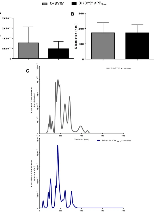
1.2. Exosomes from SH-SY5Y and SH-SY5Y APPSwe cells show similar diameter size ... 34
1.3. SH-SY5Y APPSwe cells show upregulated inflammatory-associated miRNAs, from which some of them are transferred into their derived exosomes... 36
1.4. SH-SY5Y APPSwe cells express elevated DAMPs and cytokines that are not significantly reflected in their exosomes ... 36
2. Evaluation of human microglia CHME3 reactivity to exosomes released by SY5Y and SH-SY5Y APPSwe cells ... 38
2.1. Exosomes from SH-SY5Y and SH-SY5Y APPSwe cells are collected by CHME3 microglia and co-localize with lysosomes ... 38
2.2. Treatment of CHME3 microglia with SH-SY5Y APPSwe-derived exosomes does not differently compromise autophagy but reduces cell viability in a small extent ... 40
2.3. SH-SY5Y APPSwe-derived exosomes do not induce significant changes of inflammatory-associated miRNAs in CHME3 microglial cells, but determine an increased expression of miR-21 in their derived exosomes, as compared with the exosomes from SH-SY5Y cells ... 43
2.4. SH-SY5Y APPSwe-derived exosomes trigger CHME3 increased expression of alarmins and cytokines that is not recapitulated in derived exosomes ... 46
3. Dissecting the profile of astrocytes derived from iPSCs of AD patients ... 48
3.1. Astrocytes derived from AD-iPSCs do not show loss of cell viability ... 48
3.2. Exosomes released by astrocytes derived from AD-iPSCs show different size populations ………..49
3.3. Astrocytes derived from iPSCs of AD patients show decrease of GFAP expression ... 50
3.4. iPSCs-derived astrocytes from patients with PSEN1ΔE9 mutation show a depressed RAGE/miR-155 pathway with impact in extracellular vesicle molecular cargo ... 52
IV. Discussion ... 57
INDEX OF FIGURES
Figure I.1-Alzheimer’s disease pathological hallmarks. ... 4
Figure I.2-Biological characteristics of exosomes: biogenesis, composition and cargo. ... 7
Figure I.3-Cell-to-cell communication between neurons and glial cells through exosomes and soluble factors in Alzheimer’s disease. ... 9
Figure I.4-Schematic representation of the main pathological pathways occurring in Alzheimer’s disease. ... 13
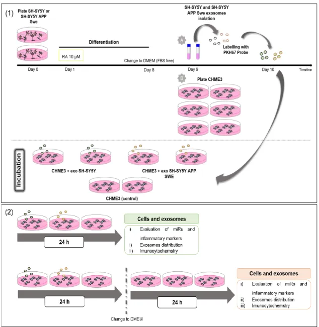
Figure II.1-Schematic representation of the experimental model for human neuroblastoma cell lines, isolation of exosomes and incubation with human microglia. ... 25
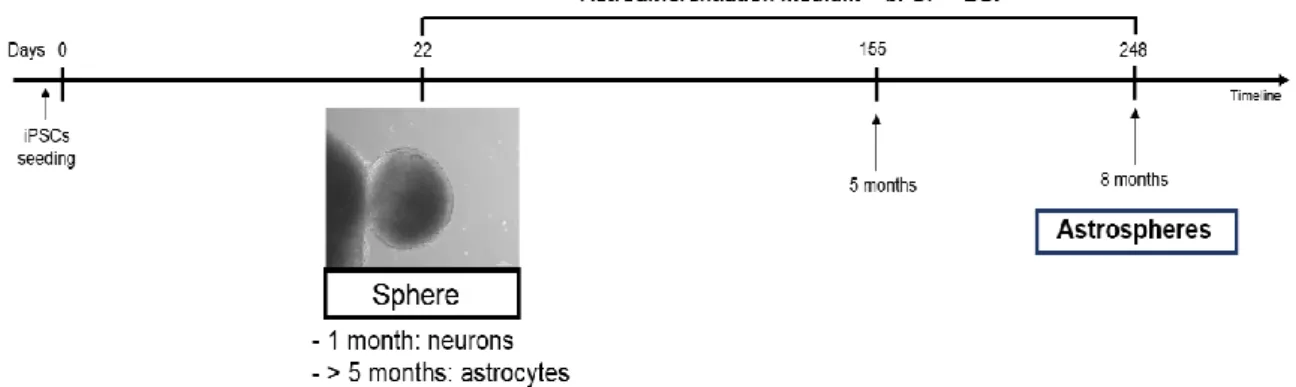
Figure II.2-Representative timeline to differentiate astrospheres from iPSCs. ... 26
Figure II.3-Schematic procedure for isolation of exosomes from cell extracellular media. ... 28
Figure III.1-Evaluation of cell viability/cell death in SH-SY5Y and SH-SY5Y APPSwe cells. ... 34
Figure III.2-Size and particle concentration of exosomes derived from neuroblastoma cells. ... 35
Figure III.3-Expression of microRNA (miR)-124, miR-155, miR-146a, miR-21 and miR-125b in SH-SY5Y and SH-SY5Y APPSwe and their derived exosomes. ... 36
Figure III.4-SH-SY5Y APPSwe cells express increased levels of S100B and HMGB1, as well as of their recepector RAGE. ... 37
Figure III.5-TNF-α and IL-10 mRNA expression is increased in SH-SY5Y APPSwe cells. ... 38
Figure III.6- SH-SY5Y and SH-SY5Y APPSwe-derived exosomes were similarly internalized by CHME3 microglia after 24 h incubation, and degraded at 48 h by an additional 24 h period of microglia incubation in a new medium. ... 39
Figure III.7-SH-SY5Y APPSwe-derived exosomes area was reduced at 48h in CHME3 cells. ... 40
Figure III.8- Co-localization of SH-SY5Y APPSwe-derived exosomes with lysosomes in CHME3 cells decrease at 48 h ... 40
Figure III.9- Fluorescence intensity of the autophagic marker LC3 in CHME3 microglial cells is reduced after 24 h exposure to both SH-SY5Y- and SH-SY5Y APPSwe-derived exosomes followed by a period of 24 h recovery ... 42
Figure III.10-Effect of exosomes delivered by SH-SY5Y and SH-SY5Y APPSwe cells on the viability of CHME3 microglial cells ... 43
Figure III.11- Exosomes from CHME3 microglia treated with SH-SY5Y APPSwe display increased levels of miR-21 when compared with cells incubated with SH-SY5Y-derived exosomes, although no changes were noticeable in the cells ... 45
Figure III.12- CHME3 microglia treated for 24 h with SH-SY5Y APPSwe-derived exosomes show upregulation of inflammatory-related molecules, from which only IL-10 is recapitulated in the secreted exosomes ... 47
Figure III.13-Evaluation of cell viability in iPSCs-derived astrocytes from controls and AD patients, as well as from AD isogenic controls ... 48
Figure III.14-Size and particle concentration of exosomes derived astrocytes differentiated from iPSCs. ... 49
xvi Figure III.15-GFAP marker is reduced in astrocytes derived from AD-iPSCs. ... 50 Figure III.16-GLT-1 marker is higher in astrocytes derived from AD-iPSCs of patient AD4. ... 51 Figure III.17-MiR-155 is downregulated in astrocytes differentiated from AD-iPSCs of patient AD5. .. 52 Figure III.18-Exosomes released by iPSCs-derived astrocytes show inflammatory-associated microRNA variability, with downregulation of miR-146a in those released by astrocytes derived from AD5-iPSCs and upregulation of miR-124 only in those from... 53 Figure III.19-RAGE is downregulated in astrocytes differentiated from iPSCs of AD patients and isogenic control... ... 54 Figure III.20-Exosomes derived from astrocytes differentiated from iPSCs of AD patients show decreased levels of S100B, HMGB1 and TNF-α. ... 55 Figure IV.1-Schematic representation of the main research contributions of this thesis. ... 63
INDEX OF TABLES
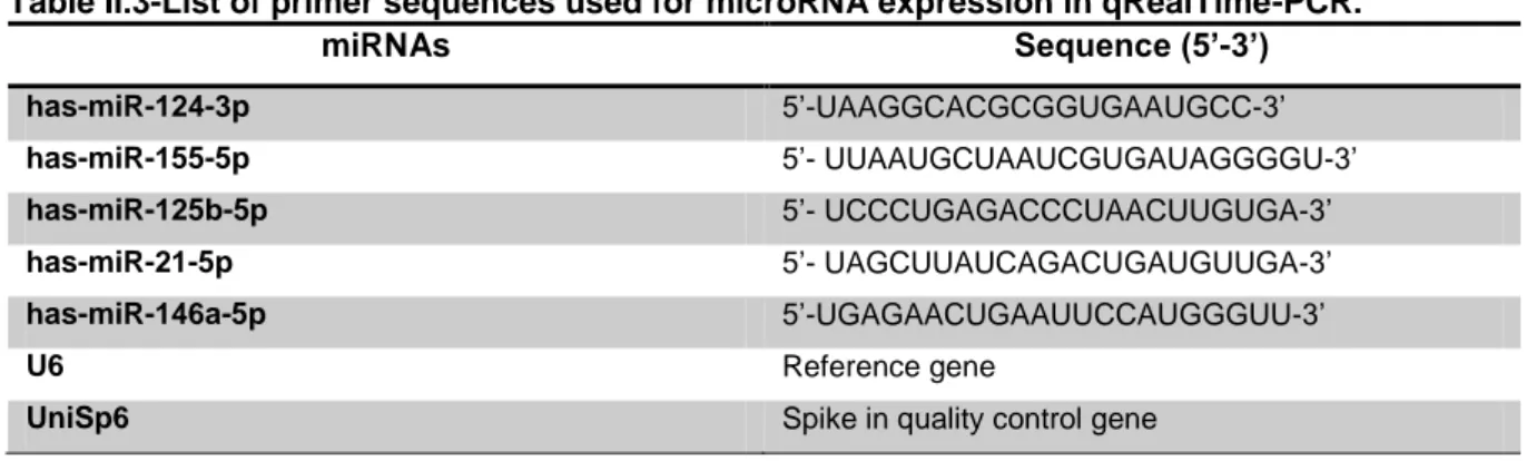
Table II.1-Genotype description of iPSCs-derived astrocytes. ... 27 Table II.2-List of primer sequences used for gene expression in qRealTime-PCR. ... 30 Table II.3-List of primer sequences used for microRNA expression in qRealTime-PCR. ... 31
ABBREVIATIONS
AB/AM Antibiotic/antimycotic
AD Alzheimer’s disease
AICD APP intracellular C-terminal domain
ALS Amyotrophic lateral sclerosis
AMPA α-amino-3-hydroxy-5-methyl-4-isoxazolepropionic acid receptor
Apo E Apolipoprotein E
APP Amyloid precursor protein
Argo Argonaut protein
Aβ Amyloid-beta
BACE1 Beta-site APP-cleaving enzyme 1 or β secretase
BBB Blood-brain barrier
BDNF Brain derived neurotrophic factor
bFGF Basic fibroblast growth factor
BMP4 Bone morphogenic protein 4
BSA Bovine serum albumin
Cas9 CRISPR associated protein-9 nuclease
CNS Central nervous system
CNTF Ciliary neurotrophic factor
COX-2 Cyclooxygenase-2
CRISPR Clustered regularly interspaced short palindromic repeats
CSF Cerebrospinal fluid
Cx Connexin
CX3CR1 CX3C chemokine receptor 1
DAMPs Danger-associated molecular patterns
DMEM Dulbecco’s modified Eagle’s medium
EGF Epidermal growth factor
ERK Extracellular signaling-related kinase
ESC Embryonic stem cells
ESCRTs Endosomal sorting complexes required for transport
EVs Extracellular vesicles
Exp5 Exportin 5
FAD Familial AD
FBS Fetal bovine serum
FDA Food and Drug Administration
FGF Fibroblast growth factor
xx
GDP Guanosine diphosphate
GFAP Glial fibrillary acid protein
GLT-1 Glutamate tansporter-1
GTP Guanosine triphosphate
HD Huntington’s disease
hiPSCs human induced Pluripotent Stem Cells
HMGB1 High-mobility group box 1
IFN- Interferon-
IL Interleukin
ILVs Intra-luminal vesicles
iNOS Inducible nitric oxide synthase
IPSCs induced Pluripotent Stem Cells
JAK/STAT Janus kinase/signal transducers and activators of transcription
JNK c-Jun N-terminal kinase
LC3 Microtubule-associated protein 1A/1B-light chain 3
L-glu L-glutamine
LPS Lipopolysaccharide
MCI Mild Cognitive Impairment
miR MicroRNA
miRNAs MicroRNAs
mRNA Messenger RNA
MVBs Multivesicular bodies
NEAA Non-essential amino acid
NEP Neprilysin
NDM Neural differentiation medium
NF-B Nuclear factor-kappa B
NFTs Neurofibrillary tangles
NMAD N-methly-D-aspartatic acid receptor
NO Nitric oxide
NPCs Neural Progenitor Cells
NTA Nanoparticle Tracking Analysis
p38/MAPK p38 mitogen-activated protein kinase
PBS Phosphate buffer saline
PD Parkinson’s disease
PFA Paraformaldehyde
PKC Protein kinase C
PSEN1 Presenilin 1
PSEN2 Presenilin 2
qPCR quantitative polimerase chain reaction
qRT-PCR quantitative real time-PCR
RA Retinoic acid
RAGE Receptor for advanced glycation end-products
RISC RNA-induced silencing complex
RT Room temperature
S100B S100 calcium-binding protein B
SAD Sporadic AD
SCI Spinal cord injury
SH-SY5Y APPSwe SH-SY5Y expressing APP695 Swedish mutation
SOCS-1 cytokine signaling-1
SOD1 Superoxide dismutase 1
TGF-β Transforming growth factor-β
TLRs Toll-like receptors
TNF-α Tumor necrosis factor-α
TREM-2 Triggering receptor expressed on myeloid cells-2
ULA Ultra-low attachment
Dissecting deregulated cell-to-cell communication in in vitro Alzheimer’s disease models
I.
Introduction
1.
Alzheimer’s disease
Initially described in 1906 by Alois Alzheimer, Alzheimer’s disease (AD) is the most common, progressive and irreversible neurodegenerative disorder, characterized by memory loss, cognitive impairment and behavioral abnormalities. AD is the leading cause of dementia in the elderly, accounting for more than 80% of cases worldwide. Currently, it is estimated that the disease affects 37 million cases (WHO, 2017) and, by the year of 2050, it is expected to achieve 13.8 million cases, with nearly a million new cases per year (Kumar et al., 2015). Curiously, a recent report compared dementia prevalence among 65 years individuals or older, between 2000 and 2012, showing that a reduction from 11.6% to 8.8% has occurred. This decreased prevalence was associated to increased educational attainment and healthier lifestyles (Langa et al., 2016).
1.1. Alzheimer’s disease etiology
The etiology of AD remains unclear. Apparently it is likely to be the combination of both environmental and genetic factors, in which older age is the strongest risk factor (Mayeux and Stern, 2006). According to the age of onset, there are two types of AD: early onset or familial AD (FAD < 65 years) and late onset or sporadic AD (SAD > 65 years). FAD is associated with a rapid progression rate and is often caused by a rare autosomal dominant mutation in genes for amyloid precursor protein (APP) or associated with its processing, such as Presenilin 1 (PSEN1) and Presenilin 2 (PSEN2). On the other hand, SAD is associated with late symptoms appearance even though the main cause is still unclear
(Dzamba et al., 2016). Polymorphisms in Apolipoprotein E (Apo E), particularly in the Apo E 4 allele,
has been considered an important risk factor across many studies (Qiu et al., 2009). Apo E has an important role in the amyloid-beta (Aβ) peptide metabolism suggesting that it affects Aβ deposition
forming senile plaques (Kim et al., 2009). This evidence is supported by studies comparing Apo E 4
carries with non-carries (Liu et al., 2013; Yamazaki et al., 2016). Recent genetic studies have identified a rare variant of the triggering receptor expressed on myeloid cells-2 (TREM-2) as an important risk factor for AD (Colonna and Wang, 2016). TREM-2 is a transmembranar receptor and is found in various tissue macrophages, including in the central nervous system (CNS) microglia. It has an important role on the Aβ clearance and phagocytosis, and in the presence of mutations leads to an increase of Aβ accumulation, as well as to an inflammatory reaction (Hickman and El Khoury, 2014).
1.2. Alzheimer’s disease symptoms and treatment
AD symptoms start with manifest symptoms related to recent memory and thinking ability called Mild Cognitive Impairment (MCI). Only 10-15% of people diagnosed with MCI develop AD (Alberdi et al., 2016), but the reason why some people do not develop dementia is still an unexplored field. The final AD stage is characterized by memory, thinking and behavioral impairments affecting patient’s ability in daily life. Neurobiological investigations have shown a reduction in the number of cholinergic neurons
Dissecting deregulated cell-to-cell communication in in vitro Alzheimer’s disease models
2 together with synapse loss, mostly in the hippocampus, entorhinal and frontal cortices, which are involved in cognitive functions like memory and language, and at the amygdala, prefrontal cortex and hypothalamus, related with emotional behavior (Whitehouse et al., 1982).
Despite problem symptoms, an accurate diagnosis of AD remains difficult to establish. Cognitive impairment in older people is frequently due to the existence of co-morbidities (Alves et al., 2012). In contrast, being AD a progressively condition, a proper diagnosis can only be achieved on an advanced stage of neurodegeneration (Kidd, 2008). A definitive diagnosis is based on the clinical and pathological hallmarks, as senile plaques and neurofibrillary tangles discussed later in this thesis. Nowadays it is already possible to identify the formation of senile plaques in patients using the Pittsburg Compound-B (PiB)-PET imaging (Cohen et al., 2012), but the clinical diagnosis realized by highly experienced and sensitive clinicians remains the most practicable approach (Alves et al., 2012). Moreover, physiological, biochemical and anatomic biomarkers, measurable in vivo, can also be considered to improve AD
diagnosis (Jack and Holtzman, 2013). Aβ deposits, together with total and phosphorylated tau, are
measurable in the cerebrospinal fluid (CSF). Additionally, extracellular vesicles (EVs), such as exosomes, containing Aβ peptide deposits can be isolated from body fluids (e.g. serum and CSF), further suggesting its utility as a novel non-invasive strategy in AD diagnosis (Vella et al., 2016). Apart from AD hallmarks content, exosomes can also carry microRNAs (miRNAs) involved in AD pathogenesis suggesting another useful and complementary tool in AD diagnosis (Gallo et al., 2012; Vella et al., 2016) Regarding the possibility of using common biomarkers to diagnose AD, usually they are only used to confirm or exclude other clinical symptoms related with neurodegenerative dementia (Leidinger et al., 2013).
Due to AD multifactorial etiology, a reliable and effective therapy that would reverse its progression has been extremely difficult to develop. Nowadays, symptom treatment is the only practicable approach to improve patient’s life quality (Khanam et al., 2016). Drugs approved by Food and Drug Administration (FDA) to AD include acetylcholinesterase inhibitors (Donepezil, Galantamine and Rivastigmine), non-competitive N-methly-D-aspartatic acid receptor (NMAD) antagonist (Memantine), and behavior signs adjuvants (antipsychotics and anticonvulsants) (Scarpini et al., 2003). In an attempt to develop new effective treatments, several studies are in progress, with some including new pharmacological compounds and innovative immunotherapy strategies against Aβ aggregates (Alves et al., 2012).
1.3. Pathological hallmarks: amyloid-beta peptide and neurofibrillary tangles
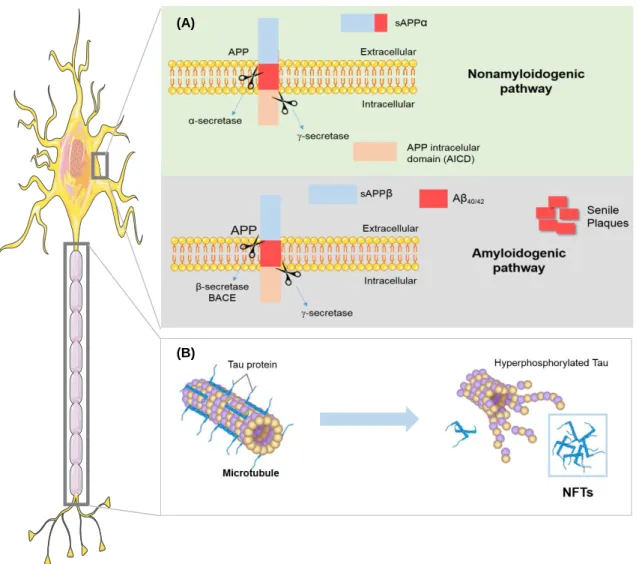
Histopathological hallmarks are represented by extracellular accumulations of misfolded Aβ peptide, known as senile or neuritic plaques, and intracellular accumulations of hyperphosphorylated tau (p-tau) protein in neurofibrillary tangles (NFTs) and neuropil threads (Rosenberger et al., 2016), in the autopsied brains of people with AD. Alterations in the metabolism of the amyloid precursor protein (APP) and tau hyperphosphorylation are two hallmarks that are believed to play a key role in AD genesis (Figure I.1). Additional changes include chronic neuroinflammation, neuronal loss and brain atrophy, as mentioned above.
The central pathological feature in AD is the deposition of Aβ peptides. APP is a membrane
Dissecting deregulated cell-to-cell communication in in vitro Alzheimer’s disease models
APP can be processed by two main pathways: non-amyloidogenic and amyloidogenic, in which APP is sequentially cleaved by proteolytic enzymes, named secretases (Gupta and Goyal, 2016) (Figure I.1).
In the non-amyloidogenic pathway, the extracellular APP domain is cleaved by α-secretase forming a
soluble extracellular fragment called sAPPα. Subsequently, a plasma protein named -secretase,
composed by the anterior pharynx defective 1, nicastrin, PSEN 1, PSEN 2 and PSEN enhancer 2, forms an intracellular fragment called APP intracellular C-terminal domain (AICD) (Murphy and LeVine, 2010). In contrast, in the amyloidogenic pathway, the extracellular APP domain is cleaved by β-secretase
(beta-site APP-cleaving enzyme 1 - BACE1), releasing the sAPPβ fragment. Afterwards, the remaining
fragment C99 is processed by -secretase forming two Aβ major isoforms of different length, Aβ40 with
40 amino acids in length, and Aβ42, with 42 amino acids in length (Bolduc et al., 2016). Physiologically,
there is a balance between production and clearance of Aβ peptides. However, in pathological
conditions there is an increase of total Aβ concentration or Aβ40/Aβ42 ratio or even a decrease in the
clearance of Aβ leading to elevated levels of Aβ42 (Gupta and Goyal, 2016). Aβ42 isoform is considered
more hydrophobic and aggregates faster, thus resulting in the formation of neurotoxic senile plaques (Peric and Annaert, 2015).
FAD is associated with several mutations in genes related to APP processing, as stated previously. Among them, the Swedish mutation was first described in 1992, when Mullan and colleagues observed two Swedish families linked by genealogy (Mullan et al., 1992). It is characterized as a double mutation in exon 16 at codons 670 and 671 (K670M/N671L) (Marques et al., 2003) leading to a new cleavage
site in APP that favors the amyloidogenic pathway increasing the total amount of Aβ and its further
accumulation.
In addition to Aβ plaques, NFTs composed by p-tau protein are the other hallmark associated with AD. Tau protein, in normal conditions, is associated to the microtubules, stabilizing cell cytoskeleton. P-tau sequesters normal P-tau away from microtubules endangering the axonal transport in affected brain regions (Figure I.1). Consequently, neurons develop NFTs, synaptic dysfunction, oxidative stress, mitochondrial impairment and DNA damage causing neuronal loss and microglia activation (Qin et al., 2016). Traditionally, AD has been considered a disorder proceeding with a dual pathway as described above. However, recent studies have shown functional interactions between Aβ and tau protein. An in
vitro study using SH-SY5Y neuroblastoma cell line, revealed that when cells were cultured in a media
supplemented with Aβ42 (and not Aβ40), tau phosphorylation was increased (Han and Shi, 2016).
Moreover, pathological phosphorylation might be mediated by activation of protein kinases dependent on Aβ, particularly GSK3 (Tatarnikova et al., 1800). Therefore, synergic interactions between these two toxic proteins accelerate AD pathogenesis
Dissecting deregulated cell-to-cell communication in in vitro Alzheimer’s disease models
4 .
Figure I.1-Alzheimer’s disease pathological hallmarks. (A) Changes in the metabolism of the amyloid precursor protein (APP). Most APP is processed through the non-amyloidogenic pathway, in
which cleavage by α-secretase generates sAPPα soluble fragment and, subsequently, cleavage by
-secretase forms an intracellular fragment called APP intracellular C-terminal domain (AICD). In the amyloidogenic pathway, APP is cleaved by β-secretase releasing sAPPβ fragment and then, the
remaining APP fragment, is cleaved by -secretase leading to the formation of the amyloid-beta (Aβ)
peptide. In the end, mediated by Aβ accumulation, there is the formation of high complex molecules known as senile plaques. (B) Tau hyperphosphorylation: tau protein is normally associated to the microtubules, thus stabilizing cell cytoskeleton. In Alzheimer’s disease (AD), tau suffers hyperphosphorylation and consequently detach from the microtubules with the formation of neurofibrillary tangles (NFTs). NFTs contribute to axonal transport impairment and neuronal dysfunction.
2. Cellular diversity of central nervous system: the interaction between neurons and
glial cells
The CNS exhibits a tremendous cell type diversity. Apart from neurons, glial cells constitute a large fraction, between 33 and 66%, of the mammalian brain. Glial cells were first identified by Rudolf Virchow, Santiago Ramón y Cajal and Pío del Río-Hortega and suggested that they solely function as so-called “nerve glue”. However, with time, scientist started to speculate about additional possible roles for these cells (Jäkel and Dimou, 2017). In fact, nowadays, glial cells are intended to be more than glue due to their involvement in many central hemostatic processes and also during development. Although many studies have been performed in order to understand glia specific roles, the full properties of these cells
(A)
Dissecting deregulated cell-to-cell communication in in vitro Alzheimer’s disease models
remain unresolved (Jäkel and Dimou, 2017). There are three types of glial cells, microglia, astrocytes and oligodendrocytes, in each one play specific roles in SNC.
Oligodendrocytes are the myelinating cells of the CNS and constitute about 5 to 10% of the total glial population. These cells are responsible for myelin production around axons, which is essential not only for the rapid and efficient conduction of the electrical impulses along the axons, but also for preserving axonal integrity (Barateiro et al., 2017). Regarding AD pathogenesis, some in vitro studies have shown that oligodendrocytes and myelin alterations occurs before the appearance of Aβ and tau pathology (Cai and Xiao, 2016). In fact, it has been suggested that myelin breakdown releases iron thus promoting the development of toxic Aβ fibrils and enhancing the formation of senile plaques (Bartzokis et al., 2007).
Microglia cells are distributed throughout the CNS and present multiple heterogeneous roles within the healthy CNS, showing diverse morphological and functional profiles, depending on their surrounding environment (Nimmerjahn et al., 2005). They are dynamic cells responsible for the surveillance of the extracellular space working as protective cells (Caldeira et al., 2014). Considered as mononuclear phagocytes, or even the CNS macrophages (Dzamba et al., 2016), the fact of sharing phenotypic characteristics and lineage-related properties with bone-marrow-derived macrophages, makes microglia capable of secreting cytokines and serving as antigen-presenting cells (Harry and Kraft, 2008), although not having the same cell origin. In the presence of diverse types of damage and stimulus, microglia becomes activated, changing their morphology from ramified to amoeboid, migrating to the lesion sites and clearing the debris of dead cells and pathogens (Brites and Vaz, 2014). During this process, microglia releases not only neurotrophic factors, but also inflammation-related molecules (e.g. pro-inflammatory cytokines and neurotoxic molecules) leading to a state of neuroinflammation (Pinto et al., 2017).
Lastly, astrocytes are the most abundant non-neural cells and are predominantly responsible for maintaining a proper chemical environment for neuronal signaling and for providing metabolic connections through the blood-brain barrier (BBB) (Purves et al., 2001). Moreover, astrocytes express metabotropic and ionotropic receptors capable of sensing neuronal activity. They express glutamate, GABA and glycine receptors involved in the tripartite synapse with pre- and post-synaptic neurons (Meyer and Kaspar, 2016). Therefore, the interplay between structure, morphology and functional characteristics allow them to exert an active influence in the neuronal signaling and protection by releasing antioxidant molecules, such as glutathione, thus preventing neurons from oxidative stress. In addition, by uptaking excess of glutamate they prevent its excitotoxicity (Osborn et al., 2015).
Over the past two decades, microglia and astrocytes have gained special attention in what concerns AD impairment, since clear evidences of their involvement have been reported in several neurodegenerative disorders (Meyer and Kaspar, 2016). Their role in AD pathogenesis will be further discussed and elucidated in this chapter.
3.
Alzheimer’s disease as a non-cell autonomous disease
3.1. Vesicular trafficking mediated by exosomes and microvesicles
Eukaryotic cells maintain contact with the extracellular compartment by receiving molecular signals (e.g. cytokines and chemokines) and by secreting proteins into the extracellular space. For
Dissecting deregulated cell-to-cell communication in in vitro Alzheimer’s disease models
6 communication, each cell has a complex network of membranes that allows them to uptake macromolecules, named endocytosis, and release biomolecules to the exterior environment, named exocytosis (Keller et al., 2006). There are three main vesicles described so far: (i) microvesicles or ectosomes (150 nm to 1 µm), which directly bud from the plasma membrane, (ii) apoptotic bodies (50-500 nm) that are released by apoptotic cells, and (iii) exosomes (40–100 nm) that derive from multivesicular bodies (MVBs) in a mechanism described next (Urbanelli et al., 2013).
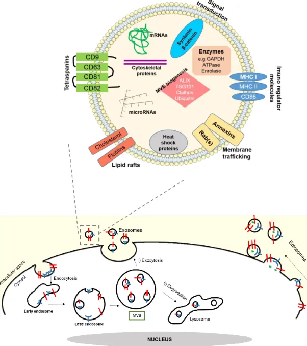
Exosomes are small vesicles that are secreted from the majority of cell types, including neurons, oligodendrocytes, microglia and astrocytes (Yuyama et al., 2014) and are retrieved in vivo in several body fluids, such as plasma, urine, saliva, seminal fluid, amniotic liquid, ascites, bronchoalveolar lavage fluid, synovial fluid, breast milk and CSF (Urbanelli et al., 2013). Exosomes are endocytic membrane-derived vesicles that are contained in MVBs in the endosomal system and secreted upon MVB fusion with the plasma membrane (Brites and Fernandes, 2015). In other words, these small vesicles have their own cytosol and are secreted by exocytosis from the MVBs, as shown in Figure I.2. MVBs result from inward budding inside an intracellular endosome forming intra-luminal vesicles (ILVs), which progressively accumulate inside lumen of the late endosome (Kowal et al., 2014). Endosomal sorting complexes required for transport (ESCRTs) and ubiquitination are involved in the biogenesis and degradation mechanisms of MVBs (Cocucci and Meldolesi, 2015).
Besides their morphology, exosomes have a particular composition due to their endosomal origin (Figure I.2). All exosomes contain membrane transporter and fusion proteins, including GTPases, annexins and flotillin, clusters of differentiation (CD9, CD63, CD81, CD82), heat shock proteins (Hsc70, Hsp90), proteins involved in MVB biogenesis (Alix, TSG101), cytoskeleton proteins, lipids, phospholipids and saccharide groups. Exosomes also exhibit a few markers from other intracellular organelles (Golgi complex, mitochondria, nucleus) proving to be a specific subcellular compartment (Kowal et al., 2014). Although these proteins are used as positive exosomes markers, there is wide variation across exosomes from different sources (Vlassov et al., 2012). Furthermore, exosomes have been reported to contain significant amounts of microRNAs (miRNAs), other non-coding RNAs, as well as messenger RNA (mRNA) (Vlassov et al., 2012). Several papers indicate that the exosomes RNA cargo is significantly different from the origin cell content. This counteracts several papers related to cancer studies, which have noted that the miRNAs content for their originating cells is similar to that found in circulating exosomes (Rabinowits et al., 2009). Regarding this, authors have postulated the feasibility of using exosomes as a basis for diagnostic marker (Vlassov et al., 2012).
Ectosomes, also called microvesicles, are quite large vesicles that bud directly from the plasma membrane (Figure I.2) (Brites and Fernandes, 2015) and are released into the extracellular space. Shedding of ectosomes often involves a budding process, in which surface pimples selectively accumulate cellular constituents that are packaged into microvesicles (Turola et al., 2012). Regulation of this process involves several enzymes such as calpain, flippase, floppase, scramblase and gelsolin (Mathivanan et al., 2010). Due to their formation process, ectosomes contain a variety of cell surface
Dissecting deregulated cell-to-cell communication in in vitro Alzheimer’s disease models
receptors, intracellular signaling proteins and genetic material derived from the cell of origin (Turola et al., 2012).
Figure I.2-Biological characteristics of exosomes: biogenesis, composition and cargo. Vesicular trafficking and cell communication are mediated by exosomes and ectosomes. Ectosomes are generated by direct budding of the plasma membrane. Exosomes are produced by exocytosis from multivesicular bodies (MVBs), in which they are produced by inward budding of the plasma membrane originating, consequently, the early and late endosome. MBVs can be i) released into the extracellular space as exosomes or ii) degraded via lysosomes. Exosome composition is mainly determined by their endosomal origin. Exosomal membranes are composed by tetraspanins, lipid rafts, proteins involved in membrane trafficking and immuno regulator molecules. Several types of cytosolic proteins and nucleic acids may be identified inside the lumen of exosomes. Cytosolic proteins include heat shock proteins, cytoskeletal proteins, enzymes, proteins involved in MVBs biogenesis and proteins involved in signal transduction. Some nucleic acids already identified in exosomes are messenger RNAs (mRNAs) and microRNAs (miRNAs).
Dissecting deregulated cell-to-cell communication in in vitro Alzheimer’s disease models
8
3.2. Neuron and glia cells interplay: cell-to-cell communication
Intercellular communication can be mediated through direct cell-to-cell communication or by the action of secreted molecules (Frühbeis et al., 2013).
As previously stated, eukaryotic cells use EVs to communicate and exchange information between cells (Figure I.3). EVs can interact with the recipient cells by three mechanisms: (i) EVs membrane proteins bind directly to the signaling receptors of target cells; (ii) EVs fuse with the plasma membrane and release their cargo inside recipient cell; and (iii) EVs are internalized into the recipient cells and have two fates (Zhang et al., 2015). In one case, some engulfed exosomes merge into endosomes and undergo transcytosis, which will move exosomes across the recipient cells and release them into neighboring cells. In the other case, endosomes will move to lysosomes and undergo degradation (Zhang et al., 2015). The communication between neurons and glia is essential to synchronize diverse functions with the brain activity. Secreted exosomes by neurons have been implicated in the synapse plasticity. Cultures of cortical neurons with enhanced glutamatergic activity show that the secreted
exosomes carrying the α-amino-3-hydroxy-5-methyl-4-isoxazolepropionic acid receptor (AMPA) may
help to adapt the efficacy of synaptic transmission by depletion of neurotransmitter receptors from the postsynaptic compartment (Frühbeis et al., 2012). EVs have been suggested as potential carriers in the intercellular delivery of pathological proteins such as misfolded proteins associated to neurodegenerative disorders. This includes p-tau and Aβ in AD, α-synuclein in Parkinson’s disease (PD), superoxide dismutase 1 (SOD1) in Amyotrophic lateral sclerosis (ALS) and huntington in Huntington’s disease (HD). Concerning AD, studies using in vivo and in vitro models suggest a role of EVs on Aβ aggregation and neurotoxicity and, on the other side, opening the possibility of their potential in AD therapy, as discussed in Joshi review paper (Joshi et al., 2015). Exosomes delivered by neuroblastoma cells are able to carry Aβ since Ranjendran observed that typical exosomal proteins, such as Alix, appears surrounding the senile plaques (Rajendran et al., 2006) and moreover, subsequent studies in vivo have demonstrated that exosomes are specially enriched with APP C-terminal fragments, a source of Aβ (Perez-Gonzalez et al., 2012). Corroborating this, in a recent paper from Yuyama and Igarashi, it is mentioned in, an in vivo APP/PSEN 1 transgenic mice model, that intracellular Aβ accumulates in abnormal endosomes, including MVBs, whose ILVs are precursors of exosomes (Yuyama and Igarashi, 2017). In the same article, authors describe that glycosphingolipids
present on the outer layer of cellular and exosome membrane are implicated in Aβ binding (Yuyama
and Igarashi, 2017). It is important to note that with those studies it was suggested that neurons have the capacity to encapsulate Aβ through exosomes followed by its release into the extracellular medium. Such evidence suggests that exosomes could be a way to neurons get rid of excessive Aβ. Through exosome secretion extracellular Aβ levels raises up and promotes its aggregation. (Joshi et al., 2015). However, regarding discussed evidences, it is still not clear whether exosomes help cells to get rid of potential detrimental components or act as a way of spreading the neurodegenerative “seeds” to other cells (Frühbeis et al., 2012).
Similar to neurons, astrocytes and microglia are able to release EVs to the extracellular space acting in physiological and pathological functions (Frühbeis et al., 2013). On one hand, exosomes delivered by astrocytes are implicated in neuroprotection carrying Hsp/ Hsc70 and synapsin I (Taylor et al., 2007;
Dissecting deregulated cell-to-cell communication in in vitro Alzheimer’s disease models
Wang et al., 2011), as well as in angiogenesis modulation (Proia et al., 2008). One the other hand, they mediate the propagation of pathogenic proteins. Wang and colleagues described that astrocytes exposed to Aβ are capable of releasing pro-apoptotic exosomes, which in turn suffer uptake by other astrocytes promoting their apoptosis (Wang et al., 2012). In addition, Goetzl and colleagues compared exosomes from astrocytes with those from neurons from plasma samples of AD patients. It was found
that levels of BACE1, -secretase, sAPPα and sAPPβ were higher in exosomes released by astrocytes.
Moreover, amyloid aggregates were not observed in the astrocyte-derived exosomes suggesting that astrocytes are more efficient in Aβ clearance. Therefore, these findings suggest that if the interplay between neurons and astrocytes is a central pathogenic pathway in AD, exosomes from astrocytes may be a valuable window for further research on the neural cell-to-cell interaction (Goetzl et al., 2016).
In what concerns exosomes released by microglia, they may act as vehicles for antigen presentation, cytokines and miRNAs secretion. Back in 2015, in a study conducted by Hirohide and colleagues, it was hypothesized that microglia may facilitate tau protein propagation between neurons by phagocytosing and exocytosing tau protein. Indeed, results have demonstrated that the depletion of microglia dramatically suppressed tau protein propagation, as well as that the inhibition of exosome synthesis significantly reduced its spreading, either in vitro or in vivo assays (Asai et al., 2015). Microglia-derived EVs also represent a way for microglia to eliminate neurotoxic Aβ when microglial degradative pathways are saturated in response to excessive phagocytosis of amyloid aggregates. Such EVs may then also contribute to the spread and seeding of neurotoxic amyloids in the brain. Increased levels of microglial EVs formation were observed in AD patients and correlated with classical markers of neurodegeneration associated with neuronal damage in the human brain (Agosta et al., 2014).
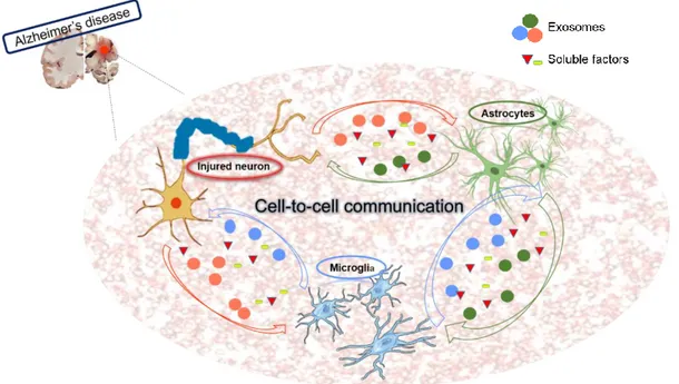
Figure I.3-Cell-to-cell communication between neurons and glial cells through exosomes and soluble factors in Alzheimer’s disease. Eukaryotic cells, including neurons and glial cells, give and receive messages from their environment proving that cells do not live isolated. In the context of Alzheimer’s disease (AD), damage neurons release exosomes containing pathological features, such as amyloid-beta (Aβ) peptide, hyperphosphorylated tau (p-tau) or even inflammatory cytokines. Microglia and astrocytes, once triggered by delivered exosomes, suffer activation changing their phenotypes. The communication can also be mediated by soluble factors, such as pro- and anti-inflammatory cytokines.
Dissecting deregulated cell-to-cell communication in in vitro Alzheimer’s disease models
10
4.
Neuroinflammation as an inducer of Alzheimer’s disease
Neuroinflammation processes are a central feature of AD and derive from the local activation of innate immune response (Rosenberger et al., 2016). This is a complex process that involves different cellular components in the CNS, such as microglia, astrocytes, ependymal cells, macrophages and mast cells (Figure I.4). Nonetheless, it is still not clear whether inflammation is a simple “watcher”, i.e. a consequence or a cause of neurodegeneration (Dá Mesquita et al., 2016).
Receptors binding cytokines on the surface of astrocytes and microglia stimulate a variety of intracellular signaling, implicated in AD pathology, including activation of protein kinase C (PKC), c-Jun N-terminal kinase (JNK), p38 mitogen-activated protein kinase (p38/MAPK), PI3 kinase, extracellular signaling-related kinase (ERK) and caspases 1 and 3 (Garwood et al., 2011).
4.1. Reactive microglia
Similar to macrophages, microglia constitute the first line of defense in the CNS sensing the neuronal environment against pathogens and host-derived ligands (Solito and Sastre, 2012). Moreover, microglia plays an important role in the inflammation resolution.
Many efforts have been done to clarify the role of microglia in what concerns inflammation and AD pathogenesis. Simultaneously with the appearance of amyloid plaques in the brain, there is a dramatic phenotype activation of the surrounding microglia, which displays high immunoreactivity. Post-mortem brain, as well as brain from transgenic APP animals, exhibit increased levels of pro-inflammatory
cytokines and chemokines, including interferon- (IFN-) and tumor necrosis factor-α (TNF-α),
interleukin (IL)-1β and IL-6 (Heneka and Banion, 2007). Additionally, IFN- and TNF-α, not only have
toxic effects on neurons, but also contribute to the reduction of insulin degrading enzyme involved in Aβ proteolysis and to the reduction of microglia ability to clean Aβ deposits. This mechanism is described as a secondary mechanism by which inflammation increases amyloid deposition (Mandrekar and Landreth, 2013).
Under pathological conditions, microglia assumes high plasticity and adopts distinct phenotypes (Perry et al., 2010). Indeed, in case of prolonged or chronic stimulation, microglia may become deleterious to the neuronal population.
The surveillance/nonpolarized stage, defined as M0, describes an alert and non-activated microglia stage in which the almost exclusively microglial fractalkine receptor, CX3C chemokine receptor 1 (CX3CR1), is highly expressed (Cunha et al., 2016). Microglia polarization M1, is mediated by activation
of toll-like receptors (TLRs) or by IFN-, with the production of pro-inflammatory mediators, such as
IL-1β, IL-4, TNF-α and transforming growth factor-β (TGF-β) (Figure I.4). Microglia can also assume a M2 phenotype when activated by IL-4 or IL-13, more associated with an anti-inflammatory state or when mixed populations of both types exist (Cameron and Landreth, 2010). Furthermore, in terms of morphology, microglia dramatically change from ramified cells to activated amoeboid cells (Kreutzberg, 1996). However, numerous studies have shown microglial activation phenotypes to be heterogeneous and the categorization as M1 and M2 is still a matter of debate.
Dissecting deregulated cell-to-cell communication in in vitro Alzheimer’s disease models
4.2. Reactive astrocytes
Astrocytes are crucial regulators and depending on the context and time, they may either promote immunosuppression and tissue repair mediated by anti-inflammatory molecules (e.g. TGF-β), or exacerbate inflammation and tissue damage (Colombo and Farina, 2016). This is corroborated by membrane expression of TLRs, being TLR3 predominantly expressed (Chen et al., 2012). TLR3 engagement triggers the production of pro-inflammatory cytokines capable of promoting inflammatory responses, such as TNF-α, IL-6 and inducible nitric oxide synthase (iNOS), which leads to nitric oxide (NO) increase. Indeed, high levels of such molecules were found in serum and brain of AD patients, compared to non-AD patients, and might exert a direct neurotoxic effect (Calsolaro and Edison, 2016). Despite cytokine toxic effect, they can also upregulate β-secretase mRNA expression and consequently
BACE1 enzymatic activity, which in turn is a key regulator of Aβ formation (Chen et al., 2012). Aβ
production seems to be increased not only because of cytokine action, but also by TNF-α activated
nuclear factor kappa B (NF-B) signal (Chen et al., 2012) and by upregulation of critical inflammatory
mediators, such as TNF-α, IL-1β and cyclooxygenase-2 (COX-2) (Figure I.4) (Medeiros and LaFerla, 2013). Extended exposure to these cytokines compromises the integrity of astrocytes and BBB composition (Minter et al., 2016). Due to their morphology, astrocytes extend thin branches allowing contact with neuronal cells bodies, dendrites and synapse terminals. Spatial contact between neurons and astrocytes allows astrocytes to sense neuronal activity (Meyer and Kaspar, 2016).
Reactive astrogliosis is currently accepted (Rodríguez-Arellano et al., 2016) as a defensive process associated with gradated continuum morphological, molecular and functional changes (Sofroniew and Vinters, 2010) in response to various signals in the extracellular space. Such signals comprehend bacterial molecules [e.g. lipopolysaccharide (LPS)], misfolded proteins and protein aggregates (e.g. Aβ),
increase of cytokines [e.g. IL-6, ciliary neurotrophic factor (CNTF), TNF-α, INF, IL-1, IL-10, TGF-β,
basic fibroblast growth factor (bFGF) 2, among others] and chemokines, or the absence of normal signals from surrounding cells, like neurotransmitters and growth factors (Haim et al., 2015). Astrogliosis results in astrocytes hypertrophy, upregulated expression of intermediated filaments like, predominantly, glial fibrillary acid protein (GFAP) and vimentin, S100 calcium binding protein B (S100B) and astrogial connexins (Cx30 and Cx43) (Yi et al., 2016), and production of inflammatory factors (cytokines, chemokines and growth factors) (Dzamba et al., 2016). GFAP is widely used, both in in vivo and in vitro studies, for identification of astrogliosis (Colombo and Farina, 2016).
In AD, astrogliosis has an important role in the pathological progression. Senile plaques and p-tau are usually surrounded by activated astrocytes, wherein this process has thought to be a neuroprotective barrier by restricting them from the rest of brain tissue. In addition, astrocytes are capable to take up Aβ peptides, through the receptor of advanced glycation end product (RAGE), for lysosomal degradation with the purpose of maintaining Aβ homeostasis (Steardo et al., 2015). However, the persistent activation and inflammation may also favor the AD progression (Osborn et al., 2015).
4.3. Danger-associated molecular patterns: the alarmins of neuroinflammation
Danger-associated molecular patterns (DAMPs), also known as alarmins, are a pleiotropic group of intracellular proteins that include, among others, the High-mobility group box 1 protein (HMGB1 or
Dissecting deregulated cell-to-cell communication in in vitro Alzheimer’s disease models
12 amphoterin) and calcium binding proteins family known as S100. Under pathological conditions, such
as chronic inflammation, DAMPs released into the extracellular space are considered as “danger
signals” triggering the activation of the receptor RAGE (Buhimschi et al., 2009), as shown in Figure I.4. RAGE is a transmembranar receptor, acting as a chief for products of nonenzymatic glycoxidation, HMGB1 and S100 family proteins, as well as Aβ, as described above. Binding of DAMPs to the RAGE
intracellular domain, results in the activation of NF-B and the recruitment of inflammatory cells, which
in turn amplify the process of tissue damage (Chavakis et al., 2004).
HMGB1 is primarily located in the nucleus of the majority of the cells. It was originally identified as a non-histone binding DNA protein involved in maintaining DNA structure and regulating gene transcription, among other physiological functions (Frank et al., 2015). Under pathological conditions, HMGB1 is released by the cell into the extracellular space. In the brain, HMGB1 is actively released by activated microglia and passively released by necrotic or damaged cells (Fonken et al., 2016). Consequently, it interacts with TLR2 and TLR4, and with RAGE, as previously mentioned. Thereby, the interaction between HMGB1 and the receptors drives the pro-inflammatory cellular responses (Fonken et al., 2016).
S100B is a member of the S100 family and is mostly expressed by astrocytes. Intracellularly, S100B promotes neuronal proliferation, oligodendrocyte differentiation, astrocyte morphology maintenance, and facilitates the cell migration of both astrocytes and microglia. Extracellularly, S100B can act either as neurotrophic or neurotoxic molecule, depending on the concentration achieved (Barateiro et al., 2016). Nowadays, S100B is considered a peripheral biomarker of brain damage since its level may increase in CSF and/or in blood by several brain pathologies, including astrocytes damage. In what concerns AD, it was reported an association between the deposition of Aβ and the presence of activated astrocytes overexpressing S100B (Chaves et al., 2010).
Dissecting deregulated cell-to-cell communication in in vitro Alzheimer’s disease models
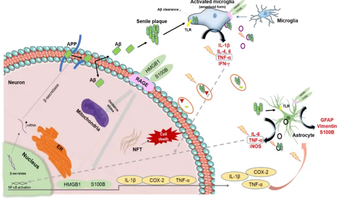
Figure I.4-Schematic representation of the main pathological pathways occurring in Alzheimer’s disease. Neuroinflammation has been described for many years as one of the central features involved in Alzheimer’s disease (AD) pathogenesis. The amyloid-beta (Aβ) peptide is generated in neurons by β-secretase proteolysis and further senile plaques are formed by Aβ aggregation and sensed by membrane receptors – toll-like receptors (TLRs) – present in microglia and astrocytes. Microglia has a crucial role in inflammation resolution being involved in Aβ clearance. Once activated, microglia change their morphology to an amoeboid shape and become reactive. Inflammatory mediators, namely pro-inflammatory cytokines, such as interleukin (IL)-1β, IL-4, IL-6, tumor necrosis factor-α (TNF-α) and
interferon- (IFN-), are involved in neuroinflammatory processes. Reactive astrocytes are also
important inflammatory mediators by the release of such pro-inflammatory cytokines to the extracellular
space. Moreover, TNF-α activates the nuclear factor kappa B (NF-B) signaling pathway leading to the
upregulation of pro-inflammatory mediators, which in turn activate microglia and astrocytes. β-secretase mRNA expression is also upregulated by pro-inflammatory cytokines. Exosomes delivered by neurons (orange circles), microglia (purple circles) and astrocytes (black circles) may contain Aβ peptides and soluble factors (e.g. pro-inflammatory cytokines), recognized to have a pathological role. High-Mobility Group Box 1 (HMGB1) and S100 calcium binding protein B (S100B) alarmins, released by activated cells, trigger inflammatory pathways by activating the receptor of advanced glycation end product (RAGE) or TLRs on the surface of neurons, microglia and astrocytes, contributing to the neuropathological process. Intracellularly accumulations of hyperphosphorylated tau in neurofibrillary tangles (NFTs) along with endoplasmic reticulum (ER) and mitochondria stress-related are involved as well in AD impairment that culminates with neuronal death.
5. MicroRNA: the emerging roles in Alzheimer’s disease
Among proteins and lipids, EVs contain additional nucleic acids, namely RNA species, in their cargo. Among RNA species, miRNAs are able to modify cellular functions and gene expression in the recipient cells (Lafourcade et al., 2016).
MiRNAs, a newly described class containing approximately 22 nucleotide of long non-coding RNAs, act as regulators of gene expression in eukaryotes. Generally, they act as post-transcriptional repressors and may be involved in epigenetic events promoting gene silencing (Breving and Esquela-Kerscher, 2010). MiRNAs can not only mediate gene silencing, but can also promote translational