Vol.58, n.2: pp. 185-191, March-April 2015 http://dx.doi.org/10.1590/S1516-8913201400040
ISSN 1516-8913 Printed in Brazil
BRAZILIAN ARCHIVES OF BIOLOGY AND TECHNOLOGY
A N I N T E R N A T I O N A L J O U R N A L
Partial Purification and Characterization of β
-glucosidase
from
Monascus
sanguineus
Rashmi Dikshit and Padmavathi Tallapragada
*Department of Microbiology; CPGS; Jain University; Bangalore - India
ABSTRACT
The aim of the present work was to studythe production and characterization of β-glucosidase from Monascus sanguineus. Agro-waste residues were screened to obtain the maximum yield of enzyme. Jack fruit seed was the best substrate for enzyme production. Studies on the optimization of pH and temperature showed acidic pH favorable for enzymatic activity, whereas the optimum temperature was 60°C. Enzyme kinetics studies with different concentration of pNPG showed the calculated value of Km approximately 0.89 mM with the non-linear regression and 0.98 mM with the linear regression techniques. The enzyme was predominantly inhibited by KCl(69.8%) and moderately inhibited by CaCl2 (14.8%). Studies on the sensitivity for glucose showed that after 100 mM concentration of glucose, inhibition in pNPG hydrolysis took place. The molecular weight of the protein was estimated as 116 and 66 kDa with SDS- PAGE and zymography was carried out to verify the specific activity.
Key words: Zymography, agro-waste, kinetics, Monascussanguineus, pNPG
*Author for correspondence: [email protected]
INTRODUCTION
β-glucosidase (β-D-glucoside glucohydrolase, EC 3.2.1.21, BGL) is an enzyme, which catalyzes the
hydrolytic cleavage of β-glycosidic linkage between two glycone residues or between glucose and an alkyl or aryl aglycone. BGL has great importance in variety of physiological as well as biotechnological processes. Its activity is mainly dependent on the nature of the glycone or aglycone moiety of the substrate. For instance, in plants, it is known to play the role in
phytohormone activation, chemical defense
against pests, lignifications, etc. It is also used for
β-glucan synthesis during cell wall development
and cell wall degradation in the endosperm during germination. As far as the mammalians are
concerned, the human acid β-glucosidase, commonly known as glucocerebrosidase, plays an
important role in the degradation of
glucosylceramide in the lysosome (Turan and Zheng 2005). Almost all the living organisms from bacteria to higher living kingdoms are
known to contain β-glucosidase enzyme. BGL is having several applications such as the enzymatic saccharification of cellulosic materials, liberation of flavor compounds in fruit juices and wines, etc. It also helps in the release of phenolic compounds with antioxidant activity from fruit and vegetable residues (Dongyang et al. 2012). In view of its efficiency to catalyze the transglycosylation
from terpenylglucoside (Kaur et al. 2007). This volatile alcohol has enormous importance in food, cosmetics and tobacco industry (Jerkovic and
Mastelic 2004). The filamentous fungus Monascus
is used for centuries as a source of colorants in
traditional foods. Monascus sp. is a subject of
constant studies, mainly due to the growing interest for natural pigments for usage in food industry. Although other metabolic products from
Monascus species, such as alcohols, organic acids,
antimicrobial agents and substances with
therapeutic activity have been described, little
information about enzymes from Monascus is
available (Daroit et al. 2007).
The aim of the present work was to study the
production and characterization of β-glucosidase
enzyme from a Monascus sp.
MATERIAL AND METHODS
Culture
Pomegranate was used to isolate the strain of
Monascus, which was identified as Monascus
sanguineus. The strain was maintained on Potato
Dextrose Agar (PDA) medium and incubated at 28-30°C for seven days. It was preserved at 4°C, and sub-cultured once every four weeks (Dikshit and Tallapragada 2013).
Inoculum preparation
The spores were scraped off from the seven day old culture plate and dissolved in 0.90% saline water to produce a spore suspension. The spore suspension was used as inoculum.
Screening of substrates for β - glucosidase
enzyme production
Different agro waste residues were chosen as solid substrates for the screening of BGL viz. wheat bran, coconut residue, tamarind seed and jack fruit seed. These substrates were purchased from a local market of Bangalore, India. Five gram of the substrate was placed in a 250 mL conical flask to which Mandels and Weber (1969) basal media salt solution was added. After cooling, these substrates were inoculated with 10% of the seed culture of
M. sanguineus and incubated at 30°C for 10 days.
Extraction of enzyme
Enzyme was extracted by adding 50 mL citrate buffer (0.05 M, pH 4.8) in each flask and kept on rotary shaker at 150 rpm for 24 h. Solution was filtered through Whatman filter-paper and
centrifuged at 5000×g at 4°C for 10 min. This filtrate was used as crude enzyme.
Partial purification of crude enzyme
Partial purification of BGL was carried out by ammonium sulphate precipitation, followed by dialysis. Cell free extract (250 mL) was saturated with ammonium sulphate up to 80%. The content was incubated over-night and centrifuged at 6000×g for 25 min. Supernatant was collected and saturated up to 90% with ammonium sulphate. Once again the content was centrifuged with above said conditions. Collected pellets were dissolved in phosphate buffer (20 mM, pH 6.5) and transferred in a dialysis bag and immersed in phosphate buffer at 4°C for 24 h. Buffer was changed several times in order to achieve proper purification (Ashwini et al. 2011).
β-glucosidase activity assay
Activity of the BGL from crude as well as partially purified enzyme was determined using 5
mM, 4-Nitrophenyl, β-D-glucopyranoside (pNPG)
as substrate. Reaction mixture contained 0.5 mL of diluted enzyme sample (either crude or partially purified), 0.5 mL of 10 mM pNPG and 1.0 mL of citrate buffer (0.05 M, pH 4.8). It was incubated at 40°C for 15 min. This reaction was terminated with 2.0 mL of cold 0.2 M Na2CO3. The activity was observed by the liberation of p-nitro-phenol and was estimated in a spectrophotometer by reading the absorbance at 410 nm. One unit of enzyme activity was defined as the amount of
enzyme required for the hydrolysis of one μmole
of pNP per minute under assay condition and was expressed in units/gram dry substrate (U/gds) (Daroit et al. 2007).
Determination of optimum pH and
temperature for enzyme assay
Effect of different range of pH on BGL activity was determined by using 0.05 M concentration of citrate buffer (pH of 3.0, 4.0, 5.0 and 6.0), sodium phosphate (pH 7.0 and 8.0) and Tris-HCl (pH 9.0). Assay was performed as described above. For optimum temperature, the reaction mixture was incubated at different range of temperatures (30, 40, 50, 60, 70, and 80°C) (Daroit et al. 2008).
Protein concentration
Poly-acryl amide gel electrophoresis and zymography
Poly-Acryl amide Gel Electrophoresis (PAGE) of the partial purified enzyme was performed according to Laemmli (1970) using 12% acryl amide gel in the presence of sodium dodecyl sulphate and 2-mercaptoethanol. Concentrated protein (1.0 mg/mL) was used for electrophoresis. Staining of the band was done with coomassie
brilliant blue, R-250 (CBB). To obtain
zymograph, PAGE was performed according to above procedure. After the electrophoresis, the gel was incubated with 20 mM of pNPG solution in citrate buffer at 50°C for 10 min to observe yellow band.
Kinetic parameters for β-glucosidase
Different concentrations of pNPG (0-25 mM) were used to estimate the kinetic parameters, Km
and Vmax using double reciprocal Lineweaver–
Burk plot. Michaelis-Menten equation was used to fit the data for the kinetic constant in non-linear manner and curve fitting was done by using MATLAB software (Daroit et al. 2008).
Effect of activators/ inhibitors on β-glucosidase
activity
Different activators/inhibitors in the concentration of 5 mM were used to investigate the enzyme activity. NaCl, sodium dodecyl sulphate (SDS), CaCl2, ethylene diamine tetraacetic acid (EDTA) and KCl were used for enzyme assay. Glucose inhibition was carried out with different concentration of glucose (100-500 mM). Enzyme was pre-incubated with above said compounds for 15 min. After incubation, the activity was
observed with above mentioned standard
protocols (Moloud et al. 2013).
RESULTS AND DISCUSSION
Screening of substrates for solid state fermentation
Among four substrates tested, maximum enzyme yield was observed with jack-fruit seed and minimum with tamarind seed (Fig. 1). The jack-fruit seeds are rich source of carbohydrates, proteins and good source of fiber and vitamin A. Wheat bran is rich in vitamin B. Coconut oil cake is also rich source of mineral and protein, which can promote fungal growth and yield of secondary metabolites. Tamarind seed is a polysachcharide
and contains three major sugars viz. glucose, galactose, and xylose in molar ratio of 3:1:2. Glucose being a major constituent in tamarind seeds, the BGL activity shown was relatively less. There has been an increased exploitation of organic residues from various sectors of agriculture and industries over the past few decades. Crop residues such as bran, husk, bagasse, and fruit seeds have been utilized as potential raw materials in bioprocesses as they provide an excellent substratum for the growth of microorganism, supplying the essential nutrients (Pandey and Soccol 1998, 2000).
Figure 1 - Screening of substrates for β-glucosidase enzyme ( depicting the crude
enzyme and the semi-purified enzyme; y-axis showing the enzyme activity in Units/gram dry substrate and x-axis various substrates viz. T- Tamarind seed J- jack-fruit seed, W- Wheat bran, C- Coconut oil cake).
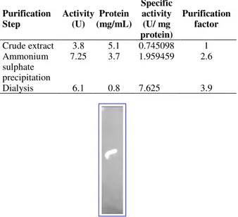
Partial purification, molecular weight and zymography of enzyme
The enzyme present in the crude extract was purified by ammonium sulfate precipitation, which resulted 2.6-fold purification and this further increased up to 3.9-fold with dialysis (Table 1). Partially purified enzyme was associated with other molecules depicting two major bands as revealed from SDS-PAGE. It has
been reported that Monascus pigments react with
amino acids present in the medium to form hydro soluble pigments (Dufosse et al. 2005; Daroit et al. 2008). The molecular weight of the BGL from
M. sanguineus was calculated by plotting a graph
between linear logarithms of relative molecular mass versus the Rf value. Calculated molecular masses of obtained bands were 116 and 66 kDa. The zymography revealed a single yellow band,
conforming BGL activity from M. sanguineus
Table 1 - Purification Summary for β-glucosidase.
Purification Step
Activity (U)
Protein (mg/mL)
Specific activity (U/ mg protein)
Purification factor
Crude extract 3.8 5.1 0.745098 1
Ammonium sulphate precipitation
7.25 3.7 1.959459 2.6
Dialysis 6.1 0.8 7.625 3.9
Figure 2 - Zymogram for β-glucosidase.
Several authors have reported different molecular mass for the BGL from different organisms; for instance, approximately 120 and 66 kDa molecular mass of the protein was observed for
the BGL from Penicillium funiculosum NCL1.
Similarly, P. purpurogenum KJS506 and P.
occitanis had shown the molecular mass of 120,
89.6 and 98 kDa, respectively (Bhiri et al. 2008; Jeya et al. 2010; Ramani et al. 2012). BGL from
A. terreus NRRL 265 exhibited molecular mass of
116 kDa (Elshafei et al. 2011). Hongzhi et al.
(2013) reported a BGL of 126.0 kDa from P.
simplicissimum identified by 12% SDS- PAGE.
As far as Monascus sp. is concerned, very few
attempts have been made for enzymatic studies. Daroit et al. (2008) had reported high molecular mass of protein (than 100 kDa) for a BGL from
M. purpureus and single yellow fluorescent band
with partially purified sample, when subjected to zymography.
Optimization of enzyme activity with
temperature and pH
β-glucosidase activity was observed at 40, 50, 60,
70 and 80°C. The results showed that the BGL activity increased from 40 to 60°C after which decrease in activity was observed (Fig. 3A). Temperature is an important factor for enzymatic activity. Activity of enzyme at higher temperature range is an advantageous factor for the saccharification of biomass and can also prevent contamination to allow the reaction to proceed at higher range of temperature. According to Daroit et al. (2008), increase in activity was observed with increase in incubation temperature. However, when the incubation temperature reached 80°C, the enzymatic activity diminished. As far as pH is concerned, the plot obtained followed the expected bell-shaped curve and the maximum activity was observed at acidic pH range (Fig. 3B). Lucas et al. (2000) had reported optimum
BGL activity from Chalara paradoxa at the range
of pH 4.0-5.0 whereas Daroit et al. (2008) had reported optimum BGL activity at pH 5.5 (acidic)
from M. purpureus. A BGL from A. foetidus also
has been reported optimally active at acidic range of pH (Hang and Woodams, 1994).
A
B
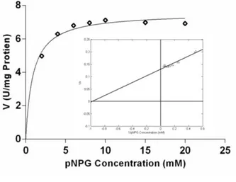
Kinetic constants of β-glucosidase
Km and Vmax (kinetic constants) of the BGL were estimated by non-linear regression technique utilizing Michaelis-Menten method. For this, the concentration of the substrate (pNPG) was plotted against the enzyme activity (V). The curve followed the Michaelis-Menten kinetics trend, which in-turn was used to calculate the value of the kinetic constant Km and Vmax (Fig. 4). The maximum value of the enzymatic activity (Vmax) was approximately 7.56 U/mg protein as extrapolated by the Michaelis-Menten graph.
Figure 4 - The non- linear regression analysis of β -glucosidase activity. Inset: Lineweaver-Burk plot (1/V vs 1[S]) for pNPG hydrolysis.
As per the theory, the value of Km is the substrate concentration required to attain half of the maximum enzyme velocity. The value of Km, thus obtained was 0.89 mM. Smaller Km value is a representative of powerful affinity towards the substrate. Similarly, to obtain these values and to further endorse the results, the Lineweaver-Burk plot was attempted (Fig. 4 inset). This calls for the inverse of the substrate concentration on the x-axis and the inverse of the enzymatic activity on the y-axis. The equation can be expressed as:
(1)
A linear fit is obtained between these two variables. The equation thus obtained can be expressed as
(2)
From Eq. (1), it is evident that the intercept at the
x axis gives the value of – 1/Km and the intercept
at y-axis gives the value of 1/Vmax. Hence, the value of Vmax obtained was 7.52 U/mg protein and the value of Km was 0.98 mM (Table 2). As the reciprocals distort the experimental errors, this double reciprocal plot does not really follow the assumptions made for linear regression. Still, the results corroborated with the results obtained from the Michaelis-Menten kinetics plot. Figure 4 showed that the hydrolysis of pNPG had not truly obeyed Michaelis-Menten kinetics and some inhibition was observed at higher concentration of the substrate. Daroit et al. (2008) had also reported inhibition in the hydrolysis of pNPG at higher
concentration from M. purpureus.
Table 2 - Kinetic parameters for β-glucosidase from
Monascus sanguineus. From Non-Linear
Regression
From Linear Regression Km
(mM)
Vmax
(U/mg protein)
Vmax /
Km
Km
(mM)
Vmax
(U/mg protein)
Vmax /
Km
0.89 7.56 8.49 0.98 7.52 7.68
Influence of chemical reagents on β-glucosidase
activity
Studies on the influence of various chemical reagents on the BGL showed reduced activities by all the experimented reagents. Minimum inhibition
was observed with CaCl2 (14.8%) and maximum
with KCl (69.9%) (Fig. 5). Certain enzymes, apart from a requirement of a coenzyme also need a metal ion for complete activity. Removal of metal ion often reduces the enzymatic activity or even results in a total loss of enzymatic activity. Metals are the common inorganic modifiers. Besides accelerating the rate of enzyme- catalyzed reactions, it can also inhibit the rate of reaction (Jain et al. 2008).
Inhibition of pNPG hydrolysis at different concentration of glucose
Tolerance of glucose is an important factor since it determines the suitability of enzyme for biomass
hydrolysis. M. sanguineus BGL tolerated the
glucose concentration up to certain level and thereafter inhibition in enzymatic activity was observed (Fig. 6). Several authors have reported that glucose acts as an inhibitor for the BGL activity. Daroit et al. (2008) has reported almost
purpureus with glucose. Glucose acts as a competitive inhibitor and is commonly observed in microbial BGL with a Ki value ranging from 0.5-14 mM (Yan et al. 1998; Yun et al. 2001). Gao et al. (2012) had reported glucose inhibition for BGL
activity by F. proliferatum and they concluded that
it might be due to the availability of variable special residuals on active site of BGL. The changes of the special residuals cause difference in extent of bonding to glucose as these residuals are not only the binding site of glucose but also the binding site for the substrate. This, in turn, makes variation in the degree of tolerance to glucose. However the mechanism of BGL tolerance to glucose is still ambiguous.
Figure 5 - Influence of chemical reagents on β -glucosidase activity (y-axis showing the percentage relative inhibition and x-axis various substrates).
Figure 6 - Inhibition of pNPG hydrolysis at different concentration of glucose (y-axis showing the enzyme activity in Units/gram dry substrate and x-axis the glucose concentration in mM).
CONCLUSIONS
Among the various substrates, jackfruit seed was the best substrate for the production of BGL. The maximum enzyme activity was in acidic range of
pH and at 60°C. Since Monascus strains are
considered as safe organisms and widely used as
food microorganisms, BGL produced by M.
sanguineus could be used for releasing of terpenes
and other aromatic compounds from wine and also for the liberation of antioxidant compounds from fruits and vegetables.
This Monascus strain needs more exploitation for
better yield of enzyme, pigments and other secondary metabolites, which can be used for industrial applications.
REFERENCES
Ashwini K, Gaurav K, Karthik L, Bhaskara Rao KV. Optimization, production and partial purification of
extracellular α-amylase from Bacillus sp. marini.
Arch Appl Sci Res. 2011; 3 (1): 33-42.
Bhiri F, Chaabouni SE, Limam F, Ghrir R, Marzouki N. Purification and biochemical characterization of extracellular β-glucosidases from the hypercellulolytic Pol6 mutant of Penicillium occitanis. Appl Biochem Biotechnol. 2008; 149:169-182.
Caldini C, Bonomi F, Pifferi PG, Lanzarini G, Galante YM. Kinetic and immobilization studies on fungal glycosidase for aroma enhancement in wine. Enzyme Microb Technol. 1994; 16(4): 286-291.
Daroit DJ, Aline S, Pinho FH, Adriano B. Purification
and characterization of extracellular β-Glucosidase from Monascus purpureus. J Microbiol Biotechnol.
2008; 18(5):933-941.
Daroit DJ, Silvana TS, Plinho FH, Adriano B.
Production of extracellular β-glucosidase by
Monascus purpureus on different growth substrates.
Process Biochem. 2007; 42: 904-908.
Dikshit R, Tallapragada P. Exploring Monascus sanguineus as a Potential Natural Source for Pigment Production. Int Res J Biological Sci. 2013; 2(5): 59-67.
Dongyang L, Ruifu Z, Xingming Y, Zhenhua Z, Song S, Youzhi M, et al. Characterization of a thermo
stable β-glucosidase from Aspergillus fumigates Z5, and its functional expression in Pichia pastoris X33.
Microb Cell Fact. 2012; 11:25.
Elshafei AM, Hassan MM, Morsi NM, Elghonamy DH. Purification and some kinetic properties of β -glucosidase from Aspergillus terreus NRRL 265. Afr J Biotechnol. 2011; 10: 19556-19569.
Gao Z, Duong VH, Le Thi HY, Katsuhiko A, Shuichi
H, Ryuichiro K. The production of β-glucosidases by
Fusarium proliferatum NBRC109045 isolated from Vietnamese forest. AMB Express. 2012; 2: 49. Hang YD, Woodams EE. Apple Pomace: A Potential
Substrate for Production of β-Glucosidase by Aspergillus Foetidus. LWT-Food Sci Technol. 1994; 27(6): 587-589.
Hongzhi B, Hui W, Junde S, Muhammad I, Mei H, Yuqian H, et al. Production, Purification and
Characterization of Novel β Glucosidase from Newly
Isolated Penicillium simplicissimumh-11 In Submerged Fermentation. EXCLI Journal. 2013; 12:528-540.
Jain JL, Jain S, Jain N. Fundamentals of Biochemistry, S Chand., New Delhi, 2008; 385p.
Jerković I, Mastelić J. GC-MS characterization of
acetylated β-D-glucopyranosides: transglucosylation
of volatile alcohols using almond β-glucosidase.
Croat Chem Acta. 2004; 77(3): 529-535.
Jeya M, Joo AR, Lee KM, Tiwari MK, Lee KM, Kim S , et al. Characterization of beta-glucosidase from a strain of Penicillium purpurogenum KJS506. Appl Microbiol Biot. 2010; 86: 1473-1484.
Kaur J, Chadha BS, Badhan AK, Kaur GS, Saini HS.
Purification and characterization of β-glucosidase from Melanocarpussp. MTCC 3922. Electron J Biotechn. 2007; 10 (2):260-270.
Laemmli UK. Cleavage of structural proteins during the assembly of the head of bacteriophage T4. Nature.
1970; 227 (5259): 680-685.
Lowry OH, Rosebrough NJ, Farr AL, Randall RJ. Protein measurement with the folin phenol reagent. J Biol Chem. 1951; 193: 265-275.
Lucas RA, Robles GA, De Cienfuegos, Galvez A. β -Glucosidase from Chalara paradoxa CH32: Purification and properties. J Agric Food Chem.
2000; 48: 3698-3703.
Mandels M, Weber J. The production of cellulases. Adv Chem Ser. 1969; 95:391-413.
Moloud GC, Seyed MA, Mohammad GMS, Vahid HN, Hadi S. Biochemical characterization of digestive carbohydrases in the rose sawfly, Arge rosae
Linnaeus (Hymenoptera: Argidae). J Crop Prot. 2013; 2 (3): 305-318.
Pandey A, Soccol CR. Bioconversion of biomass: A case study of lignocellulosics bioconversions in solid state fermentation. Braz Arch Biol Technol. 1998; 41(4): 379-390.
Pandey A, Soccol CR. Economic utilization of crop residues for value addition - A futuristic approach, J Sci Ind Res. 2000; 59 (1): 12-22.
Ramani G, Meera B, Vanitha C, Rao M, Gunasekaran P. Production, purification, and characterization of a
β-glucosidase of Penicillium funiculosum NCL1.
Appl Biochem Biotechnol. 2012; 167: 959-972. Sarry JE, Gunata Z. Plant and microbial glycoside
hydrolases: volatile release from glycosidic aroma precursors. Food Chem. 2004; 87(4): 509-521. Turan Y, Zheng M. Purification and Characterization of
an Intracellular β-Glucosidase from the Methylotrophic Yeast Pichia pastoris. Biochemistry (Moscow). 2005; 70 (12): 1363-1368.
Yan TR, Lin YH, Lin CL. Purification and
characterization of an extracellular β-glucosidase II with high hydrolysis and transglycosylation activities from Aspergillus niger. J Agric Food Chem. 1998; 46: 431-437.
Yun SI, Jeong CS, Chung DK, Choi HS. Purification
and some properties of a β-glucosidase from
Trichoderma harzianum type C-4. Biosci Biotechnol Biochem. 2001; 65: 2028-2032.