Ultrasound-guided venous cannulation in a critical
care unit
Punção venosa guiada por ultra-som em unidade de terapia
intensiva
INTRODUCTION
In the intensive care setting, ultrasound guided venous cannulation has now become an essential procedure for critically ill patients, howev-er it has risks inhhowev-erent to its inshowev-ertion as well as phowev-ermanence. To reduce risks related to insertion, the applicability of ultrasonography to aid insertion of a central and peripheral venous catheter will be discussed. This is a technology available in our daily clinical practice with some ad-vantages in intensive care units, such as absence of exposure of patients to radioactivity, reproducibility, low cost, practicality and portability. Further, the method is not invasive and provides important bedside in-formation, as well as support in invasive procedures.(1-4) Initially, its use
was restricted to medical radiologists and echocardiographers. However, universalization of the method and standardizing of training by some societies such as the American College of Emergency Physicians (ACEP),(5)
European Federation of Societies for Ultrasound in Medidicine and Bi-ology(6) and World Interactive Network Focused on Critical Ultrasound
(WINFOCUS,(7) furthered implementation in the developed countries
while it is still incipient in developing countries such as Brazil. In the last decade, applicability of this method was not merely restricted to the intensive care setting, but also to the pre-hospital emergency system, intra-operative and during cardiac arrest (CA).(8-10) Another interesting
point is the growing number of publications related to the method in indexed journals, stressing its importance as a complementary tool for care of the critically ill.(11)
Uri Adrian Prync Flato1, Gustavo
Mascari Petisco2, Fernanda Bezerra
dos Santos3
1. Physician at the Intensive Care Unit of the Centro de Referência da Saúde da Mulher - São Paulo (SP), Brazil. 2. Physician at the Intensive Care Unit of the Centro de Referência da Saúde da Mulher - São Paulo (SP), Brazil. 3. Physician at the Intensive Care Unit of the Centro de Referência da Saúde da Mulher - São Paulo (SP), Brazil.
ABSTRACT
Use of ultrasound introduced as part of intensive care therapy makes viable bedside invasive procedures and diagnosis. Due to portability, combined with team training, its use guarantees less complications related to insertion, as well as patients’ safety. It also reduces severe conditions
re-lated to the catheter, such as pneu-mothorax among others. Probably, in a near future, as purchase of ultra-sound equipment becomes easier and team training more adequate, this tool will become essential in daily clinical practice.
Keywords: Ultrasonography; Echo-cardiography; Intensive care
Received from the Intensie Care Unit of the Centro de Referência da Saúde da Mulher – São Paulo (SP), Brazil.
Submitted on November 27, 2008 Accepted on May 16, 2009
Author for correspondence:
Uri Adrian Prync Flato
Gerência Hospitalar - Centro de Referência da Saúde da Mulher Avenida Brigadeiro Luís Antônio, 683 CEP: 01317-000 - São Paulo (SP), Brazil.
BASIC pRINCIpleS Of UlTRASOUND
Sound waves are mechanical vibrations that induce al-ternate refractions and compressions in any physical me-dium they cross. Sound waves are deined by their ampli-tude and frequencies.
he classical image of general ultrasound or echog-raphy relies on echoes and is based upon the principles of relection, refraction and dispersion of energy of ul-trasound waves. he ulul-trasound equipment (US) gener-ates an electronic wave and the piezoelectric transducers transform the electric wave into a mechanical wave. US is used in frequencies between 01 MHz and 50MHz. Attenuation of the echographic wave depends on the frequency of repetition of the wave peaks and valleys. Frequency of insonation also controls axial resolution of the echographic image. he greater the frequency the smaller the distance between peaks and valleys of the wave, which is called wave length that establishes the image axial resolution. Axial direction is based upon a model of lines to display tissue isonation. Depth of US wave penetration in the body is directly related to wave length - a short length wave has a lesser penetration into tissues when compared to longer waves. A linear transducer creates parallel beams that penetrate into the tissue perpendicular to the skin, which is why they are preferred for structures such as veins and arteries. he sectorial transducer creates divergent beams, creating an angular sector, that is why linear trasducers present greater axial resolution.
UlTRASOUND gUIDeD veNOUS CANNU-lATION
Today, in the USA more than 5 million central ve-nous catheters are introduced, associated with a com-plication rate of about 15%.(12) Main complications are
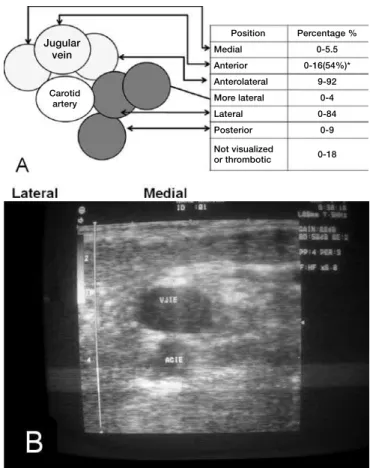
arterial puncture, pneumothorax, hematoma and to a large degree , failure in catheter insertion of up to 35%. A series of factors are related to this percentage, among them operator experience and the patient’s anatomical aspects (morbid obesity, coagulopathy, urgency of the procedure, etc.) Noteworthy about anatomy and posi-tion of the internal jugular vein in relaposi-tion to the carotid artery, discloses that 50% of the times it (jugular vein) is positioned anterior to the carotid artery.(13-17) With
use of US its location can be assessed, as well as the diameter and adverse conditions such as venous throm-bosis, facilitating insertion and or choice of puncture site (Figures 1 and 2).
Position Percentage %
Medial 0-5.5
Anterior 0-16(54%)* Anterolateral 9-92 More lateral 0-4
Lateral 0-84
Posterior 0-9
Not visualized
or thrombotic 0-18 Jugular
vein
Carotid artery
figure 1 – A - Anatomical variation of the jugular vein(13-16);
(*) 54% are located anterior to the carotid artery; B - Ultra-sound image site of the internal jugular vein.
Based on two meta analyses,(18-19) the American Agency
for Healthcare Research and Quality) published in 2001 a recommendation for use US guided cannulation. It is one of the 10 main practices of safety ands improves patients’ care.(20) US guided venous cannulation prevents one
punc-ture accident for every seven central accesses (number needed to treat (NNT=7) and prevents one case of failure in the insertion for every ive trials (NNT= 5). Similar to recommendations of the North American agency, the British National Institute for Clinical Excellence included in 2004, these recommendations in its guidelines.(21)
An-other factor related with this procedure would be the cost/ efectiveness analysis of implementing training of a team and acquisition of the equipment, Reviewing literature, we found the work of Calvert and Hind,(22) who by an
analyti-cal model of cost/efectiveness for each thousand catheters inserted, saved 2 thousand sterling pounds, when com-pared to usual insertion. Development of new equipment and cost reduction in a near future will foster utilization of US as a routine practice in the intensive care setting.
high resolution and good penetration in tissue. However, as described in literature any type of probe may be used including the transvaginal (Figures 3 and 4).(23-25)
Difer-ences between probes are based upon positioning of the quartz crystals and emission of US waves at diferent fre-quencies (Hertz) and distance between them. he higher the probe’s frequency, the greater the resolution, however lesser the depth. he lower the frequency, the greater the depth, the lesser the resolution. he possibility to change frequency and distance of the waves in the same equip-ment must be highlighted.
Insertion through the US may be guided by two wave modes: the B mode (Bright) or through the Dop-pler mode, transformation of US waves relected from a moving object, for instance, blood into an audio or color signal. Due to lack of availability of US with Doppler mode in all equipment, a longer learning curve and a
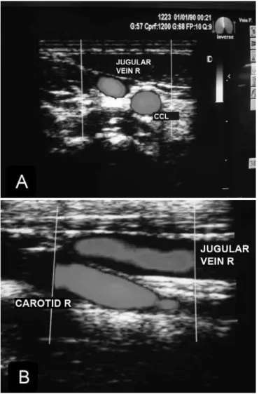
figure 2 – A - Cross sectional incidence with color doppler (short axis); B - longitudinal incidence with color doppler (long axis).
figure 3 – Types of probes, respectively: microconvex (2.5 MHz), linear (5-10MHz), convex (3.5-5.0MHz), transvaginal.
longer time of insertion, our review is restricted to the two-dimensional mode.
he technique must follow the same steps of conven-tional cannulation of the central venous access: operator and patient asepsis and antisepsis, placement of sterile drapes, use of sterile devices protecting the probe (sterile glove or speciic device for probe, placement of sterile gel between interface of the probe of patient body surface, between probe and sterile device to ease wave propaga-tion and reduce artifacts). he technique may be carried out with two operators, that is to say, one positioning the probe and the other doing the puncture or one operator performing the entire procedure.
he static puncture technique,(26) that is to say, the
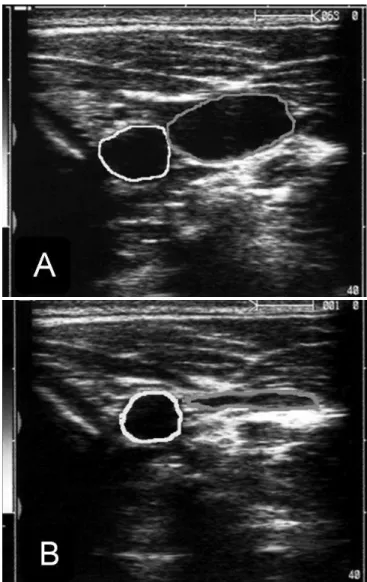
anatomical assessment of the vein in which the catheter will be inserted and outline of the puncture point are car-ried out. Once the puncture point is outlined, puncture is performed. Another way to perform puncture is the dy-namic technique, as it permits real time visualization of the catheter with immediate assessment of complications. In both techniques, orientation of the probe in relation to the anatomic structures may be with a cross-sectional axis (short axis) (Figure 4) or longitudinal axis (long axis) (Figure 5A). he diferentiation of the anatomical struc-tures in this case, the visualization of the jugular vein and carotid artery are made through compression movements, using the probe (compressibility sign of the venous sys-tem) (Figure 5B).
Diferentiation between both structures must not rely only on pulsatility, because not everything that pulsates is an artery. Other form to diferentiate these vessels is com-pression of the liver (hepatojugular relux) and by observ-ing engorgement of the venous system. his method (US) permits to assess thrombosis of the potential puncture segments, thereby avoiding complications. Some bedside maneuvers facilitate puncture: Trendelemburg position, Valsalva maneuver (increase of intrathoracic pressure), among others.
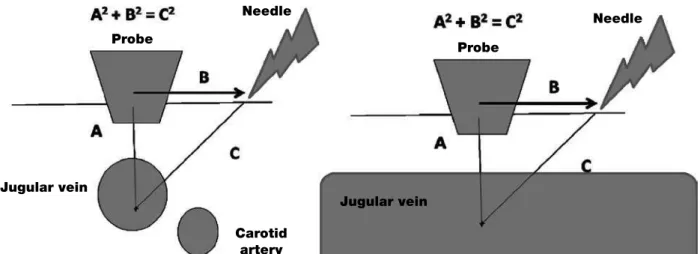
he vein must be positioned in the center of the monitor and needle insertion at a 45 degrees angle in relation to the probe and equidistant from this with the vein must be carried out, similar to the Pythagorean he-orem (Figure 6).
Once skin is overcome in the cross-sectional direction with the needle, it must progress towards the anterior wall of the vein, with its movement as a reference. Non-visualization of the needle or non-Non-visualization of the movement of structures on the monitor, probably points to non-alignment of the probe in relation to needle inser-tion. In this case, the needle should be pulled back and a
figure 5 – A - Cross sectional axis (short axis) showing the right carotid artery in yellow and the internal jugu-lar vein in green; B - Signs of collapse of the internal jugular vein upon performance of probe compression (compressibility sign).
new angulation and insertion must be made. After assur-ing that the needle is inside the vein, by blood aspiration with the plunger, the later is withdrawn and the line is in-troduced by the usual technique (Seldinger). Advantage of the cross- sectional axis (short axis) technique is lesser time of the learning curve and possibility to visualize smaller veins. Nevertheless, ACEP(5) recommends the longitudinal
figure 6 – A - Needle positioning and insertion, cross-sectional axis; B - longitudinal axis.
figure 7 – guide-line insertion longitudinal axis.
he internal jugular vein is the preferred access way for ultrasound-guided venous cannulation, however the subclavian vein has some peculiarities. Visualization of the subclavian vein by infraclavicular projection is made dii-cult by the clavicular acoustic shadow, thereby the
dynam-ic puncture technique is unviable. he statdynam-ic technique should be used, outlining the puncture site. An alternative would be puncture by the supraclavicular pathway at the emergence of the subclavian vein with the jugular vein. However, it is not often used today, due to needle orienta-tion towards the pleura and elevated chances of puncture accidents. An approach described in literature is puncture of the axillary vein as alternative to the subclavian vein, since it is easily visualized with US, more laterally located, associated with a longer distance between the probe and clavicula make dynamic insertion easier, with lower com-plication rates than the traditional method.(29-31)
Peripheral veins should be used as irst option in an emer-gency setting, however, some clinical situations, for instance, edematous patient, obesity, among others, make the insertion diicult. US, preferentially the linear ones (high resolution and discernment of structures with less than 1mm) make possible catheterization, using cross- sectional or longitudinal axes of basilica, cephalic and or axillary veins. Care must be given to the pressure applied on the probe to avoid collapse of the structure to be visualized (Figure 8).
figure 8 – A - performance of the procedure by the nursing team; B - Anatomic location cross sectional axis; C - Compressibi-lity maneuver of the venous system; D - portrait of the venous system.
Needle
Needle Probe
Probe
Jugular vein
Jugular vein
Carotid artery
CONClUSION
Utilization of ultrasound warrants precise visual-ization of the target, direct visualvisual-ization of the needle and guide-line’s progression, reduces puncture at-tempts, improves success rates of insertion, minimizes catheter related complications and reduces insertion time, mainly in patients with a difficult vascular ac-cess. Therefore, the disadvantages of equipment cost, team training time and interpersonal barriers repre-sent obstacles to be overcome in view of the method’s benefits.
ReSUMO
A utilização da ultrassonograia inserida no contexto de tera-pia intensiva possibilita a realização de procedimentos invasivos e diagnósticos beira-leito. Através da portabilidade dos equipa-mentos atuais, associado ao treinamento da equipe, sua imple-mentação assegura diminuição das complicações relacionada á inserção assim como segurança aos pacientes. Sua implemen-tação reduz complicações graves relacionada ao cateter, como pneumotórax e lesão vascular, entre outras. Provavelmente, em um futuro próximo, com a facilitação de aquisição de equipa-mentos de ultra-som e treinamento adequado da equipe, esta ferramenta se tornará indispensável na prática clinica diária.
Descritores: Ultra-sonograia; Ecocardiograia; Cuidados intensivos
RefeReNCeS
1. Lichtenstein DA. General ultrasound in the critically ill. Berlin: Springer-Verlag; 2004.
2. Beaulieu Y, Marik PE. Bedside ultrasonography in the ICU: part 2. Chest. 2005;128(3):1766-81.
3. Beagle GL. Bedside diagnostic ultrasound and therapeutic ultrasound-guided procedures in the intensive care setting. Crit Care Clin. 2000;16(1):59-81.
4. Price S, Nicol E, Gibson DG, Evans TW. Echocardio-graphy in the critically ill: current and potential roles. Intensive Care Med. 2006;32(1):48-59. Comment in: In-tensive Care Med. 2006;32(8):1283. InIn-tensive Care Med. 2006;32(1):9-10.
5. American College of Emergency Physicians. American Col-lege of Emergency Physicians. ACEP emergency ultrasound guidelines - 2001. Ann Emerg Med. 2001;38(4):470-81. 6. European Federation of Societies for Ultrasound in
Me-dicine and Biology. Minimum training requirements for the practice of medical ultrasound. Eur J Ultrasound. 2006;27(1):79 –105.
7. World Interactive Network Focused on Critical Ultra-sound. (WINFOCUS). Ultrasound in critical care medi-cine - Continuing medical education. [cited 2008 Jan 1]. Available from: http://www.winfocus.org/usccm .
8. Vignon P, Goarin JP, editors. Echocardiographie doppler en réanimation, anesthésie et médecine d’urgence. Paris: Elsevier; 2002
9. 9. Sloth E. Echocardiography in the ICU. Intensive Care Med. 2006;32(8):1283. Comment on: Intensive Care Med. 2006;32(1):48-59. Intensive Care Med. 2006;32(1):9-10. 10. Denault AY, Couture P, McKenty S, Boudreault D, Plante F, Perron R, et al. Perioperative use of transesophageal echocardio-graphy by anesthesiologists: impact in noncardiac surgery and in the intensive care unit. Can J Anaesth. 2002;49(3):287-93. 11. Vieillard-Baron A, Slama M, Cholley B, Janvier G, Vignon P.
Echocardiography in the intensive care unit: from evolution
to revolution? Intensive Care Med. 2008;34(2):243-9. Com-ment in: Intensive Care Med. 2008;34(2):215-7.
12. McGee DC, Gould MK. Preventing complications of central venous catheterization. N Engl J Med. 2003;348(12):1123-33. Review.
13. Gordon AC, Saliken JC, Johns D, Owen R, Gray RR. US-gui-ded puncture of the internal jugular vein: complications and anatomic considerations. J Vasc Interv Radiol. 1998;9(2):333-8. Comment in: J Vasc Interv Radiol. 1998;9(5):854-5. 14. Turba UC, Ulacker R, Hannegan C, Selby JB. Anatomic
relationship of the internal jugular vein and the common ca-rotid artery applied to percutaneous transjugular procedures. Cardiovasc Intervent Radiol. 2005;28(3):303-6.
15. Caridi JG, Hawkins IF Jr, Wiechmann BN, Pevarski DJ, Tonkin JC. Sonographic guidance when using the right inter-nal jugular vein for central vein access. AJR Am J Roentgenol. 1998;171(5):1259-63.
16. Troianos CA, Kuwik RJ, Pasqual JR, Lim AJ, Odasso DP. In-ternal jugular vein and carotid artery anatomic relation as de-termined by ultrasonography. Anesthesiology. 1996;85(1):43-8. Comment in: Anesthesiology. 1996;85(6):1501. Anesthe-siology. 1996;85(6):1502-3.
17. Legler D, Nugent M. Doppler localization of the internal ju-gular vein facilitates central venous cannulation. Anesthesio-logy. 1984;60(5):481-2.
18. Randolph AG, Cook DJ, Gonzales CA, Pribble CG. Ultra-sound guidance for placement of central venous catheters: a me-ta-analysis of the literature. Crit Care Med. 1996;24(12):2053-8. Comment in: ACP J Club. 1997;126(3):75.
19. Hind D, Calvert N, McWilliams R, Davidson A, Pais-ley S, BeverPais-ley C, homas S. Ultrasonic locating devi-ces for central venous cannulation: meta-analysis. BMJ. 2003;327(7411):361.
20. Making health care safer: a critical analysis of patient safe-ty practices [Internet]. 200. [cited 2008 June 12]. Available from: http\\www.ahrq.gov\clinic\ptsafety\ .
49: the use of ultrasound locating devices for placing central venous catheters [Internet]. 2004. [cited 2008 June 12]. Avai-lable from: http://www.nice.org.uk/pdf/inal_CVC_place-ment_survey_report.pdf.
22. Calvert N, Hind D, McWilliams RG, homas SM, Beverley C, Davidson A. he efectiveness and cost-efectiveness of ul-trasound locating devices for central venous access: a systema-tic review and economic evaluation. Health Technol Assess. 2003;7(12):1-84.
23. Phelan MP. A novel use of the endocavity (transvaginal) ultra-sound probe: central venous access in the ED. Am J Emerg Med. 2003;21(3):220-2.
24. Milling TJ Jr, Rose J, Briggs WM, Birkhahn R, Gaeta TJ, Bove JJ, Melniker LA. Randomized, controlled clinical trial of point-of-care limited ultrasonography assistance of central venous cannulation: the hird Sonography Outcomes Assessment Program (SOAP-3) Trial. Crit Care Med. 2005;33(8):1764-9. Comment in: Crit Care Med. 2005;33(8):1875-7. 25. Untracht SH. Progress in central venous access. Crit Care
Med. 1996; 24(1):183-4.
26. Denys BG, Uretsky BF, Reddy PS. Ultrasound-assisted
can-nulation of the internal jugular vein. A prospective compari-son to the external landmark-guided technique. Circulation. 1993; 87(5):1557-62.
27. Maury E, Guglielminotti J, Alzieu M, Guidet B, Ofenstadt G. Ultrasonic examination: an alternative to chest radiogra-phy after central venous catheter insertion? Am J Respir Crit Care Med. 2001;164(3):403-5.
28. Lichtenstein D, Mezière G, Biderman P, Gepner A. he “lung point”: an ultrasound sign speciic to pneumothorax. Intensi-ve Care Med. 2000;26(10):1434-40.
29. Sharma A, Bodenham AR, Mallick A. Ultrasound-guided infraclavicular axillary vein cannulation for central venous access. Br J Anaesth. 2004; 93(2):188-92. Erratum in: Br J Anaesth. 2004;93(5):752. Comment in: Br J Anaesth. 2004;93(2):175-7.
30. Sandhu NS. Transpectoral ultrasound-guided catheterization of the axillary vein: an alternative to standard catheterization of the subclavian vein. Anesth Analg. 2004;99(1):183-7. 31. Use of ultrasound imaging by emergency physicians.