Vol. 44, N. 3 : pp. 283 – 289, September, 2001
ISSN 1516-8913 Printed in Brazil
BRAZILIAN ARCHIVES OF
BIOLOGY AND TECHNOLOGY
A N I N T E R N A T I O N A L J O U R N A L
Coprophilous Fungi from Brazil
Michael J. Richardson
165 Braid Road, Edinburgh Eh10 6je U.K.
ABSTRACT
Thirty-two species of coprophilous fungi were recorded from seven dung samples collected from the state of Matto Grosso do Sul, Brazil, and incubated in moist chambers. Descriptions of some of the more interesting fungi are given, and aspects of their biodiversity and ecology are discussed.
Key words: Ascomycetes, coprophilous fungi, diversity, species richness
INTRODUCTION
Coprophilous fungi may be useful indicators of habitat diversity (Richardson, 2001). During a visit to Brazil in 1998, seven samples of herbivore dung were collected from the Bonito and Pantanal do Rio Negro areas (Matto Grosso do Sul) and incubated, on return to the U.K., in a damp chamber. The coprophilous fungi that developed were recorded. Lundqvist (1972) and Van Brummelen (1967), in their monographic treatments of the Sordariaceae and Ascobolus and
Saccobolus, noted some early records but, apart
from the collections of Andre de Meijer (personal communication) and Jahn (2000), there seem to be few recent records of coprophilous fungi from Brazil. Da Silva & Minter (1995), in their compilation of fungi recorded by Batista and co-workers, noted only two coprophilous ascomycetes, Sordaria fimicola and Ascobolus
scatigena (as A. notatus). Details of the fungi
found from the 1998 collection of samples are provided and discussed, together with some aspects of the diversity and species richness of the dung habitat in a small area of Brazil.
MATERIAL AND METHODS
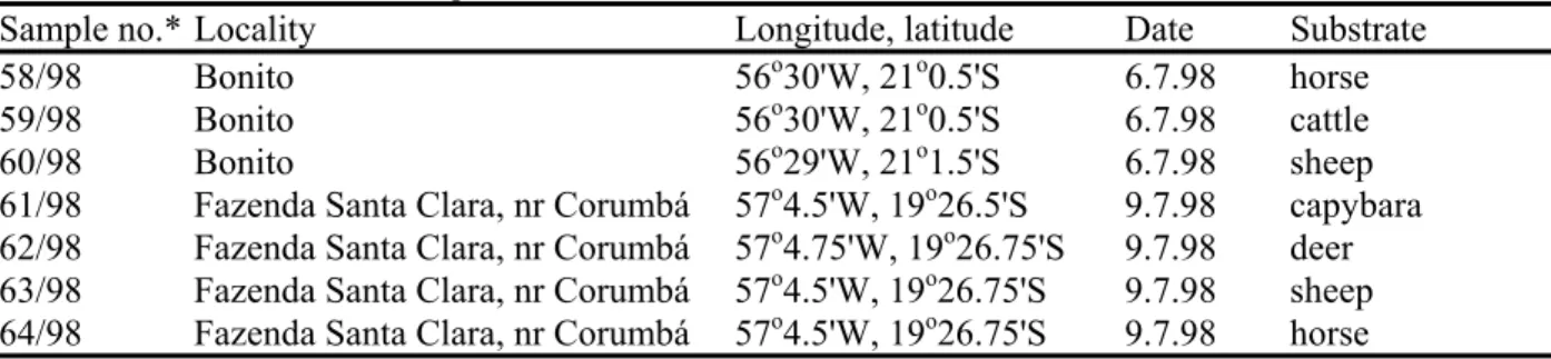
Table 1 - Details of Brazilian samples and collection localities.
Sample no.* Locality Longitude, latitude Date Substrate 58/98 Bonito 56o30'W, 21o0.5'S 6.7.98 horse 59/98 Bonito 56o30'W, 21o0.5'S 6.7.98 cattle 60/98 Bonito 56o29'W, 21o1.5'S 6.7.98 sheep 61/98 Fazenda Santa Clara, nr Corumbá 57o4.5'W, 19o26.5'S 9.7.98 capybara 62/98 Fazenda Santa Clara, nr Corumbá 57o4.75'W, 19o26.75'S 9.7.98 deer 63/98 Fazenda Santa Clara, nr Corumbá 57o4.5'W, 19o26.75'S 9.7.98 sheep 64/98 Fazenda Santa Clara, nr Corumbá 57o4.5'W, 19o26.75'S 9.7.98 horse
* MJR collection no./year
An estimate of comparative species richness was made by constructing a cumulative species curve and deriving the equation for that curve (y = axb) and solving for x = 50 samples, and comparing with values obtained from a worldwide study of a similar range of substrates (Richardson, 2001).
RESULTS AND DISCUSSION
The seven samples provided a total of 75 records of 32 species. These records, although based on a very small number of samples, point to the high diversity of Brazilian coprophilic fungi. The mean species richness of 10.7 per sample is within the range of values of 9-12 obtained for various herbivore mammalian dungs examined in other samples from a worldwide collection of over 400 samples (Richardson, 2001). That study used cumulative species curves to compare the species richness of an area or substrate by calculating the number of species expected to be recorded from a standard number (50) of samples of dung. To provide a confident estimate of species richness a minimum sample size of 40-50 is recommended, and estimates based on smaller subsets may give a less accurate value (Richardson, 2001). Bearing this in mind, and using the very small data set from the seven samples in this study, an estimate of species richness of the Brazilian coprophilous mycobiota of the groups studied here is 140 species/50 samples (Fig. 1). This value can be compared with other values from the worldwide study (Richardson, 2001) of ca 150 (from 30oS– 40oN), ca 123 (from 40o –50oN), and ca 100 (from 50o – 60oN). The worldwide study showed that coprophilous fungi demonstrate a gradient of increasing species richness with decreasing latitude, a phenomenon that is well documented
y
= 7.78
x
0.74R
2= 0.96
0
5
10
15
20
25
30
35
0
2
4
6
Sample no.
No. of species
Figure 1 - Cumulative total of taxa observed on seven successive samples of dung collected in Brazil and incubated in moist chambers.
for other faunal and floral groups. The Brazilian estimate is as expected for its latitude, and can be contrasted with a low value of 80 species/50 samples obtained from thirteen samples collected from Morocco (30oN), from an area identified subjectively as environmentally degraded (Richardson, 2001).
Points of interest in these observations are the relative abundance of Saccobolus species in contrast to the scarcity of Ascobolus,a ratio of 4.3 : 1, compared to a value of 0.7 : 1 found in a worldwide study of 418 other samples; the absence of any Schizothecium spp.; the relatively low frequency (about 80 % of the records which might have been expected, based on data from a larger study) and poor diversity of Sporormiella spp., with S. minima common, as it is generally, and only one occurrence of another species; and the high frequency of Podospora communis, P.
pauciseta and Zygopleurage zygospora, the latter
RECORDS
Zygomycotina :-
Pilobolus crystallinus(Wigg.) Tode
MJR60, 62/98.
Pilobolus sphaerosporus (Grove) Palla
MJR60, 61, 64/98.
Ascomycotina :-
‘Discomycetes’ :-Ascobolus immersus Pers.
A common and distinctive Ascobolus, especially on ruminant dung, and characterised by very large ascospores (51.5-58 x 29-32 µm in the Brazilian material). MJR 61, 62, 64/98. Recorded from Rio Grande do Sul, Brazil in 1936, and from Argentina (Van Brummelen, 1967).
Coprotus lacteus (Cooke & W. Phillips) Kimbr.,
Luck-Allen & Cain
Apothecia pure white, 200-300 µm diam. Asci long-clavate, 8-spored, 65-100 x 13-16 µm. Spores 1-2-seriate, ellipsoid, hyaline, 8-11.5 x 6.5-7 µm. Paraphyses hyaline, non-capitate. MJR 58, 64/98.
Iodophanus carneus(Pers.) Korf
One of the commonest species of Pezizaceae on dung worldwide, especially on sheep and cattle dung, and recognised by its pale fleshy apothecia, KI +ve asci, and ellipsoid, hyaline, minutely verrucose ascospores, 18-21 x 11-12.5 µm in the two Brazilian collections. MJR62, 63/98.
Ryparobius polysporus (P. Karst.) Speg.
MJR 63/98.
Saccobolus citrinus Boud. & Torrend
Apothecia yellow. Ascus 120 x 38 µm. Spore mass 42-48 x 16-17 µm. Spores in a 4 x 2 arrangement when young (sect. Saccobolus), smooth or very minutely verrucose, truncate, 17.5-19.5 x 6.5-10 µm. Paraphyses with yellow contents. MJR 58, 61, 62, 63/98. Recorded from Rio Grande do Sul, Brazil in 1936 (Van Brummelen, 1967)
Saccobolus depauperatus (Berk. & Broome) E. C.
Hansen
Apothecia hyaline violaceous, <420 µm diam. Asci 80 x 19-20 µm. Spore mass 29-32 x 9.5-12 µm. Spores in a 3+3+2 arrangement (sect.
Eriobolus), smooth to minutely verrucose, 11-11.5
x 6-6.5 µm. MJR 61/98. Recorded from Minas Gerais, Brazil in 1934 (Van Brummelen, 1967).
Saccobolus truncatus Velen.
Apothecia very small, <350 µm diam. Asci ± globular-clavate, 64-80 x 29-32 µm, with a distinct stalk. Spore mass 38-45 x 16 µm when immature, becoming shorter, ± rounded with maturity, 29 x 22.5 µm. Spores in a 4 x 2 arrangement when young, irregularly arranged at maturity, episporium smooth and violet when young, rougher brown when old, slightly fusoid-truncate, 16-19 x 9-10 µm. Paraphyses with yellowish contents, very slightly clavate. MJR 62-64/98. A widespread but overlooked species, recorded from Peru in 1945 (Van Brummelen, 1967).
Saccobolus verrucisporus Brumm.
Apothecia very small, 150-200 µm diam., with only 10-12 ripe asci present at a time. Asci 125 x 29 µm. Spore mass 32-38 x 16 µm. Spores in a 3+3+2 arrangement, coarsely warted, 12.5-16 x 8-9.5 µm. Paraphyses hyaline, very slightly clavate. MJR 62-63/98. Van Brummelen (1967) described
S. verrucisporus from deer dung from New
Guinea, and I am not aware of any other records.
Saccobolus versicolor (P. Karst.) P. Karst.
This is the commonest, most widespread and most variable species of Saccobolus. The Brazilian specimens were typical, with hyaline to violaceous apothecia, asci 110-150 x 32-38 µm, and spore masses 38-48 x 16-20 µm. Spores arranged in a 3+3+2 pattern, violaceous, becoming brown with age, rough, 14.5-19 x 7-10 µm. MJR 62-64/98.
Unitunicate ‘pyrenomycetes’: -
Cercophora mirabilis Fuckel
Perithecia schizothecioid, with small scales of thin-walled inflated cells around the neck and flexuous-hairy below. Immature vermiform spores 48-58 µm long, with caudae of equal length, 45-50 µm long. Mature spores not well differentiated, but with pigmented cell ca 19 x 8 µm and hyaline pedicel ca 26 x 4 µm. These spores are slightly smaller than those described for C. mirabilis by Lundqvist (1972) and, but for the equal length of the apical caudae, the collections are nearer to C.
anisura N. Lundq. MJR 59(M), 60-61/98.
C. mirabilis and Jahn (2000) reported Cercophora
cf. coronata from capybara dung from Rio de
Janeiro.
Phomatospora minutissima (H. Crouan & P.
Crouan) N. Lundq.
Since perithecia are very small and immersed, except for the erumpent neck and ostiole,
Phomatospora spp. are not often recorded.
Perithecia 120-180 µm diam. Asci cylindrical, 50-70 x 4-6 µm, with a KI -ve pore. Spores obliquely uniseriate, hyaline, ellipsoid, 4-6.5 x 2.5-3 µm. MJR 59(M), 62(M), 63(M)/98.
Podospora argentinensis (Speg.) J. H. Mirza &
Cain
Limited material, originally determined as P.
decipiens, of P. decipiens-like perithecia, but with
rather small ascospores, 29-32 x 16 µm, although the characteristic, but ephemeral, distal secondary appendage at the tip of the primary appendage was not noted. MJR 64/98. P. argentinensis was described from Argentina, and has also been recorded from Mexico and the USA. P. decipiens
is generally common, but especially on cattle and sheep dung in more temperate regions, and Lundqvist (1972) had not verified any material of
P. decipiens from the tropics.
Podospora communis (Speg.) Niessl
One of a small group of species that has spores with four apical secondary appendages. Spores are biseriate, 29-32 x 19-19.5 µm, with an apical germ pore and basal primary appendage, which is slightly clavate and often curved, 22-26 x 6 µm. The four apical secondary appendages are curved, claw-like, 12-15 µm long. MJR 58-61(M), 63-64(M)/98. Recorded from Minas Gerais, Brazil in 1934, and from Argentina (Lundqvist, 1972).
Podospora inflatula Cain
Perithecia with short hairs (<50 µm long), paler and very slightly swollen towards their apex. Asci 8-spored, ca 220 x 30 µm. Ascospores 1-2 seriate, 25.5-32 x 14.5-19 µm, with primary appendage 16-26 x 3-4 µm, and a simple apical secondary appendage ca 25 x 4 µm. Some spores had a single basal secondary appendage at the tip of the primary appendage, but other spores were seen with 2-3 short secondary appendages that appeared to be attached laterally to the primary appendage. MJR 61/98. The details of this collection agree
well with Mirza & Cain’s (1969) description of P.
inflatula, apart from the atypical basal secondary
appendages noted in some spores. It is recorded from Brazil, Mexico and the Society Islands (Mirza & Cain,1969).
Podospora pauciseta(Ces.) Traverso
Perithecial necks with asymmetrically arranged tapering tufts of setae, composed of fascicles of non-inflated hyphae. Asci 4-spored, fusoid-clavate, tapering to a long sinuous stalk, 190-250 x 23-29 µm. Spores 31-38 x 16-20 µm, with primary appendage 19-30 (45) x 4-5 µm. MJR 58, 60-61(M), 62, 63-64(M)/98.
A common and widespread species recorded in several south American countries, including Brazil (Mirza & Cain, 1969; Jahn, 2000, both as P.
anserina; and Lundqvist, 1972). The majority of
my records are from less temperate areas (<40o N or S), but Mirza & Cain (1969) and Lundqvist (1972) cite many records from more northerly latitudes.
Podospora sp.
Limited material of a large-spored species that could not be named. Perithecia were globose, ± immersed, some without distinctive scales, hairs or setae, others with long brown flexuous hyphal hairs and some P. decipiens-type papillae. Spores 48-54.5 x 21-27 µm, with the primary appendage slightly clavate distally, 22-30 x 6-7 µm. Secondary appendages difficult to see, either in the ascus or on free spores, but the short simple apical appendage had a fibrillose appearance, rather like the illustration of P. ostlingospora in (Mirza & Cain,1969). MJR 60/98.
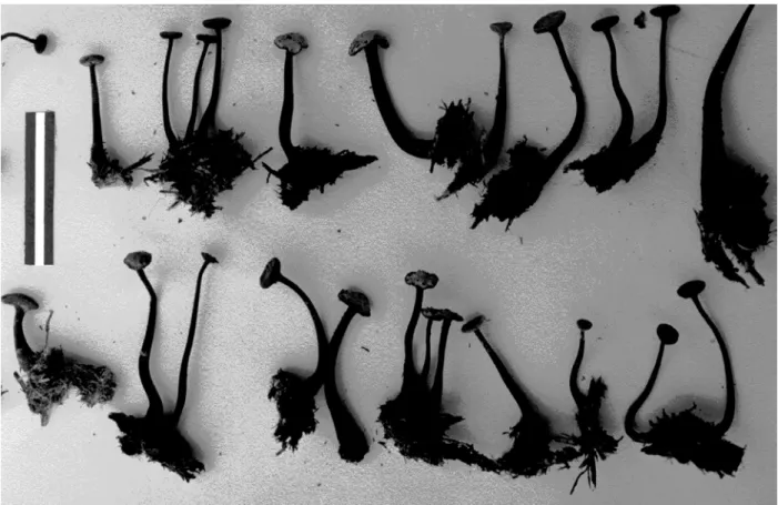
Poronia oedipus (Mont.) Mont.
A good collection of fertile stroma was found on horse dung in the field (Figs 2, 3). This Poronia
appears to be frequent in tropical areas, but a detailed description of the Santa Clara collection is given. MJR 64(M)/98.
Asci (110) 130-170 x 29 µm, widest in the lower half when the lowest 2-5 spores are biseriate, or 20
µm diam. if uniseriate, with short stalk and marked apical plug 9.5 µm diam. x 6 µm composed of 3-4 rings or discs, intense blue in KI. Spores mostly partially biseriate, black, ellipsoid, 21-29 x 12.8-18 µm (L/D 1.4-2.3), with germ slit 12-16 µm long and a 2-3 µm wide gel sheath. Paraphyses hyaline
ca 2 µm diam.
Figure 2- Stromata of Poronia oedipus on horse dung. Coin = 23 mm diam.
Selinia africana R. S. Khan & J. C. Krug
Macroscopically similar to S. pulchra, with superficial reddish orange stromata, 2-3 mm diam for stroma with a single perithecium, larger for stroma with multiple perithecia, but distinguished by its smaller ascospores, 35.3-41.7 x 16-19.3 µm. MJR 60(M)/98. S. africana was described by Khan & Krug (1989) from Africa, New Zealand and Venezuela, and I have two records from South Australia. It seems to be less frequent than S.
pulchra, but Selinia spp. generally seem to be
infrequent, and this is possibly the first record of
S. africana from Brazil.
Selinia pulchra (G. Winter) P. Karst.
Ascospores 54.5-61 x 22.5-24 µm. MJR 60/98.
Zygopleurage zygospora (Speg.) Boedijn
The particular feature of this fungus is its large spores, which have two pigmented cells (22.5-) 32-38.5(-42) x (10-)14.5-19 µm separated and joined by a long, hyaline cell (95-)110-170 µm long x 6 µm diam. at the insertion with the pigmented cells, narrowing/collapsing to 3-4 µm but slightly inflated centrally. Each pigmented cell has four short claw-shaped hyaline apical secondary appendages, 15-20 µm long. The extreme values for spore size, in brackets above, were observed in the specimens from sample 58/98, which were very variable. Perithecia are smooth, matt black, pyriform-globose, superficial,
ca 400-500 µm diam. x 750 µm high. Immature asci are clavate, ca 250 x 50 µm, and with the cluster of spores spirally arranged. Delgado et al.
(2000) reported Z. zygospora from Venezuela, and Lundqvist (1972) notes that it is not uncommon in Europe and N. America, but I have not found any other records from Brazil or S. America. MJR 58, 59-61(M), 64(M)/98.
Bitunicate ‘pyrenomycetes’: -
Sporormiella cf. megalospora (Auersw.) S. I.
Ahmed & Cain
Limited material was observed, with highly variable and distorted spores. The few ‘normal’ 4-celled spores were 80-90 x 19 µm, had round ends, germ slits almost parallel to the long axis of the spore, and were deeply constricted at the septa, readily breaking up into component cells. Asci were gradually tapered towards the base. On balance these features suggest an atypical S.
megalospora. MJR 59/98.
Sporormiella minima (Auersw.) S. I. Ahmed &
Cain
One of the commoner Sporormiella species generally, with a tendency to be more frequent at lower latitudes and on cattle dung. It is characterised by small (29-32 x 4-5 µm) cylindrical, 4-celled ascospores that tend to break into two 2-celled halves in the ascus or after liberation. MJR 58-60, 62-64/98.
Basidiomycotina:
-Coprinus cordisporus Gibbs
Basidia 4-spored, spores flattened, ellipsoid in two planes, cordate in the third, 6.5-7 x 6.5-7 x 4 µm. MJR 58, 61/98.
Coprinus curtusKalchbr.
Cap reddish, setulose, the setules brown and capitate. Spores 10-12.5 x 6 µm. MJR 64/98.
Coprinus heptemerus M. Lange & A. H. Sm.
Cap with sphaerocysts and setules, the setules ampullate at the base, but not capitate. Spores ellipsoid, 6-9 x 3.5-4.5 µm, germ pore slightly excentric. MJR 60, 62/98.
Coprinus pellucidus P. Karst.
Small setulose basidiomes. Cap setules ca 30 µm long, not or very slightly capitate. Basidia 4-spored. No facial cystidia. Spores ellipsoid, 9-10 x 5 µm. MJR 60/98.
Coprinus radiatus (Bolton) Fr.
Typical sect. Lanatuli, with a veil of long-celled inflated hyphae. Spores 9.5-10 x 6 µm. MJR 60/98. The basidiospores are intermediate in size between those of C. radiatus and C.
pseudoradiatus, but on balance are nearer those of
C. radiatus.
Coprinus stercoreus (Bull.) Fr.
This is the commonest Coprinus species developing on dung worldwide when incubated in moist chambers, and is characterised by its bright white veil of globose cells and small (7-8 x 4-4.5 µm) ellipsoid spores. MJR 58, 62, 64/98.
Coprinus sp.
views, slightly hexagonal in others. Basidia 4-spored. No facial cystidia. MJR 64/98.
Cyathus stercoreus (Schwein.) De Toni
Mature fruit bodies of this ‘bird’s nest fungus’ were present in the field. MJR 64/98.
ACKNOWLEDGEMENTS
I am grateful to my son, Andrew, for translating the Portuguese summary.
RESUMO
Sete amostras de esterco, que foram recolhidas do Estado do Mato Grosso do Sul no Brasil e incubado em câmaras humidas, renderam trinta e duas espécie de fungos estercorários. Descrições dos fungos mais interessantes são fornecidas e são discutidos aspectos de biodiversidade e ecologia destes.
REFERENCES
Da Silva, M. and Minter, D. W. (1995), Fungi from Brazil recorded by Batista and co-workers. Mycological Papers, 169, 1-585. CABI, Wallingford, U.K.
Delgado, A.; Kimbrough, J. W. and Hanlin, R. T. (2000), Zygopleurage zygospora, a new record from Venezuela. Mycotaxon, 75, 257-263
Jahn, E. (2000), Pyrenomyceten von Dungkulturen aus Gebeiten auserhalb Deutschlands. Zeitschrift für Mykologie, 66, 79-94
Khan, R. S. and Krug, J. C. (1989), A new species of Selinia. Mycologia 81, 653-655
Lundqvist, N. (1972), Nordic Sordariaceae s. lat. Symbolae Botanicae Upsalienses XX, 1-374 + pl. 1-63
Mirza, J. H. and Cain, R. F. (1969), Revision of the genus Podospora. Canadian Journal of Botany, 47, 1999-2048
Richardson, M. J. (2001), Diversity and occurrence of coprophilous fungi. Mycological Research, 105 (in press)
Van Brummelen, J. (1967), A world monograph of the genera Ascobolus and Saccobolus. Persoonia, Supplement 1, 1-260 + 17 pl.