The organization of flash electroretinography unit in Veterinary Medicine
Cristiane dos Santos Honsho1 Arianne Pontes Oriá1 Luiz Paulo da Veiga Monteiro Lazaro Júnior2
Francisco Dorea Neto1 José Luiz Laus3
Organização de uma unidade de eletrorretinografia flash em Medicina Veterinária
1PhD candidate, Veterinary Surgery Program, Faculty of Agronomic and Veterinary Sciences (FCAV), Sao Paulo State University
(UNESP), Campus de Jaboticabal.
2Ophthalmologist Physician, Oftalmoclínica Copacabana, Rio De Janeiro, Rua Xavier da Silveira, 45, sala 1006, 22061-010, RJ,
Brazil, fone: +55 21 521 78 44.
3Professor, Department of Veterinary Clinic and Surgery, Faculty of Agronomic and Veterinary Sciences, UNESP, Campus of
Jaboticabal, Via de Acesso Paulo Donato Castellane, s/n, 14884-900, Jaboticabal, SP, Brazil. phone: +55 16 3209 26 26..E-mail: [email protected]. Corresponding author.
ABSTRACT
Electroretinography is the technique that allows the evaluation of changes in electrical potential that occur when the eye is stimulated by light. Such changes reflect the response of several retinal cells including photoreceptors (cones and rods). Thus it evaluates the retinal functionality and can diagnose abnormalities in retinas seem to be normal by ophthalmoscopy. Due to the constant evolution in veterinary ophthalmology, new centers of retinal electrophysiology have been introduced around the world either for early diagnosis of retinopathies or for preoperative evaluation of animals with cataracts and glaucoma, as well as for continuing research. The Ophthalmology Unit of the “Governador Laudo Natel” Veterinary Teaching Hospital, in the Faculty of Agronomic and Veterinary Sciences (FCAV) – Sao Paulo State University (UNESP) – Jaboticabal Campus, Brazil, supported by the Fundação de Amparo à Pesquisa do Estado de São Paulo – FAPESP (Sao Paulo State Foundation for Research), set up the Unit of Ocular Electrophysiology for animals in order to provide conditions for the diagnosis of retinopathies. As a pioneering venture in Brazil, the organization of the services faced many challenges till the moment it was set up: the organization and arrangement of appropriate rooms, independent electrical distribution for the installation of separate pieces of equipment, adaptation of containment tables, training and qualification of the staff, and the elaboration and standardization of anesthetic and stimulation protocols. The wealth of information generated by our experience gave us the inspiration to write this paper, which aims to contribute to the work of researchers and veterinary ophthalmologists in this new and opportune field of specialization.
Key words: electroretinography, retina, electrophysiology, dogs.
RESUMO
Eletrorretinografia é a técnica que possibilita observar mudanças no potencial elétrico, que ocorrem quando o olho é estimulado pela luz. Tais mudanças refletem a resposta de várias células retinianas, incluindo os fotorreceptores (cones e bastonetes). Com ela, portanto, é possível avaliar a funcionalidade da retina e diagnosticar alterações, mesmo em retinas normais à oftalmoscopia. Em face do crescente desenvolvimento da oftalmologia, no âmbito da Veterinária, novos centros de eletrofisiologia da retina vêm sendo implementados em todo o mundo, tanto para o diagnóstico precoce de retinopatias, como para avaliação pré-operatória de animais com catarata e glaucoma ou, ainda, direcionados à pesquisa. A Unidade de Oftalmologia do Hospital Veterinário “Governador Laudo Natel”, da Faculdade de Ciências Agrárias e Veterinárias (FCAV) – Universidade Estadual Paulista (UNESP) – Campus de Jaboticabal, com o apoio da Fundação de Amparo à Pesquisa do Estado de São Paulo - FAPESP, visando a implementar condições diagnósticas em retinopatias, implantou a Unidade de Eletrofisiologia da Retina para animais. Por ser empreendimento pioneiro, no Brasil, sua organização passou por inúmeras intercorrências até que fosse consignado. Entre elas, a organização e montagem de salas apropriadas, distribuição elétrica independente para a instalação de equipamentos, visando a diminuir interferências externas, adequação de mesas de contenção, treinamento e habilitação de pessoal, elaboração e padronização de protocolos anestésicos e de estimulação, entre outras. A riqueza de informações e as experiência geradas obstinam a conceber este trabalho que visa a contribuir com pesquisadores e oftalmologistas veterinários neste, recente e oportuno, campo de especialização.
INTRODUCTION
The search for ophthalmology units in veterinary medicine grows every year; especially the search for cataract treatment. Except for some specific conditions, the disease commonly is concurrent with diseases of the posterior segment of the globe. The decision to extract the lenses in patients with cataracts depends on the application of a pre-surgical electroretinography. Degenerative retinopathies in dogs have been often reported in connection with cataracts. The retina electrophysiology permits the recognition and characterization of acquired, congenital or hereditary disorders. Thus, the electroretinogram (ERG) permits the obtaining of information concerning the degree of functioning of retinal cells involved in the visual process. Whenever abnormal conditions are detected, it is possible to identify involved cellular elements in order to characterize the visual deficit, its origin, as well as its evolution and prognosis.
RETINA
The retina is the photosensitive film lining of the posterior segment of the globe. As a segment of the nervous system, the retina function is to capture the light stimulus and to transform it into an electrical one, which is conducted to the brain cortex in order to be interpreted (BEDFORD, 1996; SAMUELSON, 1999). The retinal division has basically two layers: the retinal pigment epithelium and the sensory retina or neuroretina (CURTIS & LIGHTFOOT, 1993; BEDFORD, 1996; COOK, 1999; JONES et al., 2000). The neuroretina has a complex structure, composed of photoreceptors and transmission elements disposed in nine layers: the visual cell layer (rod and cone layer), the outer limiting membrane, the outer nuclear layer, the outer plexiform layer, the inner nuclear layer, the inner plexiform layer, the ganglion cell layer, the nerve fiber layer and the inner limiting membrane (SLATTER, 1990; BEDFORD, 1996; SAMUELSON, 1999; JONES et al., 2000).
There are 2 types of light-sensitive cells (photoreceptors): cones and rods (YANASE et al., 1995; SAMUELSON, 1999), which differ from each other in their function, shape and retinal distribution (OFRI, 1999). In dogs the rods predominate over cones (CURTIS & LIGHTFOOT, 1993; MILLER & MURPHY, 1995; SAMUELSON, 1999) and act in low illumination conditions (scotopic vision) (SAMUELSON, 1999). The rods are characterized by poor visual resolution (MILLER & MURPHY, 1995; OFRI, 1999;
SAMUELSON, 1999), and no color perception (OFRI, 1999). They are extremely sensitive to alterations in light levels and motion perception (OFRI, 1999) and are located in larger numbers in the peripheral retina (KOCH & RUBIN, 1972; SLATTER, 1990; SAMUELSON, 1999). Rhodopsin, a rod photo-pigment in dogs, has a peak sensitivity to light with wavelengths between 506 and 510nm, quite common to animals adapted to dim light (ACLAND, 1988; MILLER & MURPHY, 1995).
The cones represent 5% of retinal photoreceptors in dogs (CURTIS & LIGHTFOOT, 1993), and are concentrated in the center of the retina (RUBIN, 1967; KOCH & RUBIN, 1972; KOMMONEM & RAITTA, 1987; SLATTER, 1990; CURTIS & LIGHTFOOT, 1993; SAMUELSON, 1999). They are characterized by a high resolution of fine details, and by color perception; they are rapidly adapted to intense, highly repeated and long wavelength light stimuli (photopic), and present low sensitivity to small fluctuations of light intensity (CARR & SIEGEL, 1990; NARFSTRÖM et al, 1995; OFRI, 1996a; KOMAROMY et al., 1998a; OFRI, 1999). From a morphologic view, dogs are believed to have two types of cones. One has spectral sensitivity to light; in a wavelength between 429nm and 435nm, and the other is sensitive to wavelengths around 550nm (ACLAND, 1988; CHMIELEWSKI, 1995; MILLER & MURPHY, 1995).
ELECTRORETINOGRAPHY
Electroretinography is an electrophysiologic test that permits the detection and recognition of visual diseases (SCHAEPPI & LIVERANI, 1977; KOMAROMY et al, 1998a). The electroretinogram (ERG) allow for the evaluation of the functional integrity of the whole outer portion of the retina and of the pigmented epithelial layer (RUBIN, 1967), and is essential for the early diagnosis of much hereditary retinal degeneration that affects dogs (RUBIN, 1967; AGUIRRE & RUBIN, 1972; AGUIRRE, 1995; SIMS, 1999; ORIÁ et al., 2004).
functional degree of ganglion cells, which allows for the diagnosing of disorders of the innermost layers of the retina, especially those produced by glaucoma (DANTAS et al., 1995; OFRI, 1996b; KOMAROMY et al., 1998b; SIMS, 1999).
FLASH ELECTRORETINOGRAM
Electroretinography is based on both the stimulation of the retina by light, and on the registration of its response (CARR & SIEGEL, 1990; DANTAS et al., 1995). The two basic components to obtain the electroretinogram are the stimulation and the recording system (DANTAS et al., 1995; KOMAROMY et al., 1998b; SIMS, 1999). he photo-stimulator produces light pulses with known wavelength and intensity (DANTAS et al., 1995; SIMS, 1999). It can be put directly in front of a patient, at a distance of 20 or 30cm, or even attached to a dome (Ganzfeld dome) where a
diffuser distributes the light stimuli onto the inner surface of the Ganzfeld dome, with a uniform
stimulation of the retina (OGDEN, 1994; NARFSTRÖM et al, 1995; SIMS, 1999).
The electroretinogram, during retinal stimulation, registers the difference of potential captured by electrodes arranged over the cornea (active electrode), next to the lateral canthus (reference electrode) and on the occipital crest (ground electrode) (ACLAND, 1988; LESCURE, 1993; KOMAROMY et al., 1998b). According to Ohm’s Law, a voltage is produced when electrical current, generated by a potential difference, crosses a resistance (OGDEN, 1994). In this case, the retinal cells generate the electrical current that is captured in the electroretinogram. The resistance that the current has to go through is formed by the retina, cornea and vitreous extra-cellular spaces. When a part of the retina is illuminated, the retinal cells are simultaneously activated. The electrical current originating from the stimulation of retinal cells goes through the extra-cellular spaces — vitreous and cornea, which attribute resistance, generating in this way the voltage that is recorded on the ERG (LESCURE, 1993; OGDEN, 1994). Retinal potentials are known to be small (lower than 1 mV) and difficult to visualize. The signals that are captured by electrodes must go through pre-amplifiers and differential amplifiers before they are recorded (DANTAS et al., 1995; SIMS, 1999).
During electroretinography the patient has to be motionless. So, in veterinary medicine, the necessity of pharmacological containment is understood (KOMMONEM, 1988). However anesthetics attenuate the amplitude of the
ERG-analyzed components. The depressor effect of such drugs causes just a mild alteration of the amplitude of a, b and c waves, especially when retinal responses to low intensity stimulus are studied (KOMMONEM, 1988).
ERG FLASH COMPONENTS
According to ACLAND (1988), CARR & SIEGEL (1990) and LESCURE (1993), the components of the ERG showing clinical importance are: the a-wave (negative depression) and b-wave (positive deflection). The a-wave has its origin at the outer retinal layers, as a result of electrical activity of cones and rods, as well as of other cells (SCHAEPPI & LIVERANI, 1977).
The b-wave, however, originates at the inner retinal layers (SCHAEPPI & LIVERANI, 1977). The main component of the b-waves results from the activation of Müller cells and, less significantly, of bipolar cells (SCHAEPPI & LIVERANI, 1977; LESCURE, 1993; SIMS, 1999). There are also the oscillatory potentials, which are small rhythmic waves from the ascendant phase of the b-wave (SCHAEPPI & LIVERANI, 1977; LESCURE, 1993; SIMS, 1990; SIMS et al., 1991; BROOKS et al., 1992; SIMS, 1999), coming from the inner nuclear layer (SIMS, 1990; SIMS et al., 1991; BROOKS et al., 1992; SIMS, 1999). In the face of high retinal circulation dependence, the sudden increase in intraocular pressure changes the latency and amplitude of such potentials (SIMS, 1999). A third negative deflection wave, named c wave, can be observed as the result of the retinal pigment epithelium electrical activity (PARRY et al., 1953; SCHAEPPI & LIVERANI, 1977). However, under recording conditions used in veterinary medicine, the c-wave is rarely detected (ACLAND, 1988; SIMS, 1990; LESCURE, 1993).
reasons, a rigorous standardization of electroretinography protocols is considered to be of the greatest importance (MILLER, 1993; AGUIRRE, 1995; OFRI, 1996b).
ERG INTERPRETATION
In order to interpret the ERG, three parameters must be considered: latency, implicit time and amplitude. Latency is understood as the time between the stimuli and the beginning of the wave. The implicit time, measured in milliseconds (msec), refers to the time between the initial stimuli and the peak of the wave in question. The amplitude is measured in micro-volts (µv) and represents the measurement from the base to the apex of the wave (CARR & SIEGEL, 1990; LESCURE, 1993; KOMAROMY et al., 1998b; SIMS, 1999).
ENVIRONMENT
The room appointed for the application of the electroretinography should not have any windows, so that it can be easily adapted to darkroom conditions. The room temperature should be a constant 25ºC. The electrical wiring should be independent and grounded, according to the standards set by the Associação Brasileira de Normas Técnicas (ABNT)(Brazilian Association for
Technical Standards).
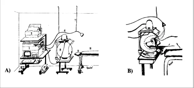
To organize these services, the following were installed in the same room: a electroretinographic unit table,
which was linked to a printer as well as an auxiliary power unit in order to guarantee security should there be a power failure. The Ganzfeld dome was placed on the other table,
and a mobile stretcher table was included in order to receive the animal to be submitted to electroretinography (Figure 1A). As part of the composition of the room, there were also two boxes for animal transportation, with the dimensions of 40 x 50 x 60cm, used to accommodate the patients during the phase of adaptation to the dark.
ELECTRORETINOGRAPHIC UNIT
A stimulation apparatus composed of a semi-automatic Ganzfeld1 dome, Grass type flash units, a
computer for data analysis and an interface unit for generation of stimulus and acquisition of data were employed. The parameters that were used included: dome with an opening 1 (one), back light with intensity of 272.7cd m-2; Grass flash with intensity of 2.35cd.s m-2, calibrated at
the manufacturer by a digital photometer (Digital
Radiometer/Photometer, model 2550). Blue gelatin filters2
- 10dB and red gelatin filters3 - 0dB, are arranged in front of
the flash, in order to allow the changing of both the intensity and wave length of the luminous stimuli.
Electrodes are connected to a preamplifier that sends the captured signals to the interface unit, and subsequently to an amplifier with both low-pass and high-pass filters, of 0.3Hz and 500Hz, respectively. After amplification and attenuation, such signals are directed to a recording unit, where they are translated to graphic form and can be represented in different scales
Figure 1 - Schematic representations of general disposition of equipments and materials inside the electroretinography room (A) and of the head positioning inside the Ganzfeld dome (B). 1 – electroretinographic unit; 2 – needles printer; 3 - no break;
(5mv/division, 10mv/division, 20mv/division or 50mv/ division).
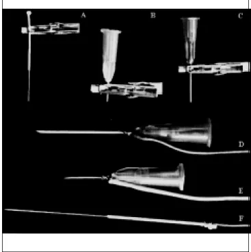
The capturing of retinal potential is possible with electrodes in ERG-jet contact lenses, left over the cornea. Needle electrodes (ground and reference) were manually produced from 25/8 and 13/4.5 hypodermic according to MUNGER’s* suggestion; and from Chinese
acupuncture needles4 (0.25 x 25mm), according to
LAZARO JR’s** suggestion. The needles were attached
to a flexible cable with a diameter of 0.35mm, using solder or “alligator claws”, or even directly to the acupuncture needle itself. In this last case, the flexible cable was rolled up or fixed by an “alligator claw”. The 6 types of electrodes are represented in figure 2.
PROTOCOLS AND METHODOLOGY
Dogs about to be submitted to electroretinography should previously undergo a
physical and ophthalmic examination, as well as a complete blood cell count (CBC), biochemistry profile (creatinine, BUN and ALT) and cardiorespiratory function evaluation.
In order to carry out the electroretinography, dogs were given levomepromazine5 intravenously at a
dose of 1mgkg-1 of body weight as a pre-anesthetic
drug. Tropicamide chloride6 was then instilled and
repeated after 15 minutes. At this point, animals were kept in transportation cages for 30 minutes to allow for adaptation to the dark. Then under room illumination arising from photophoro protected with gelatin filters, the combination of tiletamine and zolazepam7 was given intravenously, at a dose of 5mg
kg-1, in order to obtain a dissociative anesthesia (ORIÁ
et al., 2004).
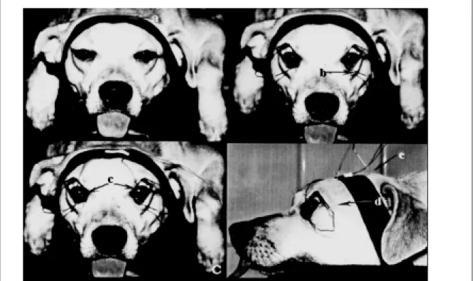
The animals were positioned in sternal recumbence under the effect of the anesthesia. In order to facilitate the fixation of electrodes and needles, the head was fixed with velcro to a head support next to the examination table. After the instillation of 0,5% proximetacaine chloride8, both eyelids were
spread with Barraquer’s lid speculums’9. Once the
entire cornea is exposed, the corneal electrodes are applied to contact lenses permeated with 2,5% hydroxypropyl methylcellulose10. The ground
electrode is fixed to the skin next to the external occipital protuberance, and the reference electrode 1 cm from the left eye lateral canthus (Figure 3). To allow a uniform illumination of the retina, the head of the animal is then introduced to the Ganzfeld dome (Figure 1B) (ORIÁ et al.,
2004).
Based on Classic protocol and Standard protocol, a mixed Classic/Standard protocol is proposed (Table 1). To make the interpretation of the electroretinogram easier, each signal is recorded and evaluated so that the 60 Hz interference occurrences are attenuated. As such, the remove interference option of the
machine software, as well as a second sub-routine, named smoothing waveforms, are used to remove
the excess of high frequency interference (USER’S manual EPIC-2000, 1999; ORIÁ et al., 2004).
COMMENTS
In order to minimize the obstacles that can be found when setting up electroretinography services, especially because of the lack of published data, a synthesis of the conditions for its installation is presented below.
Figure 2 - Photographic image of the needle electrodes (ground and reference), which were manually manufactured. In A, an electrode produced from acupuncture needle, attached to an “alligator-claw” for fixation of the flexible cable. In B, an electrode produced from 13/4.5 hypodermic needle, which is also fixed to the flexible cable through the “alligator-claw”. In C, an electrode manufactured from 25/8 hypodermic needle, fixed to the flexible cable with “alligator-claw”. In D, another electrode produced from 25/8 hypodermic needle, which was soldered to the cable. In E, an electrode manufactured from 13/4.5 hypodermic needle, also soldered to the flexible cable. In F, an electrode produced from a Chinese acupuncture needle, surrounded by the flexible cable.
Infrastructure:appropriate room for
electroretinography, with no windows, and a minimum area of 15m2 in order to facilitate the movement of
people and the arrangement of required material and equipment. The electrical circuit, as well as the grounding, should be isolated. An auxiliary power unit is required to provide security against power failures. This system should be allocated outside the exam room, to avoid interference. Another room adjacent to the exam room should facilitate the procedures of anesthesia. The communication between these two rooms should not impede adaptation to the dark. Room temperature should be controlled and kept at 25°C.
Equipment and Materials: when choosing
the electroretinography machine, one must consider the objectives and the operational conditions. Appropriate tables are needed to support the apparatus, the Ganzfeld dome and the animal. The
tables for both the dome and the animal must be equipped with lockable wheels, which should allow for the alignment of the head of the patient inside the dome. As such, a head support must be adapted to the table where the animal will be placed with devices for fixing its head. Furthermore, in order to avoid interference, tables must not have a large metal surface. It is also suggested to fix hooks and elastic cables on the ceiling, in order to keep the interface cable hanging,
thus facilitating the exam. Lanterns should have lamps protected with gelatin red filters, which will allow for better vision during the exam, however with no interference in adaptation to the dark. The choice of electrodes depends basically on the animal and where they will be used. For small and medium sized animals, contact lenses electrodes could be used, while needle electrodes could be used for large animals. Amongst needle electrodes, those made from Chinese acupuncture needles are recommended.
Human Resources: it is recommended that
three people take part in the exam. These people should be knowledgeable in retinal electrophysiology, as well as anesthesiology and also the functioning and manipulation of the electroretinographic unit.
Protocols: it is expected that a variation of
protocols of stimulation and registration, as well as anesthesiology, will occur from service to service, and from one laboratory to another. It is important, however, to standardize protocols that could be used in the majority of the cases.
CONCLUSIONS
According to the experience accumulated since the implantation of the electroretinography services, it is possible to conclude and advise: the
necessity of an exclusive room for the application of the electroretinogram. This room should be provided with adequate electrical wiring, as well as an adjacent room in order to allow animals to adapt to the dark environment. The choice of an easily operated electroretinographic unit, which should be able to perform several electrophysiological tests, and the use of the Ganzfeld dome should be considered, which
permits a stimulation of the entire field. The standardization of protocols that are possible to carry out and to reproduce with the utilization of secure anesthetic protocols. The availability of non-traumatic registration electrodes, which could be applied to different animals, moreover the necessity of qualified staff, with the knowledge of retinal electrophysiology, and precision expertise in the functioning of the available equipment.
SOURCES AND MANUFACTURERS
1-2503B - LKC Technologies Inc. 2-Kodak Wratten nº.98, 450nm, 1,1Hz 3-Kodak Wratten nº.29, 630nm, 5,1Hz
4-Suzhou Huanqiu Acupuncture Medical Appliance Co. Ltd. 5-Neozine® - Rhodia farma Ltda.
6-Mydriacil - Alcon Laboratórios do Brasil Ltda. 7-Zoletil® - Virbac do Brasil Ind. e Com. Ltda. 8-Anestalcon® - Alcon S/A.
9-Blefarostato Barraquer-N.J.Petrovich instrumentos
Cirúrgicos.
10-W-GPS® - Wilson Ophthalmic.
*MUNGER, R. J. (Animal Ophthalmology Clinic, Dallas- Texas
- USA). Personal comunication, 1999
**LAZARO JÚNIOR, L.P. (Oftalmoclínica Copacabana, Rio
de Janeiro - RJ - Brazil). Personal Comunication 2000
ACKNOWLEDGMENTS
FAPESP – research grants process 98/03153-0.
CAPES – Master in Science scholarship. Dr. DANIEL KAN HONSHO – drawings
Dr. MARLOS GONSALVES and JOHN BOLICIAN - English version
REFERENCES
ACLAND, G.M. Diagnosis and differentiation of retinal diseases in small animals by electroretinography. Semin Vet Med Surg Small Anim, v.3, n.1, p.15-27, 1988.
AGUIRRE, G.D.; RUBIN, L.F. Progressive retinal atrophy in the miniature Poodle: an electrophysiologic study. J Am Vet Med Assoc, v.160, n.2, p.191-201, 1972.
AGUIRRE, G. Electroretinography – Are we misusing an excellent diagnostic tool?. Vet Comp Ophthalmol, v.5, n.1, p.2-3, 1995. BEDFORD, P. The anatomy of the canine fundus. In: CONGRESS OF THE SMALL ANIMAL VETERINARY ASSOCIATION, 21., 1996, Jerusalem. Proceedings...
Jerusalem : Small Animal Veterinary Association, 1996. p.24.
BROOKS, D.E. et al. Changes in oscillatory potentials of the canine electroretinogram during acute sequential elevations in intraocular pressure. Progr Vet Comp Ophthalmol, v.2,
n.2, p.80-89,1992.
CARR, R.E.; SIEGEL, I.M. Electrodiagnostic testing of the visual system: a clinical guide. Philadelphia : F.A. Davis, 1990. 187p.
CHMIELEWSLI, N.T. Special boards. Progr Vet Neurol, v.6, n.2, p.73-75, 1995.
COOK, C.S. Ocular embryology and congenital malformations. In: GELATT, K.N. eterinary ophthalmology. 3.ed. Philadelphia : Lippincott Williams & Wilkins, 1999. p.3-30. CURTIS, R.; LIGHTFOOT, R.M. The canine fundus. In: PETERSEN-JONES, S.M., CRISPIN, S.M. Manual of small animal ophthalmology. London : Bristish Small Animal Veterinary Association, 1993. p.237-258.
DANTAS, A.M. Eletrorretinografia. In: DANTAS, A.M. et al. Eletrofisiologia ocular. Rio de Janeiro : Cultura Médica, 1995. 322p.
DAWSON, W.W.; KOMMONEM, B. The late positive retinal potential in dogs. Exp Eye Res, v.60, n.2, p.173-179, 1995. JONES, T.C.; HUNT, R.D., KING, N.W. Retina. In: _ _ _ _ _ _ . Patologia veterinária. 6.ed. São Paulo :
Manole, 2000. p.1334-1341.
KOCH, S.A.; RUBIN, L.F. Distribution of cones in retina of the normal dog. Am J Vet Res, v.33, n.2, p.361-363, 1972.
KOMAROMY, A.M.; SMITH, P.J.; BROOKS, D.E. Electroretinography in dogs and cats. Part I. Retinal morphology and physiology. Comp Cont Educ Pract Vet, v.20, n.3, p.343-350, 1998a.
KOMAROMY, A.M.; SMITH, P.J.; BROOKS, D.E. Electroretinography in dogs and cats. Part II. Technique, interpretation, and indications. Comp Cont Educ Pract Vet, v.20, n.3, p.355-365, 1998b
.
KOMMONEM, B. The DC-recorded dog electroretinogram in ketamine-medetomidine anaesthesia. Acta Vet Scand,
v.29, n.1, p.35-41, 1988.
KOMMONEM, B.; RAITTA, C. Electroretinography in labrador retrievers given ketamine-xylazine anesthesia. Am J Vet Res, v.48, n.9, p.1325-31, 1987.
LESCURE, F. Électrophysiologie oculaire. Encyclopédie Vétérinaire, Ophthalmologie 1700, p.1-13, 1993. MILLER, T.R. The uses and limitations of the electroretinogram in veterinary practice. Brit Vet J, v.149, n.3, p.3-4, 1993.
MILLER, P.E.; MURPHY, C.J. Vision in dogs. J Am Vet Med Assoc, v.207, n.12, p.1623-1634, 1995.
OFRI, R. Applied retinal physiology. In: CONGRESS OF THE SMALL ANIMAL VETERINARY ASSOCIATION, 21., 1996, Jerusalem. Proceedings... Jerusalem : Small Animal
Veterinary Association, 1996a. p.27.
OFRI, R. The electroretinogram - a powerful, yet often ignored, tool in the diagnosis of retinal diseases. In: CONGRESS OF THE SMALL ANIMAL VETERINARY ASSOCIATION, 21., 1996, Jerusalem. Proceedings...
Jerusalem : Small Animal Veterinary Association, 1996b. p.28. OFRI, R. Optics and physiology of vision. In: GELATT, K. N. Veterinary ophthalmology. 3.ed. Philadelphia : Lippincott Williams & Wilkins, 1999. p.183-216. OGDEN, T. E. Clinical electrophysiology. In: RYAN, S. J.; OGDEN, T. E. Retina. 2. ed. St. Louis: Mosby, 1994. p 321-332. ORIÁ, A.P. et al. Considerations about electroretinography in dogs. Ciência Rural. v.34, n.1, p.323-328, 2004.
PARRY, H.B.; TANSLEY, K.; THOMSON, L.C. The electroretinogram of the dog. J Physiol, v.153, n.120, p.28-40, 1953.
RUBIN, L.F. Clinical electroretinography in dogs. J Am Vet Med Assoc, v.151, n.1, p.1456-1469, 1967.
SAMUELSON, D.A. Ophthalmic anatomy. In: GELATT, K. N. Veterinary ophthalmology. 3.ed. Philadelphia :
Lippincott Williams & Wilkins, 1999. p.31-150.
SCHAEPPI, U.; LIVERANI, F. Procedures for routine electroretinography (ERG) in dogs. Agents Actions, v.7,
n.3, p.347-351, 1977.
SIMS, M.H. Partial masking of the canine electroretinogram by oscillatory potentials - to problem of frequency bandwidth.
J Vet Int Med, v.4, n.1, p.40-42, 1990.
SIMS, M.H. et al. Effects of stimulus intensity and conditioning on the electroretinogram and oscillatory potentials in dark-adapted cats. Progr Vet Comp Ophthalmol, v.1, n.3, p.177-185, 1991.
SIMS, M.H. Electrodiagnostic evaluation of vision. In: GELATT, K.N. Veterinary ophthalmology. 3.ed. Philadelphia: Lippincott Williams & Wilkins, 1999. p.483-510. SLATTER, D. Retina. In:______. Fundamentals of veterinary ophthalmology. 2.ed. Philadelphia: Saunders, 1990. p.400-436.
USER’S manual EPIC-2000. Gaithersburg : LKC Technologies, 1999. 160p.
YANASE, J.; OGAWA, H.; OHTSUKA, H. Rod and cone components in the dog electroretinogram during and after dark adaptation. J Vet Med Sci, v.57, n.5, p.877-881,