Universidade de Lisboa
Faculdade de Ciências
Departamento de Biologia Vegetal
Digested raspberries polyphenols for brain health:
Attenuation of neuroinflammation
Gonçalo Filipe Rodrigues Garcia
Dissertação
Mestrado em Biologia Molecular e Genética
Universidade de Lisboa
Faculdade de Ciências
Departamento de Biologia Vegetal
Digested raspberries polyphenols for brain health:
Attenuation of neuroinflammation
Gonçalo Filipe Rodrigues Garcia
Sob a orientação de:
Maria Helena Caria, PhD
(Prof. Auxiliar FCUL; Prof. Coordenadora ESS-IPS)
Maria Paula Marinho Pinto, PhD
(Investigadora convidada ITQB; Prof. Coordenadora ESA-IPS)
Dissertação
Mestrado em Biologia Molecular e Genética
Epigraph
“Sage isn't the man who gives the true answers; is the one who formulates the true questions”
Acknowledgments
I would like to start by expressing my acknowledgement to Maria Paula Marinho Pinto, my supervisor. My gratitude for all the support, guidance and encouragement given during this incredibly intensive year. Also, Cláudia Nunes dos Santos, who gave me alternatives to the problems and always helped me to clarify and rethink the way how I was working. Thanks to both, who always had patience to listen to me, always with willingness.
My special thanks go also to Professor Ricardo Boavida Ferreira, who allowed my inclusion in the workgroup at his laboratory. Thanks for the opportunity, and for all the knowledge and experience. A note of thanks is also due to Dr. Teresa Pais (IMM, Lisbon), and Dr. Derek Stewart (JHI, Dundee) who, respectively, provided the N9 microglial cell line and the five quasi-isogenic digested raspberry polyphenolic fractions I used at this work. I would like to express my appreciation to Dr. Regina Menezes, especially for introducing me to the yeast model of neuroinflammation, but also for the critical reading of this thesis, providing numerous suggestions for its improvement. Additionally, my sincere thanks to Dr. Maria Helena Caria for having accepted to be my supervisor and for the support offered during all my work.
I cannot forget all my Disease and Stress Biology laboratory colleagues, who had a critical role in my progress over the year. Thanks to Andreia Gomes for her hospitality and readiness to help; to Carolina Jardim for her friendly disposition and comprehension; to Lucélia Tavares for her good and wise advices; to Inês Figueira for her authenticity and critical rigor; to Rui Pimpão for sharing his experience and social skills. Without them, I would not know how hard it is the world of science. You gave me strength. Thank you for all the time spent discussing results, for all the encouragement and support.
A very special acknowledge goes to the other trainees, who also started their stages at the same time as I did. Many thanks to Tânia Silva for sharing great moments in the laboratory, and for her good humor; to Vitor Hugo Gonçalves for being my laboratory brother, and for all the jokes and comprehension given; to Inês Costa for being always an exceptional person and an example in the organization and work. All of you were part my closest family during the past year, many thanks. I will never forget all the moments we spent together.
Also, I want to thank my family. To my parents and brother, without them would be impossible to keep studying and reaching my objectives. Many thanks! To Carmen Santos who had a crucial role in my stage since the beginning, when I had many doubts about what I would like to do during the year. Thanks for all the discussions we made about this thesis and the practical work; and many thanks for your comprehension and sensitive capacity. To Daniel Gaspar, my “praxis brother”, who distracted me when necessary, always providing a good company. Many thanks to all of you!!
Index
INDEX ... V INDEX OF FIGURES ... VI INDEX OF TABLES ... VI ABBREVIATIONS ... VII ABSTRACT ... IX KEYWORDS ... IX RESUMO ... XPALAVRAS-CHAVE ... XII
1. OBJECTIVES ... 1
2. THEORETICAL FUNDAMENTS... 2
2.1.MICROGLIA: THE INNATE IMMUNE CELL OF CENTRAL NERVOUS SYSTEM ... 2
2.2.NEUROINFLAMMATION ... 3
2.2.1 – Markers of microglial activation ... 4
2.2.2 – Common Inflammatory triggers ... 8
2.3.ATTENUATION OF NEUROINFLAMMATION BY DIETARY FACTORS ... 8
2.3.1 – Attenuation of neuroinflammation by polyphenols ... 9
2.3.2 – Polyphenols metabolites ... 10
2.3.3 - Raspberry: a fruit with potential anti-neuroinflammatory properties ... 10
3. MATERIALS AND METHODS ... 12
3.1.PLANT MATERIAL, IN VITRO DIGESTION AND FRACTIONS PREPARATION ... 12
3.2.MICROGLIAL MODEL OF NEUROINFLAMMATION ... 13
3.2.1 – Cell culture and treatments ... 13
3.2.2 – Protein extraction and quantification ... 13
3.2.3 – Determination of pro-inflammatory and activation markers ... 14
3.2.4 – Cytotoxicity assays ... 16
3.3.YEAST MODEL OF INFLAMMATION ... 16
3.3.1. Characterization of S. cerevisiae model of inflammation ... 16
3.3.2 – β-galactosidase assays ... 17
3.4.STATISTICS... 18
4. RESULTS AND DISCUSSION ... 19
4.1.ATTENUATION OF NEUROINFLAMMATION IN MURINE MICROGLIAL CELL LINE N9 ... 19
4.1.1 – Implementation of N9 microglial line as a neuroinflammation model ... 19
4.1.2 – Cytotoxicity of the digested raspberry polyphenolic fractions ... 21
4.1.3 – Model optimization for attenuation of neuroinflammation ... 22
4.1.4 – Attenuation of neuroinflammation by raspberry digested fractions ... 23
4.2.MECHANISTIC STUDIES OF ANTI-INFLAMMATORY PROPERTIES OF DIGESTED RASPBERRY POLYPHENOLS IN SACCHAROMYCES CEREVISIAE MODELS ... 25
4.2.1 – Digested raspberry polyphenols modulate the Crz1/calcineurin pathway ... 25
5. CONCLUSIONS AND FUTURE PERSPECTIVES ... 27
6. REFERENCES ... 29
Index of figures
Figure 1... 2 Figure 2... 3 Figure 3... 4 Figure 4... 6 Figure 5... 14 Figure 6... 19 Figure 7... 20 Figure 8... 21 Figure 9... 22 Figure 10... 23 Figure 11... 24 Figure 12... 26Index of tables
Table 1 ... 9 Table 2 ... 12 Table 3 ... 17 Table 4 ... 18 Table 5 ... 22 Table 6 ... 25 Table 7 ... 27Abbreviations
Abs: Absorbance
ABTS: 2,2'-azino-bis(3-ethylbenzothiazoline-6-sulphonic acid) AD: Alzheimer ’s disease
ANOVA: Analysis of variance APC: Antigen presenting cell ATP: Adenosine tri-phosphate Aβ: Beta-amyloid
BBB: Brain Blood Barrier
CD-(number): Cluster of differentiation, (number) CDRE: Calcineurin-Dependent Response Element CN: Calcineurin
CNB1: Calcineurin regulatory B subunit CNS: Central Nervous System
COX-2: Ciclo-oxigenase-2
CR3: Complement receptor type-3 CSM: Complete supplement mixture DHE: Dihydroethidium
DMEM: Dulbecco’s modified Eagle’s medium DMSO: Dimethyl sulfoxide
DNA: Deoxyribonucleic acid DNAse: Deoxyribonuclease
ECL: Enhanced chemiluminescence
ELISA: Enzyme linked immuno sorbent assay FACS: Fluorescence-activated cell sorting FBS: Fetal Bovine Serum
FcγR: Fragment crystallizable-gamma receptor FDW: Fruit dry weight
FIC: Fluorescence intensity counts FITC: Fluorescein isothiocyanate FSC: Forward scatter
GAE: Gallic Acid Equivalent HRP: Horseradish Peroxidase
Iba-1: Ionized calcium binding adapter molecule-1 IKK: IκB kinase
IL(number): Interleukin, (number) iNOS:inducible Nitric oxide synthase IκB: Inhibitor of nuclear factor kappa B JHI: James Hutton Institute
LPS: Lipopolysaccharide
MAC1: Macrophage antigen complex 1 MAP: Mitogen-Activated Protein
MAPK: Mitogen-Activated Protein Kinase MBA: Membrane blocking agent
MCP-1: Monocyte chemoattractant protein-1 MFI: Median fluorescence intensity
MHC (I and II): Major Histocompatibility Complex (class 1 or class 2) MS: Mass spectrometry
NADPH: Nicotinamide adenine dinucleotide phosphate NEAA: non-essential amino acids
NFAT: Nuclear factor of activated T-cells NF-κB: Nuclear factor kappa B
NLS: Nuclear localization signals NO: Nitric oxide
NOS: Nitric oxide synthases
ONPG: Ortho-Nitrophenyl-β-galactoside
PAMP: Pathogen-associated molecular pattern PBS: Phosphate Buffer Saline
PD: Parkinson’s disease PGE2: Prostaglandin E2
PHOX: Phagocyte NADPH oxidase PRR: Pattern recognition receptor
qPCR: Quantitative/real-time Polymerase Chain Reaction RIPA: Radioimmunoprecipitation assay
ROS: Reactive Oxygen Species SCM: Synthetic Complete Medium SD: Standard Deviation
SDS: Sodium dodecyl sulfate
SDS-PAGE: Dodium dodecyl sulfate - Polyacrylamide gel electrophoresis SE: Standard Error
SSC: Side scatter
TBS: Tris-buffered saline
TBST: Tris-buffered saline with Tween20 TLR: Toll-like receptors
TNFR: Tumor necrosis factor receptor TNF-α: Tumor necrosis factor - alpha YNB: Yeast nitrogen base
Abstract
Microglia are the resident innate immune cells in the central nervous system (CNS), representing the first defense line of the neural parenchyma. However, its chronic activation is implicated in neurodegenerative disorders by the uncontrolled release of diverse molecular mediators such as inflammatory cytokines.
Among all dietary phytochemicals, polyphenols are the major anti-inflammatory molecules provided by some plants. As example, raspberries are an enriched natural source of polyphenols such as ellagic acid, flavanols; and phenolic acids. However, digestion strongly modifies the structure of polyphenols, producing metabolites with different bioactivities. Thus, it is imperative the study of digested polyphenols for a better elucidation about their effects.
The main goal of this study was to evaluate the bioactivity of raspberry polyphenols as neuroinflammatory attenuators. The fractions studied were composed by polyphenols that are bioaccessible to blood serum, obtained from in vitro digestion of five different quasi-isogenic raspberries. Furthermore, N9 murine microglial cell line was implemented as model of neuroinflammation and the range of non-cytotoxic concentrations of each fraction was determined. Then, the neuroinflammatory attenuation induced by each digested fraction in LPS-stimulated microglia was evaluated and compared. As result, some of the fractions attenuated microglial pro-inflammatory activation, significantly decreasing the expression of membrane protein CD-40 (marker of microglial activation) and the production of the pro-inflammatory markers, nitric oxide (NO), tumor necrosis factor-α (TNF-α) and intracellular superoxide (O2•-).
Additionally, a yeast model of inflammation was used as a mechanistic tool to elucidate the molecular mechanisms underlying the anti-inflammatory activity of the digested raspberry fractions. The results showed that these compounds inhibit the yeast Crz1/calcineurin pathway, which is homologous to the mammalian nuclear factor of activated T-cells (NFAT)/calcineurin, suggesting that they may prevent microglial neuroinflammation through this pathway.
Keywords
Resumo
Quando exposto a diversas agressões tais como infeções, toxinas ou traumatismos, o sistema nervoso central medeia uma resposta inflamatória complexa e ao mesmo tempo dinâmica. Esta resposta inclui uma curta e eficiente ativação do sistema imunitário, que geralmente é mediada pela população de células imunitárias residentes – a microglia.
A microglia representa cerca de 12% do tecido cerebral e participa na primeira linha de defesa do parênquima neural. Num estado não ativado apresenta uma morfologia ramificada, tendo como função a continua monitorização do espaço que rodeia os neurónios. Uma vez exposta a um estímulo pro-inflamatório, a microglia entra num estado ativado que, tipicamente, é acompanhado de uma transição da morfologia para uma forma ameboide, o que favorece a fagocitose. Neste estado, a expressão de muitos recetores de superfície relacionados com a resposta imunitária é aumentada. São também induzidas muitas vias de sinalização celular que conduzem à secreção de diferentes compostos pro-inflamatórios. Por exemplo, compostos como o fator de necrose tumoral – α (TNF-α), a interleucina - 1β (IL-1β), ou as espécies reativas de oxigénio, são regulados por diferentes vias de sinalização celular tais como a via do fator nuclear – κB (NF-κB), a via do fator nuclear das células T ativadas (NFAT) ou a vias das cinases MAP (MAPK).
Muitos dos agentes pró-inflamatórios conhecidos e descritos são pesticidas como o paraquato, dieldrina, lindano ou rotenona; outros são toxinas como os lipopolissacarídeos (LPS) provenientes da membrana exterior de bactérias gram-negativas. Geralmente, estes agentes estão associados a casos patológicos, nos quais a microglia adquire um estado de sobreativação crónica. Nestes casos, muitos compostos mediadores da inflamação são continuamente e abundantemente secretados pela microglia, causando lesões e prococando ativação das vias de apoptose nas células neuronais vizinhas. Elevados e contínuos níveis de produção de óxido nítrico, de espécies reativas de oxigénio e de citocinas estão descritos como patológicos em várias doenças neurodegenerativas, tais como Alzheimer, Parkinson, Huntington, Esclerose Múltipla ou Esclerose lateral amiotrófica. Por sua vez, proteínas como a α-sinucleína ou a β-amilóide, que estão descritas nas doenças de Parkinson e Alzheimer, respetivamente, atuam como agentes pro-inflamatórios, perpetuando e intensificando a resposta inflamatória. Nos casos mais severos, ocorre uma fragilização da barreira hematoencefálica, o que permite a entrada de macrófagos periféricos para a progressão e intensificação do processo inflamatório.
Os hábitos alimentares atuais também promovem uma ação pró-inflamatória que contribui para a progressão de algumas das doenças crónicas anteriormente citadas. A dieta
atual envolve o elevado consumo de carnes vermelhas, doces, alimentos ricos em gorduras e bebidas gaseificadas, enquanto há um baixo consumo de fruta fresca, nozes, vegetais, cereais e fontes naturais de ómega-3 como o peixe.
Muitos estudos com vista à atenuação da neuroinflamação comprovaram que a dieta, e em especial, o consumo de algumas classes de polifenóis provenientes da fruta e dos vegetais, parece ter um efeito positivo nesse contexto. No entanto, é importante mimetizar os diversos processos de transformação que ocorrem nos polifenóis durante a digestão in vivo, uma vez que essas transformações alteram drasticamente a sua função biológica. Uma das formas de mimetizar a digestão é o recurso à utilização de modelos de digestão in vitro, a partir dos quais se obtêm as frações digeridas dos polifenóis. Os metabolitos constituintes destas frações são semelhantes àqueles que estariam bioacessíveis às células alvo in vivo.
A procura de polifenóis cujos efeitos sejam benéficos face às mais diversas patologias, como o cancro ou as doenças neurodegenerativas, levou ao aumento do estudo dos pequenos frutos. Estes frutos possuem uma vasta gama e um elevado teor em polifenóis, o que propicia a sua utilização na investigação científica. Em particular, as espécies do género Rubus têm sido referenciadas como altamente benéficas para a saúde.
No presente estudo, avaliou-se a eventual atenuação da neuroinflamação promovida por cinco frações digeridas de diferentes framboesas em células de microglia. As cinco cultivares de framboesa utilizadas provêm de um banco de germoplasma do James Hutton Institute, caracterizando-se por diferirem entre si na constituição nas diferentes classes de polifenóis. Para a obtenção das frações digeridas, as diferentes framboesas quasi-isogénicas foram liofilizadas, solubilizadas em água e submetidas a um modelo de digestão in vitro. De cada tipo de framboesa resultaram duas frações distintas, uma fração digerida bioacessível ao cólon e uma fração digerida bioacessível ao soro do sangue. Para o presente trabalho apenas foram utilizadas as frações bioacessíveis ao soro do sangue. Todo o processo da digestão in vitro até à obtenção das frações foi desenvolvido pelo Dr. Derek Stewart, do James Hutton Institute.
A primeira tarefa deste trabalho foi a implementação do modelo de neuroinflamação com a linha celular N9 de microglia de ratinho. Para tal, as células foram estabelecidas em cultura in vitro e estimuladas com LPS. Devido à complexidade inerente à resposta inflamatória mediada pela microglia, foi necessário identificar e aceder a diferentes marcadores pró-inflamatórios típicos da microglia ativada, através de diferentes técnicas. Para a determinação do óxido nítrico libertado pelas células, foi quantificado o teor em nitritos no meio de cultura através da reação de Griess. O fator de necrose tumoral – α (TNF-α) foi quantificado no meio de cultura, recorrendo à técnica de Enzyme Linked Immuno Sorbent Assay (ELISA) quantitativa. O radical superóxido e a proteína de membrana CD-40 foram ambos quantificados recorrendo à técnica de citometria de fluxo.
A tarefa seguinte foi a avaliação da citotoxicidade das frações de framboesa digerida, como forma de determinar, dentro da gama de concentrações descritas como fisiológicas, quais as concentrações não toxicas adequadas para a sua utilização na linha celular N9 de microglia de ratinho.
A última tarefa foi a avaliação da atenuação da neuroinflamação promovida pelas frações digeridas na linha celular N9 de microglia de ratinho. Para tal, as células foram pré-incubadas com as diferentes frações digeridas de framboesa e seguidamente estimuladas com LPS. De seguida, foram quantificados os diferentes marcadores previamente validados na primeira tarefa (óxido nítrico, TNF-α, CD-40 e radical superóxido). Como resultado, três das frações digeridas revelaram grande consistência na significativa redução da produção e expressão dos diversos marcadores na linha de microglia N9, o que evidência o seu forte contributo para a atenuação da neuroinflamação. Por outro lado, as restantes duas frações não exibiram um efeito anti-inflamatório consistente para todos os marcadores inflamatórios, sendo que em alguns desses marcadores não se observaram reduções significativas na produção/expressão relativamente aos controlos positivos da inflamação.
Adicionalmente, como ferramenta mecanística foi utilizado um modelo de inflamação construído em levedura. A utilização deste modelo revelou que, de alguma forma, os metabolitos presentes nas frações de framboesa digerida interferem na regulação da via celular calcineurina / Crz1, reprimindo a sua indução pelo dicatião cálcio (Ca2+). Esta via de
sinalização é regulada de uma forma semelhante nos mamíferos, sendo que CRZ1 é ortólogo de NFAT, que, por sua vez, está intrinsecamente relacionado com a inflamação. Assim, os resultados obtidos com o modelo de inflamação em levedura sugerem que uma das vias pela qual os metabolitos presentes nas frações digeridas de framboesa atenuam a neuroinflamação na microglia é pela via da calcineurina - NFAT.
Por último, os resultados da atenuação da inflamação na microglia e nos ensaios em levedura foram comparados com as diferentes composições químicas em polifenóis de cada uma das frações digeridas utilizadas. Por conseguinte, algumas classes de polifenóis, tais como os conjugados de ácido elágico, foram identificadas como relacionadas com o poder anti-inflamatório das frações.
Palavras-chave
1. Objectives
Neuroinflammation can be initiated in response to a variety of injuries, including infection, traumatic brain injury or toxic metabolites1. In the central nervous system, including
the brain and spinal cord, microglia are the resident innate immune cells that mediate the inflammatory response to these injuries1.
In the present study, the main objective was the evaluation of the neuroinflammatory attenuation that comes from the treatment with five different digested quasi-isogenic raspberry fractions. These fractions were obtained from the in vitro digestion of germplasm raspberry lines, in James Hutton Institute.
The first task was the implementation of N9 murine microglial cell line as a model of neuroinflammation, by accessing diverse pro-inflammatory and activation markers typical of LPS-stimulated microglia. Different techniques such as ELISA, griess reaction, flow cytometry and western blot were used for that purpose. Secondly, the cytotoxicity of the digested raspberry polyphenolic fractions was assayed in order to guarantee the range of non-toxic concentrations while using also physiological concentrations.
As the final task, the inflammatory attenuation from the treatment with each fraction in LPS-stimulated microglia was evaluated by the analysis of the markers previously validated in the model implementation. Also, a yeast model of inflammation was used as mechanistic tool for the evaluation of the anti-inflammatory bioactivity of the digested raspberry fractions as inhibiters of Crz1/calcineurin pathway.
2. Theoretical fundaments
2.1. Microglia: the innate immune cell of central nervous system
Microglia are the resident innate immune cells in the central nervous system (CNS) and comprise approximately 12% of the brain tissue2. They are involved in the first defense line for
the neural parenchyma, releasing diverse molecular mediators such as inflammatory cytokines.
Generally, it is accepted that the original microglia population differentiates from cells of the myeloid lineage, which occurs in early embryonic development. This could justify the common expression of the majority of surface markers of monocytes and macrophages: CD1a, CD2, CD4, CD16, CD18, CD40, CD45, major histocompatibility complex (MHC) class I and II,
among many others3.
Microglia can vary between two different morphological phenotypes: ramified, characteristic in cells unexposed to inflammatory triggers (surveillant cells); and amoeboid, the typical physiology of cells reacting to inflammatory triggers (activated cells)4
(Figure 1). Ramified cells are constantly surveying the surrounding environment with their retractile processes and their cell bodies are spaced within the CNS, avoiding overlap5. At this “resting” state, several key surface
receptors involved in the innate immune response are expressed at low levels, such as MHC molecules6; CD45
(the leucocyte common antigen)7 and CD-11b8. In
addition, cell surface receptor-ligands, such as, CD-200 are present, contributing to maintenance of neuron-microglia communication in the CNS9.
Once exposed to pro-inflammatory triggers such as pathogens, brain injuries, dead or dying cells and immunological stimuli, microglia cells respond by modifying its ramified morphology to the amoeboid shape, favoring phagocytic activity10. These alterations are also
followed by changes in signaling cascades, which promote upregulation in the expression of many cell surface receptors, as well an increase in the production of other pro-inflammatory mediators11, 12. As example, the release of tumor necrosis factor-alpha (TNF-α) is also a
reliable marker of microglial activation13, 14.
Although, during the development of some neurodegenerative disorders, microglia is reported to acquire a prejudicial overactivated stage that persists chronically. At this stage, the continuous high release of many of the pro-inflammatory mediators such as cytokines15, 16,
Figure 1 – Microglia are morphologically
and functionally dynamic cells. They are able to change from highly ramified shapes (non-activated cells) to completely lacking processes cellular bodies (activated cells)4.
reactive oxygen species (ROS)17 or nitric oxide (NO)18 are described as implicated in the
pathology of Alzheimer’s disease (AD), Parkinson’s disease (PD), Huntington’s disease, Multiple sclerosis, Amyotrophic lateral sclerosis and other neurodegenerative disorders. In addition, those mediators can also strongly contribute for cerebrovascular damage in diverse neurodegenerative disorders such as AD and PD19.
2.2. Neuroinflammation
The CNS mediates a dynamic immune and inflammatory response when exposed to infections, trauma, stroke, toxins and other stimuli capable of inducing an activation of the innate immune system. This response is usually mediated by microglial activation. Notwithstanding, microglial activation can have either positive or detrimental effects on neurons, according to the duration and amounts of cytokines and growth factors secreted20.
Within activation, microglia produce inflammatory mediators that are essential to protect the CNS against injuries. Also, damaged or dead cells increase the phagocytic activity of activated microglia21. However, it is typically a short-lived activation with no harmful effects
for other neuronal cells. It is believed that this acute microglial activation is beneficial for CNS, contributing for host protection and tissue repair22.
On the other hand, chronic neuroinflammation persists long time after the initial injury or trigger and it is often a self-perpetuating event. Many pro-inflammatory mediators such as cytokines are highly increased and sustained15. Also, it occurs a notable augmentation of
oxidative and nitrosative stress that persists chronically, injuring surrounding neuronal cells and promoting diverse neurodegenerative disorders23.
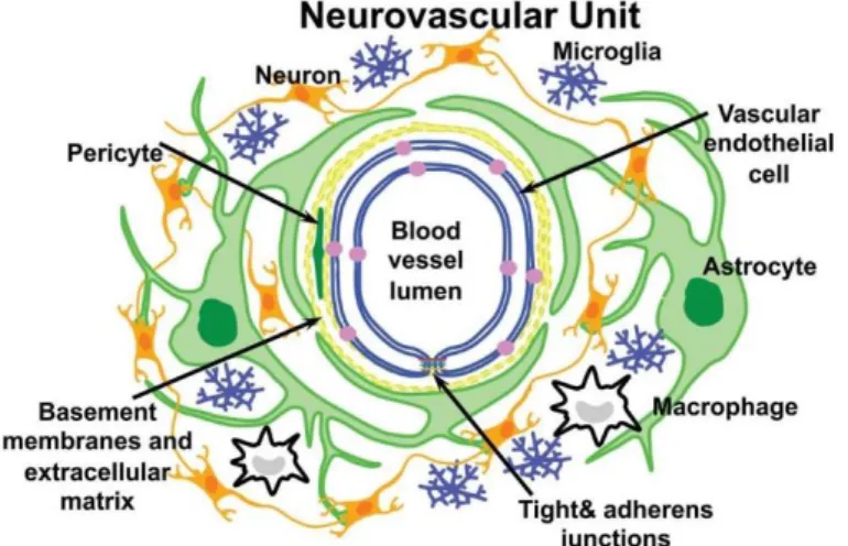
With chronic and sustained states of neuroinflammation, there is commonly a compromise of the blood brain barrier (BBB) which increases infiltration of peripheral macrophages into the brain neural parenchyma to further perpetuate the inflammation24
(Figure 2).
Whether neuroinflammation is beneficial or harmful to the brain, critically depends on the duration and intensity of the inflammatory response25, 26.
Figure 2 – Schematic representation of the neurovascular unit that
constitutes the Blood-brain barrier (BBB). Vascular endothelial cells form the blood vessel and associate with astrocyte and pericytes forming the basal lamina. Other components of the neurovascular unit are neurons and microglia. Macrophages can also infiltrate during severe neuroinflammatory events. Figure adapted from C. L. Willis, Toxicol Pathol 39, 172 (2011).
2.2.1 – Markers of microglial activation
Because microglial activation is a complex and dynamic process, it is not simple to identify and conjugate the different molecular players. However, several pathways27-29 are
implicated and some resultant molecules are described as reliable activation markers.
Nuclear factor kappa B (NF-κB) pathway
Nuclear factor kappa B (NF-κB) is a transcription factor that regulates the expression of inducers and effectors at many points in the complex networks related to the response to pathogens30, 31. However, this protein extends its transcriptional regulation of the immune
response, by influencing also gene expression pathways that impact cell survival, differentiation and proliferation. Because of these wide spectrum implications, the dysregulation of NF-κB is normally described in various pathological situations, including neuroinflammatory disorders32-34.
The NF-κB dimers are present in the cytoplasm in an inactive state, sequestered by inhibitory IκB proteins, which mask the nuclear localization signals (NLS). The activation process of this transcriptional factor is mediated by the IκB kinase (IKK) complex. When activated by pro-inflammatory stimulus, such as lipopolysaccharide (LPS)35, ROS36, TNFα37 or
interleukin 1-beta (IL-1β)38, the IKK complex promote phosphorylation and degradation of IκB
proteins and subsequent release and activation of NF-κB39. Then, NF-κB activation does not
require protein synthesis, once the interaction with diverse pro-inflammatory mediators can directly induce the activity of this transcription factor.
Nuclear factor of activated T cells (NFAT) pathway
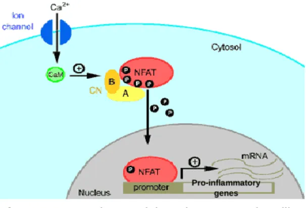
NFAT is a DNA binding protein required for pro-inflammatory gene expression, which is modulated by calcineurin phosphatase activity40.
In its phosphorylated state, NFAT localizes to the cytoplasm, where it remains inactive. Once stimulated, calcineurin dephosphorylates cytosolic NFAT allowing it’s translocation to the nucleus where it binds promoters that activate transcription of a large number of genes during an effective immune response41, 42(Figure 3). Despite the close
involvement of NFAT in the course of pro-inflammatory events, recent findings confirmed also that this transcription factor regulates microglial phenotype, as well as the expression of TNF-α and MCP-127 .
Figure 3 - The Calcineurin-NFAT signalling
pathway. Increase in the intracellular calcium activates the cellular phosphatase Calcineurin (CN) through interaction with Calmodulin (CaM). Activated CN dephosphorylates NFAT, allowing it’s translocation for the nucleus were it binds to the promoters of pro-inflammatory genes. Adapted from: (http://www.angiobodies.com/figuras/uam_fig2.gif)
Pro-inflammatory genes
Mitogen-activated protein kinases (MAPK) pathway
Mitogen-activated protein kinases (MAPK) contitute a family of serine/threonine/tyrosine-specific protein kinases. These proteins modulate cellular response to diverse external stimuli, such as osmotic stress, heat shock, pro-inflammatory cytokines or LPS. In the CNS, this cellular response in usually mediated by microglia43, 44.
Generally, MAPK proteins form a chain of proteins that transduce signals from a cellular surface receptor to the DNA in the nucleus. When the receptor on the cell membrane binds to a signaling molecule, a signal is produced and redirected to the nucleus via MAPKs which communicate by adding phosphate groups to a neighboring protein. The pathway culminates with the signal reaching the nucleus, triggering the expression of proteins that specifically promote alterations in the cell, such as mitosis or inflammatory response.
Nicotinamide adenine dinucleotide phosphate-oxidase (NADPH oxidase) pathway
NADPH oxidase is a membrane-bound enzyme that catalyzes the production of superoxide from oxygen in response to various stimuli, including pathogen-associated molecular patterns (PAMPs), inflammatory peptides45 and multiple neurotoxins46. Superoxide
aims to eliminate bacteria and fungi, mainly by interrupting their metabolic activity and promoting lipid peroxidation. This radical can spontaneously produce hydrogen peroxide that is submitted to further reactions, generating additional ROS. This also represents the main mechanism through which microglia produce neurotoxic ROS in response to stimuli47.
A recent research suggests that during neuroinflammation, NADPH oxidase plays a critical role in modulating the microglial phenotype towards a pro-inflammatory activated state
48. In the same study, it was demonstrated that inhibition of NADPH oxidase increases
production of anti-inflammatory cytokines such as IL-4, reducing the production of pro-inflammatory cytokines such as TNF-α48. Finally, this enzyme is described as an essential
player for pro-inflammatory events that occur in some disorders such as AD49.
Macrophage antigen complex 1 (MAC1)
This complex, also known as complement receptor type-3 (CR3) or integrin CD-11b/CD-18 is bifunctional, acting as an adhesion molecule, and as a pattern recognition receptor (PRR) which recognizes a diverse set of stimuli, mediating the activation of phagocytes50, 51.
The MAC1 receptor is highly expressed in post-mortem brains of patients with AD8,
matching the microglial activation that occurs in this neurodegenerative disorder. Moreover, MAC1 has been described as a player in NADPH oxidase activation in response to oxidative insults51-53. In addition, when MAC1 receptor receptor recognizes a ligand, an induction of the
transcription factor NF-κB signaling pathway and the resulting production of inflammatory factors were verified54.
A model family of pattern recognition receptors: Toll-like receptors (TLRs)
TLRs are one of the most studied families of PRRs as they contribute for the immunocompetent cell activation in the CNS and the subsequent pro-inflammatory cascade. In microglia, the expression of TLRs is regulated by signaling cascades during embryonic development and by the exposure to inflammatory triggers, such as pathogens55.
There are twelve members of the TLR family identified in mammals that recognize PAMPs from bacteria, fungi, viruses and the host itself56. The recognition of PAMPs by these
receptors initiates innate immune responses on interaction with infectious agents. In microglia, which expresses the TLRs from 1 to 957, 58, TLR4 is accepted as the primary LPS receptor59
and has been reported to be a crucial mediator of the inflammatory response to LPS60, 61.
Additionally, TLR4 also respond to damage associated molecular patterns, released by the injured tissues, and is upregulated upon brain inflammation61, 62, strongly suggesting a role of
this receptor as pro-inflammatory mediator.
Cell surface immune-associated receptors
Many cell surface receptors associated with the innate immune system response such as MHC molecules are also upregulated during microglial activation through an inflammatory stimulus (Figure 4). The abundance of those molecules during microglial activation allows these cells to act as antigen presenting cells (APC) to T-cells that will then be able to enter the brain in the course of an active infection6. As example, CD-40, which is a typical T-Cell surface
receptor and a member of the tumor necrosis factor receptor (TNFR) family63, is
over-expressed by activated microglia64, 65. Also, CD-45, the leukocyte common antigen, is highly
expressedin constitutively activated microglia from OX2-deficient mice9.However, CD-45 level
of expression is lower when compared to macrophages7, 66, and the same occurs with many
other immune-associated receptors.
Figure 4 – Markers of microglial activation. When exposed to an inflammatory stimulus, membrane immune
receptors are highly expressed (A) and inflammatory cytokines are secreted, accompanying the shift for the amoeboid morphology (B). Adapted from R. N. Dilger, R. W. Johnson, J Leukoc Biol 84, 932 (2008)67.
Inflammatory stimulus (e.g. LPS, TNF-α, IFN-γ) NO ROS Surveillant microglia cell Activated microglia cell A B
Expression of cytokines and chemokines
Important cytokines and chemokines such as IL-1β, TNF-α13, 14 (Figure 4) and also
monocyte chemoattractant protein-1 (MCP-1) are molecules that constitute good markers of microglial activation. Those molecules are characteristic of macrophage and chemoattractant cells, whereas its abundance in microglia indicates a shift for the phagocytic phenotype68.
Chronic releasing of those cytokines by pro-inflammatory microglia may also have a deleterious role, inducing neurodegenerative complications by binding to specific cell surface receptors expressed in neurons and further activating apoptotic pathways69. As example,
TNF-α binds to tumor necrosis factor receptor-1 which promotes apoptosis in neurons70.
Dysregulation of this cytokine is implicated in AD71 and cancer72, as well.
Production of ROS and NO
As previously described, NADPH oxidase activation is directly involved in the synthesis of superoxide, which is the main responsible for ROS production in activated microglia. ROS have a crucial role by enhancing host defenses against pathogens; acting also as mediators of cellular signaling for cytokine synthesis73; and contributing for the cellular homeostasis. On
the other hand, increased ROS production by chronically pro-inflammatory activated microglia can directly damage the surround tissues, contributing for further pro-inflammatory events74, 75.
For instance, O2•- is a good example of immediate ROS released by microglia in
response to stimulus76. Even so, microglial LPS-induced production of O
2•- is not mediated
through the traditional LPS receptor TLR4; instead, MAC1 is the responsible for NADPH oxidase activation and the subsequent production of O2•-
77.
Also, another important marker excessively released by overactivated microglia during pro-inflammatory events is NO, which have been reported as inducing neuronal death by damaging the mitochondrial electron transport chain, and therefore resulting in neuronal ATP synthesis disruption, increasing the generation of ROS78, 79.
Other markers: Ion channels
Another important pro-inflammatory marker that has been suggested as playing an important role in regulation of microglial activation is ionized calcium binding adapter molecule-1 (Iba-molecule-1)80. It is a calcium-binding protein that is specifically expressed in macrophages and
microglia81, being upregulated during the activation of these cells. In microglia, Iba-1
expression is up-regulated in response to nerve injury, which occurs in several neurodegenerative disorders82.
2.2.2 – Common Inflammatory triggers
An extensive list of pro-inflammatory stimuli, including LPS; pesticides such as paraquat, dieldrin, lindane or rotenone; known disease proteins like beta-amyloid (Aβ) or alpha-synuclein (α-SYN); damaged neurons and even air pollution are capable of inducing pro-inflammatory microglial activation21. Some authors have proposed that all these stimuli develop
a toxic microglia response due to its misinterpretation as a pathogen46.
LPS is one of the most effective stimuli described to promote an inflammatory response. It consists of a cell wall component from gram-negative bacteria and is known by inducing activation of many pathways such as protein kinase C, protein-tyrosine kinases, MAPK, and NF-κB47. These pathways are implicated in the release of pro-inflammatory cytotoxic factors,
such as NO and some cytokines83-85. Its systemic administration in wild-type mice activates
microglia and increases expression of pro-inflammatory factors such as TNF-α, MCP-1, IL-1β, and NF-κB86.
Another common neuroinflammatory agent is rotenone87. It is a lipophilic compound
vulgarly used as a natural pesticide (herbicide and insecticide), and it can easily cross the BBB88. Due to its high inflammatory efficiency, it is also used to induce Parkinsonism in
rodents89 and has been reported to increase superoxide (O
2• -) production by stimulating the
microglial phagocyte NADPH oxidase (PHOX)87.
2.3. Attenuation of neuroinflammation by dietary factors
As a major aspect of the environment, diet plays a crucial role in the modulation of inflammation. Certain dietary components such as polyphenols, found in fruits, vegetables, nuts, whole grains; and omega-3 fatty acids found in many sea origin foods, seem to promote attenuation of chronic pro-inflammatory processes associated with chronic diseases90.
However, since industrial revolution, dietary habits of the Western civilization have changed with the development of agriculture and food processing industry. Nowadays diet is based on high intakes of red meat, sugary desserts, high-fat foods, refined grains, and carbonated beverages, accompanied by a low intake of fresh and dried fruits, nuts, vegetables, whole grains, insoluble fiber, fish, and walnuts. These dietary habits contribute for the increasing of chronic inflammatory diseases verified in the mentioned civilizations91, 92. The
Table 1 - Dietary factors that are described as pro- and anti-inflammatory. Adapted from X. Wu, A. G. Schauss, J
Agric Food Chem 60, 6703 (2012).
Pro-inflammatory dietary factors Anti-inflammatory dietary factors
High-fat diet, including hydrogenated
unsaturated plant fats (“artificial” trans fats)
Diets with high glycemic index
Diets low in fruits, vegetables, raw nuts,
and whole grains
Sugar-sweetened carbonated and
non-carbonated beverages
Insufficient intake of fruits, vegetables,
nuts, whole grains and omega-3 enriched food
Hidden or delayed food allergies promoting
inflammation
Diets rich in monounsaturated and
omega-3 fatty acids
Diets with a greater variety of fruits,
vegetables, raw nuts, and whole grains
Diets high in soluble and insoluble fibers
Diets low in refined grains or minimally
processed whole grains
Diets rich in polyphenols including tea,
cocoa, red wine, berries and fruits.
2.3.1 – Attenuation of neuroinflammation by polyphenols
Among all dietary phitochemicals, polyphenols are considered the major anti-inflammatory molecules provided by berries, vegetables, tea, coffee, grains and legumes93.
These important molecules are characterized by one or more aromatic rings with one or more hydroxyl groups, and are derived from plant’s secondary metabolism. More than 8000 polyphenols at the whole plant kingdom have been reported, many of them present in food94.
The molecular structure of most abundant polyphenols found in the human diet is presented in Attachment 1.
The way polyphenols act as anti-inflammatory agents is not straightforward, but it is accepted that they can regulate enzymes involved in the role of inflammatory events. Some of those enzymes are glutathione peroxidase95,nitric oxide synthase (iNOS), cyclooxygenase-2
(COX-2), and lipooxygenase (LOX), which are involved in the production of many mediators of inflammation, such as NO, arachidonic acid or prostaglandins96, 97.
Recently, the hormesis theory has been associated with some polyphenols as their biological mechanism. Some studies revealed that they can actually promote cellular toxicity and stress at high concentrations98. However at the physiologic cellular level, polyphenols are
usually found in very low concentrations which promotes a hormetic response, stimulating adaptive cellular stress pathways98. Precondition with polyphenols can stimulate those
adaptive pathways, becoming more effective in the prevention and attenuation of severe neuroinflammatory processes99, 100. As example, LPS-induced NF-κB activation in microglia is
inhibited by the pre-treatment with polyphenols101.
Increasing evidences suggest that flavonoids inhibit the production of pro-inflammatory cytokines such as TNF-α, IL-6 and IL-1, suggesting its close involvement in pathways such as
NF-κB or MAPK102, 103. Catechins, the major polyphenolic components of green tea, are a group
of flavonoids with several anti-inflammatory properties. According to clinical trials and animal studies, chronic tea drinking leads to inhibition of low-level inflammation due to alterations in various inflammatory markers104. In addition, there is strong evidence that blueberry
polyphenols inhibit production of NO, IL-1β and TNF-α in activated microglial cells105.
Additionally, many studies show that polyphenols delay and slow the progression of neurodegenerative disorders, such as AD, by inhibiting neuronal apoptosis promoted by release of neurotoxic species and pro-inflammatory mediators106, 107.
2.3.2 – Polyphenols metabolites
Despite all the beneficial effects achieved with undigested polyphenols, its utilization is limited because, in vivo, those compounds result in metabolites with different biological properties due to digestion, absorption and metabolization.
For example, whereas quercetin exhibits strong anti-inflammatory activity by attenuating NOS production108 and preventing the release of inflammatory cytokines in
microglia109, 110, one of its largest metabolites, quercetin-3’-sulfate, failed in evidencing such
anti-inflammatory activity111.
Conversely, many polyphenol metabolites have stronger anti-inflammatory effects than their precursor molecules. A study comparing 45 different polyphenolic compounds showed that both flavanols (+)-catechin and (-)-epicatechin did not inhibit NADPH oxidase unlike their most common methylated metabolites, which exhibited strong NADPH oxidase inhibition112.
Many studies converge at suggesting that polyphenols have positive effects against neurodegenerative disorders, thus these particular molecules are crucial to keep under study. However, the study of polyphenols metabolites is imperative to get conclusions that can be more trustful about its real health benefits.
2.3.3 - Raspberry: a fruit with potential anti-neuroinflammatory properties
Raspberries are edible berries that represent an important commercial crop, widely grown in all temperate regions of the world. These berries belong to genus Rubus, from Rosaceae family. An important species is the red raspberry (Rubus idaeus), which is widely cultivated in Europe and Asia113; another is the eastern North American black raspberry (Rubus
occidentalis)114.
Raspberries and other small fruits are known to represent a great source of natural antioxidants115, including polyphenols such as ellagic acid, flavanols; and phenolic acids116.
Therefore, these fruits have increased their popularity in the human diet. Certainly, one of the most famous antioxidant sub-classes found on these berries are anthocyanins. Although the interest in these compounds first aroused due to its antioxidant properties, most recent studies suggest that the beneficial health effects are also associated with their chemopreventative and anti-inflammatory properties. As example, the beneficial biological activity of black raspberry against esophageal, colon, and oral cancers has been demonstrated117. In addition, these fruits
are also an enriched natural source of other phytochemicals such as flavonols, phenolic acids, ellagic acids and β-sitosterol, as well as the vitamins C, E and folate.
In respect to anti-inflammatory capacity, anthocyanins118 and ellagitannins are
independently described as biological molecules acting as anti-inflammatory agents. Ellagitannins and their colonic derived metabolites have shown anti-inflammatory capacity in rats, reducing the levels of prostaglandin E2 (PGE2), NO production, iNOS and many other markers119. Similarly, another study concluded that ellagitannins and their colonic metabolites
also suppressed arthritis incidence and inflammatory markers in the arthritic joints120.
Red raspberry polyphenolic-enriched extract, typically a complex mixture, mainly composed by ellagitannins and anthocyanins but also by ellagic acid glycosides and flavonol conjugates121, has demonstrated anti-inflammatory properties in vivo118, 122.
3. Materials and methods
3.1. Plant material, in vitro digestion and fractions preparation
For this research, 5 different digested raspberry polyphenolic fractions were obtained and provided by Dr. Derek Stewart, from the Division of Enhancing Crop Productivity and Utilisation, at James Hutton Institute, England.
The raspberries were originally harvested from the different germplasm lines (quasi-isogenic cultivars) generated by artificial selection in James Hutton Institute (JHI), Invergowrie, Dundee, Scotland, in 2012 (Table 2). The harvest of each quasi-isogenic raspberry was mixed, then freeze-dried. A chemical characterization of the polyphenolic content present in the fruits was also performed and presented in Attachment 2. The freeze-dried powder was rehidratated by shaking water (2 g in 20 mL) prior to submission to an in vitro digestion model, previously described by McDougall et al, 2005123.This model mimics gastric digestion under pH 1.7 by in
the presence of pepsin incubated with shaking at 0.56 x g at 37ºC for 2h; and small intestine digestion by the interaction with pancreatin and bile salts. At the end of the digestion process, the resulting digested fractions were dialyzed for 2h at 37ºC with a cellulose tube containing NaHCO3 to neutralize titratable acidity. The solution that entered the dialysis tubing (fraction
“IN”) and the solution outside the dialysis tubing (fraction “OUT”) were both collected, representing respectively, the bio-accessible fractions to serum and to colon. Both fractions were applied to a C18 solid phase extraction (SPE) column (GIGA tubes, 1000 mg capacity, Phenomenex Ltd.) for the removal of possible interfering compounds from the in vitro digestion model124. In the present work, only the “IN” fractions were used (Table 2).
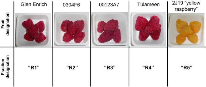
Table 2 – Different raspberry crops obtained from each of the five quasi-isogenic cultivars. The berries from each
cultivar were submitted in vitro digestion. The final digested fractions (specifically, the bio-accessible fractions to serum – fractions “IN”) were used at the present work. The correspondent designations attributed during this work are presented bellow each fruit.
Frui t de s ig n a tio n Glen Enrich Fra c tio n de s ig n a ti on “R1” “R2” “R3” “R4” “R5”
0304F6 00123A7 Tulameen 2J19 “yellow raspberry”
3.2. Microglial model of neuroinflammation
3.2.1 – Cell culture and treatments
The N9 murine microglial cell line was a gift from Dr. Teresa Faria Pais (Institute of Molecular Medicine, Lisbon). Cells were cultured in EMEM (Eagle Minimum Essential Media) supplemented with 10% fetal bovine serum (FBS) (Gibco®), L-glutamin (200 mM), 1% (v/v)
non-essential amino acids (NEAA) (Sigma–Aldrich® - Poole, Dorset, UK) and maintained at
37ºC in an humidified atmosphere of 5% (v/v) CO2. No antibiotics were used. Cells were
detached by agitation before suspension of the culture media with pipette. (No detaching agent, like trypsin, was used). Cell confluence was also monitored, avoiding cells to reach confluences higher than 80%.
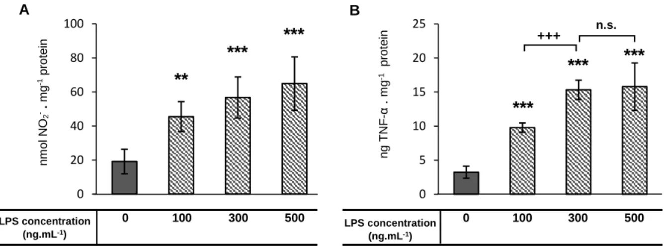
For the neuroinflammatory model establishment, N9 murine microglial cells were plated onto 6-well plates (5 x 10⁵ cells . mL-1) and cultured overnight to reach a confluence of
50-60%. Then, cells were stimulated with LPS (Sigma–Aldrich® - Poole, Dorset, UK) [100 to 500 ng . mL-1] during 24 hours, in order to release pro-inflammatory mediators, such as NO18 and
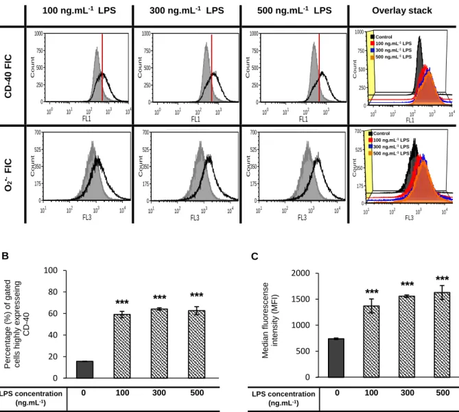
TNF-α15 in the media (see 3.2.3 section). CD-4064 and superoxide76, whose
expression/production is increased upon microglia activation, were quantified by flow cytometry (see 3.2.3 section). Also, Iba-180 was assayed by western blot (see 3.2.3 section).
To test the capacity of the different fractions (see chapter 3.1) in attenuation of neuroinflammation, N9 microglial cells were plated onto 6-well plates (5 x 10⁵ cells . mL-1) and
cultured overnight to reach a confluence of 40-60%. Then, cells were pre-incubated with the digested raspberry polyphenolic fractions [1 to 1.25 µg GAE . mL-1] during 2 to 24h. At this
stage, FBS concentration was reduced to from 10% to 0.5% (v/v), avoiding protein-polyphenol interactions and further precipitation. It was assured that the reduction of the FBS concentration did not affected the viability and functionality of the cells. After incubation with the fractions, media was discarded and cells were washed once with phosphate buffer saline (PBS) 500 µL prior to addition of fresh media with 10% FBS with LPS [100 to 500 ng . mL-1].
The inflammatory mediators validated in the model establishment (NO, TNF-α, CD-40 and superoxide) were assessed by using same methodologies.
3.2.2 – Protein extraction and quantification
Protein extraction and quantification was assayed for the normalization of the results and for western blotting. Cell media was removed and 150 µL of RIPA buffer [50 mM Tris (CarlRoth® – Schoemperlenstr, Karlsruhe, Germany); 150 mM NaCl (Sigma–Aldrich® - Poole,
Dorset, UK); 0.1% (w/v) Sodium dodecyl sulfate (SDS) (Merck® - Frankfurter Straße,
UK); 1% (v/v) NP-40; 0.05% (v/v) cocktail protease inhibitors (AppliChem Inc - Mary Avenue, Missouri, USA) ; and 0.4% (v/v) DNAse (Roche® – Basel, Switzerland)] were added. After
incubation for 15 minutes at room temperature, the lysate was scrapped, transferred into microtubes and centrifuged (10 minutes; 4ºC; 8000 x g). The lysate was stored at -80ºC and protein determination was performed by Lowry protein assay125.
3.2.3 – Determination of pro-inflammatory and activation markers
Nitric Oxide measurement by Griess Reaction
Because of the relatively short half-life of NO in aqueous solution, its quantitative measurement usually is indirectly accessed by the quantification of its oxidized products, nitrite and nitrate, which are regarded as suitable markers of NO release. The choice of the detection method depends on the type of cell and on the released quantity126, 127.
For this experiment, it was used the Griess Reagent (Sigma–Aldrich® - Poole, Dorset,
UK), which measures nitrite (Figure 5). For analysis, 100 µL of cell culture medium were quickly removed from each well of the culture plate and added to a 96-well plate for reading. For nitrite quantification, a standard curve of sodium nitrite [0 to 25 µM] was prepared. Equal volumes of Griess reagent were added to each well. The plate was incubated for 15 minutes at room temperature and the absorbance (Abs) was read at 540 nm.
Figure 5 - Griess reaction scheme. This reaction allows the detection of organic nitrite compounds and was first
described in 1858 by Peter Griess. This image belongs to the public domain, and contains no original authorship.
TNF-α quantification by ELISA
Cell supernatants were harvested after 24h and stored at -80ºC until analysis. TNF-α release was assayed by sandwich ELISA (Enzyme-Linked Immunosorbent Assay) according to the manufacturer’s instructions (PeproTech®; Princeton Business Park, Rocky Hill NJ,
United States)128. All the reagents and plates used were provided in the kit. For the standard,
recombinant murine TNF-α was diluted from 2 ng . mL-1 to zero in diluent. Antigen-affinity
purified goat anti-murineTNF-α diluted 1 µg . mL-1 in PBS was used as capture antibody. As
detection antibody, it was used a biotinylated antigen-affinity purified goat anti-murineTNF-α diluted 0.25 µg . mL-1 in diluent. As substrate, Avidin-HRP conjugate diluted 1:2000 (v/v) in
Finally, as liquid substrate, 2,2'-azino-bis(3-ethylbenzothiazoline-6-sulphonic acid) (ABTS) was used in the stock concentration. The plate was incubated at room temperature in a Synergy HT microplate reader, from Biotek® for 35 minutes, with 5-minute intervals Abs
405
readings.
CD-40 and superoxide (O2•-)quantification by flow cytometry
Culture media was discarded and 1 mL PBS was added to detach N9 adherent cells, which were then incubated with 1 mL mouse anti-FcγR (same as CD-16/32, from E-Biosciences) in FACS buffer (PBS containing 2% fetal calf serum and 0.01% NaN3) for 30 minutes at 4ºC before staining. Anti-FcγR was required for the blocking of Fc-mediated reactions with other specific antibodies129. Cells were spun down at 1000 x g, washed once
with 500 µL FACS buffer and doubly stained with 5 µg . mL-1 mouse anti-CD40 conjugated
with fluorochrome FITC (clone 3/23, from BD Biosciences®); and with 5 µg . mL-1 DHE probe
(Dihydroethidium, Invitrogen™, Carlsbad, CA, USA) as superoxide indicator. Events were acquired using CUBE 6 cytometer, from Partec®. Post-acquisition analysis was done with the
software FSC express 4 flow research edition®.
Iba-1 determination by Western Blot
For Western Blot analysis, 40 μg of total protein lysate from each treatment were separated by SDS-PAGE on a 12% (w/v) acrylamide gel. After electrophoresis, proteins were transferred to a polyvinylidene difluoride membrane, during 90 minutes at 70 Volt (V), 4ºC. The membrane was blocked in a solution of 5% (w/v) of membrane blocking agent (MBA) (GE Healthcare™, Wilmington, MA, USA) diluted in TBST (50 mM Tris; 150 mM NaCl; 0.05% Tween 20), and incubated for 1h with agitation at room temperature. Then, the membrane was incubated primary antibody [rabbit anti-iba1 (0.7 µg . mL-1) from WAKO] overnight, at 4ºC. After
washing 3x with TBST for 5 minutes, secondary antibody was added [goat anti-rabbit IgG HRP-conjugated (1:300), from Millipore] and the membrane incubated for 2h, with agitation at room temperature. Membrane was washed 3x during 5 minutes with TBST and additionally washed 1 minute with TBS (50 mM Tris; 150 mM NaCl) prior to enhanced chemiluminescence substrate (ECL) addition (PhemtoMax Super Sensitive Chemiluminescence HRP Substrate, Rockland, Gilbertsville, USA). Proteins were visualized by chemiluminescent detection using Molecular Imager ChemiDoc XRS (Quantity One™ software v.4.6.6; BioRad®, Amadora,
3.2.4 – Cytotoxicity assays
Cytotoxicity of raspberry digested polyphenolic fractions was tested with cell viability assay (CellTiter-Blue Cell Viability Assay, Promega®). This assay uses the indicator dye
resazurin to measure the metabolic capacity of cells as an indicator of cell viability. Viable cells retain the ability to reduce resazurin into resorufin, which is highly fluorescent. Nonviable cells rapidly lose metabolic capacity, do not reduce the indicator dye, and thus do not generate a fluorescent signal130.
Cells were plated into 96-well plates (5 x 10⁵ cells . mL-1 for final volumes of 100 µL per
well). After 24 hours of growth, four concentrations (0.25; 0.5; 1 and 2 µg GAE . mL-1) of the
five digested raspberry fractions were applied. The fractions were dissolved in culture media with 0.5% FBS to avoid major protein-polyphenol interactions and further precipitation. After 21h of incubation with fractions, 20µL of CellTiter-Blue reagent was added to each well. The plate was briefly shaken and incubated again with standard cell culture conditions for 3 hours (performing 24h of total incubation time with fractions). Fluorescence values were recorded in a Synergy HT microplate reader, from Biotek® and normalized for viability percentage relatively
to control.
3.3. Yeast model of inflammation
3.3.1. Characterization of Saccharomyces cerevisiae model of inflammation
As previously described in the Theoretical Fundaments section, NFAT modulates microglial activation. This transcription factor has an orthologous gene in yeast – CRZ1. Crz1 is also regulated by calcineurin, a calcium dependent enzyme, in a very similar manner as NFAT does in mammalian cells131. When dephosphorylated by calcineurin, Crz1 translocates
from the cytosol to the nucleus and binds to the calcineurin-dependent response element (CDRE)132, 133. While in mammals NFAT binds to diverse promoters that activate
pro-inflammatory gene expression40, 41, in yeast, Crz1 binding to CDRE promotes activation of
genes related to the response against cellular stress, such as ion pumps134, 135. These
regulatory similarities are the basis for the use of this microorganism as an eukaryote model of inflammation.
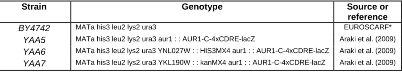
YAA5, a S. cerevisiae transgenic strain obtained from the wild-type BY4742, makes use of β-galactosidase expression as a reporter system to infer the anti-inflammatory potential of compounds. Additionally, the strains YAA6 and YAA7 are both used as negative controls. While YAA6 carry a deletion of CRZ1, YAA7 is devoid of CNB1, which encodes the regulatory subunit of calcineurin. The genotypes of each strain are presented in the Table 3 136.
Table 3 - S. cerevisiae strains used in this work. (From, R. T. Ferreira et al., Microbiology 158, 2293 (2012)).
Strain Genotype Source or
reference
BY4742 MATa his3 leu2 lys2 ura3 EUROSCARF*
YAA5 MATa his3 leu2 lys2 ura3 aur1 : : AUR1-C-4xCDRE-lacZ Araki et al. (2009)
YAA6 MATa his3 leu2 lys2 ura3 YNL027W : : HIS3MX4 aur1 : : AUR1-C-4xCDRE-lacZ Araki et al. (2009)
YAA7 MATa his3 leu2 lys2 ura3 YKL190W : : kanMX4 aur1 : : AUR1-C-4xCDRE-lacZ Araki et al. (2009)
* EUROpean Saccharomyces Cerevisiae ARchive for Functional analysis.
Calcium is the most common used inducer of Ca2+-signaling pathways137, since it is
captured and accumulated by calmodulin, which can effectively stimulate calcineurin phosphatase activity138. However, other molecules/ions are described as indirect inducers of
the system139, such as Mn2+ or Li+.
3.3.2 – β-galactosidase assays
Quantification of β-galactosidase activity was performed using different colorimetric substrates. Qualitative measurements were carried out in agar plates where yeast colonies were overlaid with an agarose solution containing 5-bromo-4-chloro-3-indolyl-β-D-galactopyranoside (X-gal) (ImmunoSource® - Ruiterslaan 29, Zoersel, Belgium). Quantitative
measurements were performed in a 96-well plate with cell lysates exposed to the colorimetric substrate Ortho-Nitrophenyl-β-galactoside (ONPG) (Sigma–Aldrich® - Poole, Dorset, UK).
Qualitative assay
Yeast strains were grown overnight at 30ºC with agitation in synthetic complete media (SCM) (Table 4) to the exponential growth phase. In the following day, the Abs600 was
measured and 4 x 10⁷ cells were centrifuged at 900 x g for 3 minutes and resuspended in 1
mL SCM. The anti-inflammatory potential of fractions was tested by incubating cells with 100 µg GAE . mL-1 of the five digested raspberry polyphenolic fractions (R1, R2, R3, R4 and R5).
Immunosuppressant macrolide drug FK506 (Sigma–Aldrich® - Poole, Dorset, UK) dissolved in
dimethylsulfoxide (DMSO) was used as positive control (6 µg . mL-1). Cells were incubated for
90 minutes at 30ºC with agitation. After incubation, cells were centrifuged at 900 x g for 3 minutes and the supernatant was discarded. Cells were washed with three time with PBS to remove polyphenols. The supernatant was completely removed and the cells were resuspended in 5 uL of SCM. Cells were spotted onto agar-SCM supplemented or not with 1.8 mM MnCl2. Cells were incubated for 90 minutes at 30ºC before X-Gal overlay - Agarose
solution was applied [0.2% (w/v) SDS; 2 mg . mL-1 X-Gal (solubilized in dimethilformamide);
0.5% (w/v) agarose; 50% (v/v) Lac Z buffer (8.5 g . L-1 Na
2HPO4; 5.5 g . L-1 NaH2PO4 ●H2O;
0.75 g . L-1 KCl; 0.246 g . L-1 MgSO
activity start to develop the indigo color, which is proportional to the activation of Crz1. Images were recorded each 10 minutes for 2h (Menezes et al; 2003; with modifications).
Table 4 - Synthetic complete media constitution.
Constituent Final concentration
Complete supplement mixture
(CSM) (QBiogene®) 0.79 g . L-1
Yeast nitrogen base (YNB)
(Difco®, USA) 0.67% (w/v)
Glucose (Sigma–Aldrich® - Poole,
Dorset, UK) 2% (w/v)
Quantitative assay
Yeast strains were pre-cultured in SCM overnight at 30ºC with agitation to obtain cells in exponential growth phase. In the following day, cells were diluted in fresh SCM and cultured for 8h at 30ºC. Then, Abs600 was measured and cells were again diluted to obtain cultures with
Abs600 1 after an overnight incubation. Once reached Abs600 1, cells were diluted in fresh SCM
to a final Abs600 0.1 and 300 L of cultures were transferred into microtubes. As a
pre-treatment, 100 µg . mL-1 of the five digested raspberry polyphenolic fractions (R1, R2, R3, R4
and R5) were applied. As positive control, 6 µg . mL-1 of FK506 dissolved in DMSO were used.
Final volumes of each pre-treatment were equalized in order to avoid interference in the Abs600.
Pre-treated cells were incubated for 90 minutes at 30 ºC. After incubation, cells were thoroughly resuspended and 150 µL were transferred to a second microtube containing 1.8 mM MnCl2. Induced and control cells were incubated for 90 minutes at 30ºC. After incubation,
microtubes were thoroughly resuspended and 10 µL of the cell suspension from each treatment were transferred onto a 96-well plate containing 20 µL Y-PER cell lysis reagent (Pierce® Protein Research, USA) per well. Three technical replicates were generated from
each microtube. The plate was incubated for 20 minute at 30ºC for cell lysis. For the β-galactosidase activity quantification, 240 µL of a solution containing 1 mg . mL-1 ONPG
dissolved in Lac-Z buffer (8.5 g . L-1 Na
2HPO4; 5.5 g . L-1 NaH2PO4 ●H2O; 0.75 g . L-1 KCl; 0.246
g . L-1 MgSO
4 ● 7H2O) were added to each well. The plate was incubated in a Synergy HT
microplate reader, from Biotek® for 2h at 30ºC and absorbance readings at 420 nm were
performed every 10 minutes.
3.4. Statistics
Data are presented as mean values ± standard errors (SE) or standard deviations (SD). Statistical differences were tested using unpaired one-way ANOVA with Tukey post hoc comparison, and considered significant when p<0.05.