Faculdade de Farmácia
Analogues of the antimicrobial peptide
BP214
Catarina Maria Boto Freire
Mestrado Integrado em Ciências Farmacêuticas
Faculdade de Farmácia
Analogues of the antimicrobial peptide
BP214
Catarina Maria Boto Freire
Monografia de Mestrado Integrado em Ciências Farmacêuticas apresentada à Universidade de Lisboa através da Faculdade de Farmácia
Orientador: Professora Doutora Maria de Jesus Perry Rocha
Co-Orientador: Professor Doutor Paul Robert Hansen
5
Abstract
One of the most concerning health problems present in the twenty-first century is antibiotic-resistant infections. The growing number of multi-resistant infections has led to complicated public health problems that require the most consideration. Simultaneously there are economic and regulatory barriers. Having all of these difficulties in mind, some researchers are trying to find new therapeutic classes that may became reliable alternatives to the therapeutics use nowadays. One of these new classes may be antimicrobial peptides. AMPs have demonstrated antimicrobial activity which makes them a good choice for research. Their cationic amino acids confer the molecule an attraction towards some components of the bacteria, this leads to the possibility of destroying the microorganism due to membrane rupture.
The AMP studied on this work was BP214, which was characterised in some previous studies. BP214 has shown a similar antimicrobial activity to colistin (a last-line therapeutic) and a reduced hemolytic activity compared to other AMPs. Therefore, on this study, eleven different BP214s analogues were synthesised and studied in order to discover which amino acids are essential to its antimicrobial and hemolytic activities. Each of these analogues differs from BP214 only in one amino acid that was replaced by an alanine.
The results demonstrated that only the peptides that had a lysine or an arginine replaced showed an improved antimicrobial activity but also an increase in their hemolytic activity. The remaining replacements resulted in a loss of antimicrobial and hemolytic activities of the peptide. It was also possible to associate the loss of activity to the low hydrophobicity of the molecule, which resulted from the amino acids replacement. The peptides where the lysine or the arginine were replaced, were also the ones that demonstrated the higher hydrophobicity. Both amino acids are basic and possess high values of pKa. This characteristic can be closely related with higher antimicrobial activity.
Key-words: Gram-negative infections; Antimicrobial peptides; Antimicrobial
6
Resumo
Um dos maiores problemas do século vinte e um tem sido a resistência aos antibióticos. O crescente número de infeções multirresistentes desencadeou um problema de saúde pública, o que requer uma enorme preocupação por parte da sociedade. Aliados a esta problemática estão os obstáculos a nível económico e regulatório. Tendo todas estas dificuldades em consideração, alguns investigadores estão a tentar encontrar novas classes terapêuticas que possam conferir alternativas às terapêuticas existentes de modo a podermos combater estas infeções com armas inovadoras e contra as quais os patogénicos ainda não possuam resistências.
Uma das novas classes emergentes podem ser os péptidos antimicrobianos. Estes péptidos têm demonstrado uma atividade antimicrobiana que faz deles uma boa opção contra as bactérias resistentes. Os seus aminoácidos catiónicos conferem-lhes uma atração específica a determinados componentes das bactérias, levando depois à possibilidade de as eliminar por rotura da membrana. Estes péptidos têm também atividades imunomodulatórias em que são capazes de aumentar a resposta imunitária contra o patogénico, aumentar a quimioatração e ativar a resposta inata e adaptativa, antibiofilme em que conseguem impedir a sua formação mesmo abaixo da sua concentração mínima inibitória e anticancerosas pela sua atividade citolítica específica para tecido tumoral que, tal como as bactérias está carregado negativamente.
Entre as várias vantagens dos péptidos antimicrobianos está a facilidade de síntese. Através de uma síntese bastante simples é possível manipulá-los aminoácido por aminoácido de modo a obter um híbrido o mais potente possível em comparação com o péptido original. Deste modo, é viável controlar a sua relação estrutura-atividade. De uma maneira tão simples, é também possível adicionar novos aminoácidos à sequência ou mutar o péptido de modo a torná-lo mais estável ou menos agressivo para o ser humano. Esta síntese pode ser feita em pequena escala no laboratório, mas também pode ser transferida para ser produzida numa grande escala de modo automatizado.
Apesar de entre si poderem ser bastante diferentes, os péptidos antimicrobianos possuem algumas características que são transversais a todos eles. Por exemplo, possuem um comprimento médio de menos de 60 aminoácidos, são geralmente carregados positivamente (com carga entre +2 a +9) e as suas estruturas são flexíveis e anfipáticas, ou seja, parte da molécula é hidrofóbica e outra é hidrofílica. Vai ser esta última característica que vai permitir ao péptido passar da sua conformação em solução para a conformação que permite a sua entrada na membrana da bactéria. Esta particularidade vem dos seus aminoácidos catiónicos e hidrofóbicos. São estes aminoácidos catiónicos que desencadeiam uma atração electroestática para as moléculas aniónicas da membrana da bactéria, como os lipopolissacáridos das bactérias gram-negativas e os ácidos lipoteicóicos das gram-positivas. É também através desta peculiaridade que mantém um elevada especificidade para as células procariotas ao invés das eucariotas.
A estrutura catiónica vai então levar a uma interação do péptido com a membrana da bactéria e também com os lípidos da membrana citoplasmática, ambos carregados negativamente. A sua carga positiva vai então estabilizar a carga dos fosfolípidos da membrana e causar a sua perturbação. A estrutura anfipática vai depois permitir a sua
7 inserção na bicamada da membrana resultando na sua disrupção e posterior morte da bactéria.
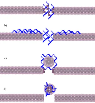
O mecanismo exato de como esta disrupção acontece ainda não é totalmente conhecido, sendo que existem diversas teorias para o explicar mas ainda nenhuma foi globalmente aceite. As teorias existentes até ao momento são o modelo “barrel-stove” (i) em que se assume que a inserção do péptido se dá com a orientação das regiões hidrofóbicas no core dos lípidos, levando à formação de um poro que causa a disrupção da membrana; o modelo “toroidal-pore” (ii) que supõe que a inserção do péptido na bicamada leva a que esta se dobre e forme um poro que permite a associação do péptido às cabeças polares dos fosfolípidos; o modelo “aggregate” (iii) em que se crê que vai ocorrer a formação de agregados péptido-lípido, agregados esses que levam a flutuações na condutância e translocações de péptidos na membrana; e, por último, o modelo “carpet” (iv) em que se supõe que a conformação anfipática do péptido leva à sua acumulação na membrana da bactéria formando uma espécie de carpete, causando a disrupção (Figura 1).
Para além desta capacidade de disrupção da membrana da bactéria que os péptidos antimicrobianos possuem, eles possuem ainda alvos intracelulares. Apesar de precisarem de estar a uma determinada concentração para serem capazes de causar a lise das bactérias, com apenas concentrações muito baixas conseguem atingir os seus alvos no interior da bactéria, provando assim que estes fenómenos acontecem por mecanismos distintos. Estes péptidos vão então ser capazes de inibir o material genético da bactéria ao nível do ADN, do ARN, da síntese proteica e também da atividade citosólica das suas enzimas.
O péptido estudado neste trabalho foi o BP214 que já foi estudado em trabalhos anteriores e que demonstrou uma atividade antimicrobiana semelhante à colistina (uma terapêutica de última linha para o tratamento de infeções bacterianas multirresistentes) e uma atividade hemolítica reduzida quando comparado com outros péptidos antimicrobianos, tendo assim menos efeitos indesejados. Tendo esses estudos em conta, neste trabalho foram sintetizados onze análogos do péptido BP214 e, posteriormente, estudados ao nível da atividade antimicrobiana e hemolítica. Este trabalho foi realizado no sentido de poder descobrir quais os aminoácidos da sequência deste péptido que são essenciais para a sua atividade antimicrobiana e hemolítica e quais podem vir a ser alterados de modo a podermos aumentar a atividade antimicrobiana mas diminuir a atividade hemolítica, de modo a torna-los viáveis ao nível comercial. Cada um dos análogos vai diferir do BP214 apenas num único aminoácido que é consecutivamente substituído por uma alanina, um aminoácido bastante simples.
Os resultados demonstraram que apenas os péptidos em que uma lisina ou uma arginina foram substituídas resultaram num aumento da atividade antimicrobiana mas também num aumento da atividade hemolítica. As restantes substituições levaram à perda de atividade antimicrobiana e também hemolítica. Foi também possível associar essa perda de atividade à diminuição do carácter hidrofóbico da molécula, resultado da substituição que foi feita. Este parâmetro foi avaliado pela percentagem de eluente B, o eluente hidrófobo, utlizado no HPLC. Os péptidos em que se trocaram a lisina ou a arginina eram também aqueles que tinham maior hidrofobicidade e nos quais ocorreu um
8 aumento das atividades, quer hemolítica quer antimicrobiana. Quer a lisina como a arginina são aminoácidos básicos que conferiram um carácter mais básico à molécula. Esta característica pode estar intimamente relacionada com o aumento das suas atividades antimicrobianas e hemolíticas.
Palavras-chave: Infeção gram-negativa; Péptidos antimicrobianos; Atividade
9
Acknowledgments
First I want to thank Professor Maria de Jesus Perry Rocha for her guidance, patience and supervision. I would also like to thank Professor Paul Hansen for all the support through the three months of laboratorial work and for his knowledge and friendship.
Thanks to all my colleagues at Copenhagen University: Bala Prabhala, Wafaa Al-Mansour, Abdullah Ben Naim Lone, Sabbah Ahmed and Natalia Mocchanova. You made me feel at home, so thank you for all the fun moments we shared. A special thanks to Birgitte Simonsen for all her patience and knowledge, who made me feel comfortable and safe in the laboratory.
Thank you to Thomas T. Thomsen, who helped me with the microbiology, both with material and, specially, knowledge.
I would also like to thank my family and friends for making my trip to Copenhagen possible and for being so supportive through all the hardest moments.
At last I want to thank my colleague Catarina Coelho for doing this research with me. We shared a lot of time on this trip to Copenhagen, with some great and hard moments. I would not have shared this adventure with anyone else. I couldn’t be more proud of both of us for succeeding on developing this project.
10
Abbreviations
ACP Anticancer Peptides
AMP Antimicrobial Peptides
BOP (benzotriazol-1-yloxy)tris(dimethylamino)phosphonium hexafluorophosphate BOP (benzotriazol-1-yloxy)tris(dimethylamino)phosphonium hexafluorophosphate
CLSI Clinical and Laboratory Standards
Institute DCC N,N'-dicyclohexylcarbodiimide DCM Dichloromethane DIC N,N′-diisopropylcarbodiimide DIEA N,N-diisopropylethylamine DMF N,N-dimethylformamide
EC50 Half Maximal Effective Concentration HA Hemolytic Activity HATU N-[(dimethylamino)-1H-1,2,3- triazole[4,5-b]pyridine-1-ylmethylene]-N-methylmethanaminium hexafluorophophate N-oxide HF Hydrogen Fluoride HOAt 7-aza-1-hydroxybenzotriazole HOBt 1-hydroxybenzotriazole HOBT Benzotriazol-1-ol
HPLC High Performance Liquid
Chromatography
LPS Lipopolysaccharides
MALDI-TOF-MS Matrix-Assisted Laser Desorption Ionization time-of-flight mass spectrometry
MHB Mueller-Hinton Broth
MIC Minimal Inhibitory Concentration
PEG Polyethylene Glycol
RBC Red Blood Cells
SDS Sodium Dodecyl Sulphate
SPPS Solid-Phase Peptide Synthesis
TFA Trifluoroacetic Acid
TIS Triisopropylsilane
11
Table of Contents
Abstract ... 5 Resumo ... 6 Acknowledgments ... 9 Abbreviations ... 10 1. Introduction ... 13 2. Objectives ... 163. Materials and Methods ... 18
3.1 Solid-phase peptide synthesis ... 18
3.1.1 Resin and Linkers ... 19
3.1.2 Coupling Reagents ... 20
3.1.3 Protecting groups and Cleavage Reagents... 21
3.2 Analysis ... 22 3.3 Purification ... 23 3.4 Antimicrobial activity ... 23 3.5 Hemolytic activity ... 24 4. Results ... 25 5. Discussion ... 28 6. Conclusion ... 29 7. References ... 30 8. Appendices ... 32
List of Figures
Figure 1 - Schematic of different mechanism of AMP's membrane disruption ... 14Figure 2 - Coupling of the first amino acid to the resin linker scheme ... 18
Figure 3 - Activation of the carboxy function of the second amino acid scheme ... 20
Figure 4 - Fmoc group ... 21
Figure 5 - N-deprotection scheme ... 22
Figura 6 - %HA vs %B ... 27
12
List of Tables
Table 1 – MALDI and HPLC results of all analogues and BP214………..……25 Table 2 – Antimicrobial and hemolytic activities and hydrophobicity of all analogues and controls………..………26
13
1. Introduction
Nowadays it is rather common to see health campaigns regarding antibiotic resistance and the hazard associated with non-adherence to the antibiotic therapy. Despite all of this, studies prove that this is a matter of public health and it is still not under control. [1, 2] Since these campaigns have not increased public awareness, we now stand at a time which requires the existence of new alternatives to fight the everyday antibiotic resistance within the community. This problem has motivated the pursuit for new classes of antibiotics which are able to attack the microorganisms with a different pathway from the existing ones, from which the pathogens already can defend themselves. Therefore, some of the focus has been on antimicrobial peptides (AMPs). AMPs are peptides with a demonstrated antimicrobial component but also with immunomodulatory, antibiofilm and anticancer mechanisms. In addition, they are also known to enhance the activities of antibiotics through synergistic effects, since their incorporation into the patients’ treatment creates lower Minimal Inhibitory Concentrations (MIC) when compared to the one antibiotic alone. [3, 4]
AMPs have an advantage which is the easiness of synthesis. AMPs can be manipulated, each amino acid at a time, in order to make them more powerful than the natural analogue and also manipulating the relation structure-activity. Likewise, it is possible to insert new amino acids or mutate the AMP in order to improve its biological activity and stability. This synthesis can be made in a laboratory and then be transferred to a much higher scale by automatization of each step. It is also possible to make a library and screening of all manipulated and synthesised peptides in order to have a place with all the important and updated information about them. [3]
AMPs have diverse structural and functional attributes, their variety is something they are known for. Depending on their amino acid composition and structure they can be divided into five subgroups. The first subgroup includes the anionic peptides (i) that are negatively charged and are present in surfactant extracts, bronchoalveolar lavage fluids and airway epithelial cells and they are active against positive and gram-negative. The second subgroup, in which one the analogues of this study is included, comprises linear amphipathic α-helical peptides (ii), which are cationic. These are usually disordered in aqueous solutions, but in the presence of trifluoroethanol, sodium dodecyl sulphate (SDS) micelles, phospholipid vesicles and liposomes or Lipid A they are converted to a α-helix, either totally or at least part of the molecule. It is believed that increased α-helical content correlates with a stronger antimicrobial activity. The third subgroup includes the cationic peptides enriched in specific amino acids (iii), which have a linear structure and a lack of cysteine residues, although some can form extended coils. On the fourth subgroup of AMPs cationic peptides fragments (iv) are included, these peptides contain cysteine residues and establish disulphide bonds and stable β-sheets. The fifth and last subgroup are the peptides with cysteines (v) that form intramolecular bonding which are anionic. They may also form cationic peptides with fragments of larger proteins that will ensure the antimicrobial activity. They are similar to the others subgroups in terms of composition and structure, they establish disulphide bonds but it is still unclear about their innate immunity activity. [5, 6]
14 Despite their diversity, AMPs have a few characteristics in common. They have an average length of less than 60 amino acids (from 12 to not more than 100 amino acids), are overall positively charged (from +2 to +9) and also possess flexible and amphipathic structures in which clusters of hydrophobic and hydrophilic amino acids are segregated. This latest attribute will allow the AMP to move from the solution conformation to one that will permit membrane interaction. [6, 7] The other characteristics are what ensures the antimicrobial activity. Their ability to possess an amphipathic structure comes from its cationic and hydrophobic amino acids and its primary sequence, creating a membrane bound conformation. These cationic amino acids will create an electrostatic attraction towards the anionic molecules within the bacterial membrane, especially towards the lipopolysaccharides (LPS) of negative and the lipoteichoic acids from gram-positive, therefore creating its specificity towards prokaryotic cells and not eukaryotic ones. As a consequence of this affinity with the bacterial membrane, the AMP will interact with the negatively charged lipids within the cytoplasmic membrane. Since AMPs have the cationic residues, they will stabilize the phospholipids, resulting in the disturbance of the membrane. In addition, its amphipathic structure will allow their insertion inside the bilayer with consequential alterations of the membrane structure. [3]
The specific mechanism of the membrane disruption is still to be confirmed, although there are some theories about it. Different models have emerged to explain this phenomenon (Figure 1), such as the “barrel-stave model” which assumes the insertion of AMPs inside the bilayer using the orientation of the hydrophobic regions in its lipid core, resulting on the formation of a pore with a barrel-like shape, therefore the name.
Figure 1 - Schematic of different mechanism of AMP's membrane disruption
a) Toroidal-pore model b) Carpet model c) Barrel-stave model d) Aggregate model
15 The “toroidal-pore model” supposes that the insertion of the AMP will cause the bilayer to bend and form a peptide-lined pore and permit the AMPs association with the polar heads of the phospholipids. Another theory is the “aggregate model” which adopts the assumption of the formation of transient peptide-lipid aggregates that allows rapid fluctuations in transmembrane conductance as well as translocation of peptides across the membranes. The “carpet model” supposes that the AMPs with its amphipathic conformation will accumulate on the membrane surface and form a carpet-like cover. Then the bilayer structure will disrupt with the accumulation of AMPs. Due to the lack of information, it is still impossible to know which theory is the most similar to what happens in reality. [3]
Additionally to this ability to disrupt the membrane, AMPs also possess intracellular targets. These peptides need a certain concentration to be able to disrupt the membrane. However, even at a lower concentration they can translocate across the bilayer and cause damage into the pathogen. For instance, AMPs can inhibit DNA, RNA, protein synthesis and cytosolic enzymatic activity. [3]
Regarding the immunomodulatory activity of AMPs, studies have shown they are skilled on modulating and enhancing the host immune response. They are capable of modelling pro- and anti-inflammatory responses (such as stimulate prostaglandin release and recruiting immune cells), enhance chemoattraction, activate innate and adaptive immunity, induce wound healing and modulate autophagy, apoptosis and pyroptosis. [3, 8]
About the antibiofilm activity, that is the ability that AMPs possess to inhibit the formation of aggregates of bacteria in a key state of growth, when they can be extremely resistant to antibiotics. This is a key-benefit from the AMPs. It is also important to mention that this capability has been demonstrated at concentrations below the MIC. This means that AMPs don’t need to achieve MIC to inhibit growth and to develop antibiofilm activity. This suggests that the pathway for the antimicrobial activity of AMPs is different from the one that originates the antibiofilm action. There is also evidence that AMPs can not only prevent biofilm formation but also inhibit biofilm progression once it is already established, becoming an important weapon against biofilms. [3, 9]
Another property is the anticancer activity, explained by the cytolitic action of AMPs towards tumor cells specifically. This happens because of the electrostatic interactions between the positively charged AMPs and the negatively charged surface of tumor cells. On the other hand, healthy cells are slightly anionic and contain a lot of cholesterol resulting in a much more rigid membrane and harder to penetrate. Since they hold this aptitude, they can also be called anticancer peptides (ACP), being able to provide a new defence against cancer cells, as shown on some previous studies. [3, 10]
As for its disadvantages, AMP´s have quite high nephrotoxicity and there have been some evidence on clinical trials of immunogenic properties, therefore the immune system would create defences against them. These are some of the reasons why AMPs fail in human trials. In addition, they also possess an important hemolytic activity which must be tested before going into human trials. [3]
16
2. Objectives
This work was performed based on the newly reported peptide, BP214, by Oddo, A., et al. (2016) [11] which showed only slightly reduced antimicrobial activity compared to colistin (last-line therapeutic on many different gram-negative infections) and a hemolytic half maximal effective concentration (EC50) of >150 µM, with bactericidal characteristics and high activity against colistin-resistant strains featuring mutated lpxC, pmrA and pmrB genes. [11]
BP124 with the sequence kklfkkilryl-NH2 is an analogue of BP100, which is a hybrid of cecropin-α-melittin. This family of peptides has shown activity against colistin-resistant strains of A. baumannii and so, the interest in studying different analogues that might show promising results. [11]
Cecropins are antibacterial peptides that were initially isolated from the silk moth Hyalophora cecropia, thus the name. These peptides with 31-39 amino acids have basic amphipathic N-terminal and hydrophobic C-terminal domains. An organization of the amphipathic N-terminal helix connected to that hydrophobic C-terminal helix by a flexible hinge region is essential to a robust and broad-spectrum activity. Studies indicate that all D-enantiomers form left-handed helices, are protease resistant and possess equal levels of antimicrobial activity as the L-enantiomers. This is one of the reasons why the analogues used in this study are all D-enantiomers. Also because by adding D-amino acids the helicity, meaning the AMP’s ability to form a spin structure, is reduced, which is essential to reduce its hemolytic activity. [4, 12]
Melittin is also an antimicrobial peptide, with 26 amino acids. It was discovered in bee venom, from Apis mellifera. Just as cecropins, it has hydrophobic and hydrophilic domains but on the opposite order as the previous. Even though it is a broad-spectrum antimicrobial peptide it also has a high hemolytic activity, since it comes from a bee venom. [12]
Cecropin-α-melittin hybrids such as BP100 incorporate the cationic N-terminal domain from cecropin and the hydrophobic amino acids from N-terminal of the mellitin. Since all D-enantiomers retain the antimicrobial activity, the analogue BP 214 was synthesised with all D-amino acids. [12]
Having this results in mind, we went on to study different analogues of BP214 in order to identify the amino acids of this peptide which are essential to its activity, changing one amino acid at the time and replacing it for an alanine (D-Alanine scan). Glycine might also be an option but since its structure is very flexible, it might change the secondary structure of the molecules. The chosen amino acid was alanine since it is one of the simplest amino acids, being the only ramification a CH3 group, which will provide the necessary stability. [11]
The analogues were named: kklfkkilrya-NH2 as CF1, kklfkkilral-NH2 as CF2, kklfkkilayl-NH2 as CF3, kklfkkiaryl-NH2 as CF4, kklfkkalryl-NH2 as CF5, kklfkailryl-NH2 as CF6, aklfkkilryl-kklfkailryl-NH2 as CC2, kalfkkilryl-kklfkailryl-NH2 as CC3, kkafkkilryl-kklfkailryl-NH2 as CC4, kklakkilryl-NH2 as CC5 and kklfkailryl-NH2 as CC6. These peptides were synthetized, analysed, purified and tested with the aim of comparing their antimicrobial and hemolytic activities to BP214s. Despite only the first six analogues (CF1-CF6) were synthesized by
17 me, the reaming five were synthesized by my co-worker and are also referred to on a different assay, the conclusions include the results of all of the analogues, in order to compare all analogues. These peptides are expected to have a α-helical structure.
18
3. Materials and Methods
3.1 Solid-phase peptide synthesis
Peptide synthesis may be achieved using different kinds of methods. On this work the solid-phase peptide synthesis (SPPS) was the method of choice. This is a method that has different benefits such as the fact that all reactions happen in a single vessel (reactor). Moreover the excess of amino acids and reagents is used to carry the reactions to completion and is then filtered and washed away, preventing from having to purify the intermediates at each step. SPPS is also a quite quick synthesis and has improved yields as well. [13]
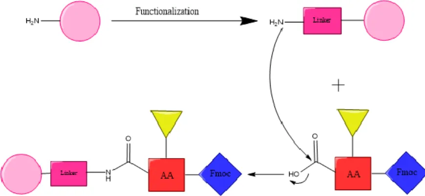
The SPPS is a method that consists of the use of an insoluble resin that will be able to anchor one amino acid at a time to permit the construction of the peptide. The amino acids will have the α-amino group and the side chain protected to avoid any unwanted reactions. This will allow the C-terminal amino acid to couple with the resin bifunctional linker at the Cα-carboxylic acid group (Figure 2).
With sequential protections and deprotections, it will be possible to add each amino acid through a nucleophilic attack of the amino group of one residue at the carboxylic group of the other residue that has been activated by an electron withdrawing group, until the peptide is finished with the N-terminus amino acid. And so, these peptides are belt by an amide bond formation that may allow the loss of the chiral integrity to happen. The mechanisms of this racemization are explained at 3.1.2 Coupling Reagents. [13-15]
The amount of each amino acid is calculated using equation 1. 𝑚𝑔 𝐴𝑚𝑖𝑛𝑜 𝐴𝑐𝑖𝑑 =
𝑀𝑜𝑙𝑒𝑐𝑢𝑙𝑎𝑟 𝑤𝑒𝑖𝑔ℎ𝑡 𝑜𝑓 𝐴𝑚𝑖𝑛𝑜 𝐴𝑐𝑖𝑑 × 3 × [𝑠𝑦𝑛𝑡ℎ𝑒𝑠𝑖𝑠 𝑠𝑐𝑎𝑙𝑒(𝑚𝑚𝑜𝑙)] (1) After this happens, it is necessary to cleave the peptide from the resin to be able to analyse and purify it and then test its antimicrobial and hemolytic activity. [13]
19
3.1.1 Resin and Linkers
To begin this synthesis it is necessary to start with the correct resin, which requires chemical, mechanical and physical stability. This is a polymer that is either poly-styrene based, a polyethylene glycol (PEG) -grafted poly-styrene, or a PEG without any styrene. In this case, it was used a PEG-grafted poly-styrene. This is the commonly used resin when using a SPPS with Fmoc strategy. The Fmoc (9-fluorenylmethyloxycarbonyl) (Figure 4) is the chosen protecting group that is used to protect the α-amino group. This is the most usually used protective group since the mainly alternative group for standard SPPS, the Boc (tert-butyloxycarbonyl) group, requires the use of hydrogen fluoride (HF) for the side chain deprotection. Besides, it has lower specificity for the cleavage of the N-terminus amino acid instead of the protecting groups of the side-chain, which must remain until the end of the synthesis to avoid unwanted reactions. On the other hand, Fmoc is a strategy that allows an orthogonal approach, being the Fmoc group base-labile and the side chain group acid-labile, overpassing those disadvantages. Therefore, we used the PEG-grafted poly-styrene resin, which is a graft PEG coupled to a low cross-linked, by an ether linkage, polystyrene matrix. An example of this resin is the TentaGel, the one used for this work. Its pressure stability allows the use in batch and continuous flow conditions. [3]
This resin has to be swollen on a solvent, in this case we used N,N-dimethylformamide (DMF), a polar aprotic solvent. After this functionalization of the resin, it was be able to reach the bifunctional linker. The linker is the portion of the resin that will allow the coupling of the first amino acid and then its cleavage from the resin. The linker for this resin is the Rink linker, which is the most used for peptide amides. The docking of the first amino acid in this case is achieved by the same method as a typical amino acid coupling. That is why this was the linker chosen in this study, for the easiness of the process. [13]
These resins often come along with a loading value. That is the quantity (mmol) of functional groups it contains for every gram. Once again, it is important to say that the resin used was a low-loading resin, which means it has a loading of ≤0,25 mmol/g. This is usually associated with a low bead size, as the one used, with 100-200 mesh (equivalent to a particle size of 74-149 µm). These characteristics will avoid peptide aggregation and undesired intermolecular reactions. [13]
The amount of resin used is calculated by equation 2. 𝑔 𝑜𝑓 𝑅𝑒𝑠𝑖𝑛 =𝑚𝑚𝑜𝑙 𝑜𝑓 𝑃𝑒𝑝𝑡𝑖𝑑𝑒
20
3.1.2 Coupling Reagents
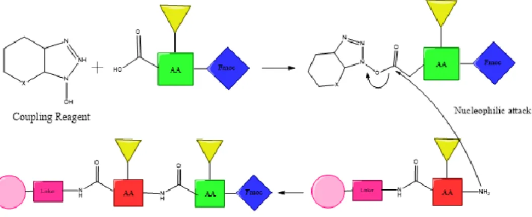
To ensure the coupling of each amino acid to the resin linker and to the previous amino acids it is necessary to use coupling reagents. They will ensure the connection between the free N-terminus and carboxyl function of the upcoming amino acid.
These can be carbodiimides such as DCC (N,N'-dicyclohexylcarbodiimide) and DIC (N,N′-diisopropylcarbodiimide), phosphonium salts such as BOP ( (benzotriazol-1-yloxy)tris(dimethylamino)phosphonium hexafluorophosphate) or amonium salts such as HOBT (Benzotriazol-1-ol) and HATU ( (N-[(dimethylamino)-1H-1,2,3-triazole[4,5-b]pyridine-1-ylmethylene]-N-methylmethanaminium hexafluorophophate N-oxide). The latest was the one used in this study. As explained, this reagent will allow amide bond formation reactions by offering a favourable leaving group, leading to the activation of the carboxy function (Figure 3).
The amount of HATU necessary is calculated using equation 3. 𝑚𝑔 𝐻𝐴𝑇𝑈 = 380,23 × 3 × [𝑆𝑦𝑛𝑡ℎ𝑒𝑠𝑖𝑠 𝑆𝑐𝑎𝑙𝑒(𝑚𝑚𝑜𝑙)]
× [𝑛𝑢𝑚𝑏𝑒𝑟 𝑜𝑓 𝑐𝑜𝑢𝑝𝑙𝑖𝑛𝑔𝑠] (3)
Nevertheless, these reactions may lead to racemization of the chiral α-carbon since the amide bond happens between optically active monomers. There are two pathways for this to happen and both of them are base-catalyzed. One is through direct enolization and the other is the formation of 5(4H)-oxazolone. [14, 15]
To minimize this kind of racemization, it is common to use coupling “additives” such as HOBt (1-hydroxybenzotriazole) or HOAt (7-aza-1-hydroxybenzotriazole). In this case we used HOAt as it is more effective than HOBt because it holds a nitrogen in the aromatic ring. [3, 13]
The amount of HOAt necessary is calculated using equation 4.
𝑚𝑔 𝐻𝑂𝐴𝑡 = 135,12 × 3 × [𝑆𝑦𝑛𝑡ℎ𝑒𝑠𝑖𝑠 𝑆𝑐𝑎𝑙𝑒(𝑚𝑚𝑜𝑙)] × [𝑛𝑢𝑚𝑏𝑒𝑟 𝑜𝑓 𝑐𝑜𝑢𝑝𝑙𝑖𝑛𝑔𝑠] (4) Also with these coupling reagents it was used DIEA (N,N-diisopropylethylamine), a non-nucleophilic base, since they all require the addition of a base. The amount of DIEA necessary is calculated using equation 5.
21 µ𝐿 𝐷𝐼𝐸𝐴 =
[129,24𝑚𝑜𝑙𝑔 × 6 × [𝑆𝑦𝑛𝑡ℎ𝑒𝑠𝑖𝑠 𝑠𝑐𝑎𝑙𝑒 (𝑚𝑚𝑜𝑙)]]
0,742𝑚𝐿𝑔 (5)
The coupling reactions are alternated with washes of DMF. Dichloromethane (DCM) is more commonly used, however it is less appropriate for the next coupling reaction that will happen quicker in more polar solvents. Therefore, DMF was the selected solvent, a good choice since both Fmoc group protected amino acids and the triazole-based reagents are soluble in it and can be washed away between every coupling along with other soluble impurities. [3, 13]
The volume of DMF necessary to dissolve HATU and HOAt before adding them to the reaction is calculated using equation 6.
𝑉 𝑆𝑜𝑙𝑢𝑡𝑖𝑜𝑛 (𝑚𝐿) =3 × [𝑆𝑦𝑛𝑡ℎ𝑒𝑠𝑖𝑠 𝑆𝑐𝑎𝑙𝑒 (𝑚𝑚𝑜𝑙)] × [𝑛𝑢𝑚𝑏𝑒𝑟 𝑜𝑓 𝑐𝑜𝑢𝑝𝑙𝑖𝑛𝑔𝑠]
0,4𝑀 (6)
3.1.3 Protecting groups and Cleavage Reagents
To avoid lateral reactions between the carboxylic acid and other nucleophilic species that are not the pretended group, it is necessary to use protecting groups. These groups can be used in an orthogonal way, meaning there are two different protecting groups on two different functional groups and one can be removed without removing the other. This is possible by one being acid labile and the other base labile, allowing a selective removal of the group protecting the α-amino group without removing the one on the side chain. [3]
For the α-amino group was used the Fmoc group (Figure 4), as it was explained above (3.1.1. Resin). The removal of this group is possible using a base-induced β-elimination.
This happens with the presence of a secondary amine, such as the one used, piperidine. The process is also called N-deprotection (Figure 5). Piperidine will have a role as a base, as a scavenger for the dibenzofulvene that is released during the Fmoc exclusion and as
22 an obstacle for the attachment of the dibenzofulvene to the amino group, being a good choice as a removal group. [3]
For the side chain the protecting group will depend on which amino acid is added. For the amino acids Ser, Thr, Tyr, Glu and Asp the used group is tert-Bu. If the amino acids used are Cys, Asn, Gln or His it is used the trityl group. [13]
After the synthesis is completed, these groups can be cleaved. This often happens at the same time as the peptide is cleaved from the resin itself, using a cleavage solution mostly composed by trifluoroacetic acid (TFA). This solution may have different concentrations of this strong acid diluted in, for example, water, triisopropylsilane (TIS), thioanisole or phenol. On this study it was used what is normally called reagent B’ that consists of TFA, water and TIS with a ratio of 95:2,5:2,5 (v/v). [13]
3.2 Analysis
After the synthesis of the peptides, it is mandatory that they go through an analysis. Doing so will guarantee that the desired peptide is present on each sample. For this, the methods of choice were analytical reverse-phase High Performance Liquid Chromatography (analytical HPLC) and Matrix-Assisted Laser Desorption Ionization time-of-flight mass spectrometry (MALDI-TOF-MS). [3]
Regarding the analytical HPLC, this consists of the passage of the samples through a column that will work as a stationary phase using different liquid buffers as the mobile phase, allowing the separation of the sample into different bands, depending on their retention time, that can then be detected by the mass spectrometer and create a chromatogram. On this chromatogram each band is seen as a peak that can be quantified by its intensity and identified by its retention time. On this work the stationary phase used was a C-18 (octadecyl carbon chain) column and the mobile phase consisted on 3 different buffers. Buffer A which was a solution of 99,9% water and 0,1% TFA, buffer B was a solution of 10% water, 90% acetonitrile and 0,1% TFA and buffer C was 100% acetonitrile. [3]
MALDI-TOF-MS is a method which requires the dilution of a portion of the sample into a highly absorbing laser light matrix and letting it dry on a MALDI-target plate. When the sample is dry into a crystalline deposit it can be analysed. It is placed into the machine and trough the sample, a projected a laser pulse will create desorption and ionisation of the sample. After this, the ions are accelerated by a strong electric field and then separated and detected. This separation happens depending on each ion time-of-flight, therefore by their mass-to-charge ratio (m/z). By calculating the expected mass of each peptide synthesized it is possible to know if it matches to the mass present on the
23 sample estimated by the MALDI-TOF-MS. The chosen matrix for this study was α-cyano-4-hydroxycinnamic acid (ACCA). [3]
3.3 Purification
The purification of the synthesized peptides is an important step, allowing the elimination of possible impurities that may contaminate the samples. Therefore, the purification on this work was performed using preparative HPLC. The principle behind this is very similar to the one used for analysis. The HPLC will allow the separation in different bands, just the same, but this time each band can be collected as it is being separated by its retention time. Thus, it is possible to collect the chromatogram peak of the peptide, which usually corresponds to the higher peak, meaning is the main compound present in the sample, since its quantity will correspond to its intensity. After that, it is possible to confirm that the peak corresponds to the wanted peptide, by doing a MALDI-TOF-MS and an analytical HPLC, same principle as for analysis. [3]
3.4 Antimicrobial activity
The determination of the peptides antimicrobial activity will find the MIC of each peptide synthesized. Therefore, it will be possible to compare the different MIC´s with BP214’s MIC [11] discovering which amino acids can be replaced without losing BP214´s activity or which can even improve its antimicrobial activity.
MIC test was performed using the broth microdilution (≤500µL/well) technique in Mueller-Hinton broth (MHB) used for cultivation of a wide variety of nonfastidious microorganisms, in accordance with the Clinical and Laboratory Standards Institute (CLSI) guidelines. The MIC was defined as the lowest concentration of peptide which inhibited visible growth of bacteria on the plates (sediment) under defined conditions and was performed in triplicate. [16]
The peptides and antibiotics were added to polystyrene 96 well plates. The first well concentration of the antibiotics were chosen according to the CLSI guidelines and for the peptides it was chosen according to previous results. [11] The chosen strains for this work were the gram-negatives Escherichia coli ATCC 25922, Acinetobacter baumannii ATCC19606,
Pseudomonas aeruginosa ATCC 27853 and Klebsiella pneumoniae ATCC
13883 with inoculums of about 5 x 10⁵ colony forming units (cfu) ml-1. This was ensured by spectrophotometer, being the absorbance proportional to the concentration of cells. They were isolated in pure culture and were identified at the genus and species level. [16] For a matter of control of the test values for each strain, routinely used antibiotics were added to the plate in order to compare the obtained MICs to the published ones. Only if the MIC of those antibiotics was between the published ranges, the MIC for tested peptides would be taken into consideration. Therefore it was used Ciprofloxacin and Colistin as controls and also RW-BP100 and BP214 whose MIC was already determinated previously. [11, 16]E. coli is one of the most common microorganisms responsible for Urinary Tract Infections (UTI) which are quite common within the hospital environment since there is a high recourse of urinary catheters. It is of most importance that this pathogen is included
24 on this work, since it would be of great assistance if there could be added a new therapeutic weapon for these patients treatment. [17]
A. baumannii infections are really difficult to treat, particularly because of extensively drug-resistant (XDR) and for the lack of treatment options available, making it also an important pathogen for this study. [18]
Pseudomonas aeruginosa is one of the pathogens responsible for the majority of nosocomial pneumonias. In addition, this pathogen has a strong ability to develop carbapenem resistance, probably because of a mutational inactivation of the oprD gene. Being carbapenems the last-line therapy used on hospitalized patients with Pseudomonas aeruginosa infections, this leads to a very difficult situation where there isn’t an alternative treatment. Therefore, this is an important microorganism to have into account while doing this work. [19]
The same problem happens with Klebsiella pneumoniae which can also develop a resistance to carbapenems, mainly by the presence of the blaKPC gene but this also happens through different other mechanisms and even different associations of mechanisms. Consequently, this pathogen is also of great importance for this study. [20]
3.5 Hemolytic activity
The hemolytic activity (HA) is a high importance parameter to take into consideration, since only peptides with low activity against erythrocytes can be used as a therapeutic alternative. This method is commonly used since it is inexpensive and fast to perform. It will provide a quick idea on the toxicity of the peptide before going too further and investigating on more complicated tests, even though a non-hemolytic compound can still be toxic through different mechanisms. [21]
This parameter was evaluated using human red blood cells (RBC) and a percentage of hemolysis was assigned for each peptide, being the 100% hemolysis induced by a mellitin stock solution of 1,25 mM mellitin in phosphate buffer saline (PBS) and the 0% hemolysis by a PBS solution. Mellitin is the “gold-standard” positive control because of its broad spectrum of lytic activity. [21, 22]
Each peptide was tested in triplicate on 96-wells microtiter polypropylene plates with different concentrations using two-fold dilutions with PBS. Their hemolytic activity towards the type-0 blood was determined by spectrophotometry at 414 nm. The absorbance will vary as the haemoglobin is released during RBC lysis, meaning that the more haemoglobin is released the higher the absorbance. The percentage of HA is determined by equation 6.
%𝐻𝐴 = 𝐴𝑏𝑠(𝑃𝑒𝑝𝑡𝑖𝑑𝑒) − 𝐴𝑏𝑠(𝑁𝑒𝑔𝑎𝑡𝑖𝑣𝑒 𝐶𝑜𝑛𝑡𝑟𝑜𝑙)
𝐴𝑏𝑠(𝑃𝑜𝑠𝑖𝑡𝑖𝑣𝑒 𝐶𝑜𝑛𝑡𝑟𝑜𝑙) − 𝐴𝑏𝑠(𝑁𝑒𝑔𝑎𝑡𝑖𝑣𝑒 𝐶𝑜𝑛𝑡𝑟𝑜𝑙)× 100 (6)
The HA is presented as the peptide concentration that results in 10% or 50% of erythrocytes lysis, HC10 or HD50 respectively. [22]
25
4. Results
The antimicrobial activities and the hemolytic activities of all analogues of BP214 and controls are presented in Table 2. For consistency with previous literature, the “BP” designation was maintained for BP214. The new analogues were named CF1-CF6 and CC2-CC6, designating all the eleven analogues possible to obtain from changing one amino acid of BP214 to an alanine. Each peptide sequence is presented in Table 2 associated with the correspondent name. Each amino acid is represented by a letter, being k for lysine, l for leucine, f for phenylalanine, i for isoleucine, r for arginine, y for tyrosine, a for alanine and w for tryptophan.
All peptides were purified until a purity of at least 95% was obtained; their retention time and mass is presented in Table 1.
For the antimicrobial activity test, the controls used were RW-BP100, Ciprofloxacin and Colistin, which MICs are previously known. MIC is represented by the concentration (µg/mL) of peptide that inhibited completely the formation of bacteria in the wells.
For the hemolytic activity the controls were Ciprofloxacin and Colistin, since their hemolytic activities were previously known. The hemolytic activity is presented both in HA10, where it is represented the concentration (µg/mL) that causes 10% of the red blood cells to lyse, and in %HA, being this the percentage of lysis observed at a concentration of 500µg/mL of peptide.
The molecule’s hydrophobicity was associated to its percentage of Acetonitrile used on the HPLC elution, since Acetonitrile is the hydrophobic eluent. This percentage was designated as %B (Acetonitrile was determined to be eluent B). The higher the percentage of B at the maximum peak of elution of the peptides, the higher the hydrophobicity.
27 The p-value for variable MIC is 0,000147.
For the variable %HA, its p-value is 1,84x10⁻⁵.
(n=11; r2=0,852343)
(n=11; r2=0,778005)
Figura 7 – Average MIC vs %B Figura 6 - %HA vs %B
28
5. Discussion
The MICs were similar to BP214s for analogues CF3, CF6, CC2, CC3 and CC6. This means that, these analogues have a comparable antimicrobial activity to BP214. Analogues CF1, CF2, CF4, CF6, CC4 and CC5 presented MICs considerably higher than the other analogues and also than BP214, meaning their antimicrobial activity is much lower.
Analogues CF1, CF2, CF4, CF6, CC4 and CC5 demonstrated low %HA and higher values of HA10, meaning that these are the analogues with lower hemolytic activity. On the other hand, CF3, CF6, CC2, CC3 and CC6 had inferior HA10 and higher %HA, making them harder to try-out on clinical trials.
Analogues with better MICs and higher HA presented higher %B, meaning they are the more hydrophobic. The analogues with higher MICs and lower HA presented lower %B, therefore being the more hydrophilic ones.
Both MIC and HA are related to the %B since their p-values are lower than 0,05. It is clear that MIC is inversely proportional to %B, meaning that lower MIC is related to higher values of %B, when the molecule is more hydrophobic. In contrast, HA is directly proportional to %B, being that when the molecule is more hydrophobic, HA is also higher.
29
6. Conclusion
In conclusion, the analogues with antimicrobial activities as promising has BP214 were CF3, CF6, CC2, CC3 and CC6. On the other hand, those were the analogues with higher hemolytic activities.
For all peptides CF6, CC2, CC3 and CC6 the changed amino acid was the lysine. The change from a lysine, a basic amino acid, to an alanine will enhance the molecules hydrophobicity, creating a molecule with better antimicrobial characteristics but also with higher hemolytic activities. Lysine’s pka (negative decadic logarithm of ionization constant) is 2,18 for its carboxyl group, 8,95 for the ammonium ion and 10,53 for its side chain group.
For peptide CF3 the substituted amino acid was arginine, also a basic amino acid. Once again, we can conclude that the change from an arginine confers the molecule higher antimicrobial activities but, then again, also higher hemolytic activity. Arginine’s pka is 2,17 for its carboxyl group, 9,04 for the ammonium ion and 12,48 for its side chain group. No other amino acid of the synthesized analogues, besides lysine and arginine, have a side chain passable of ionization.
Our analogues (with alanine amino acids for substitution) present the same properties as BP214: occurrence of a dual mode of action both specific and unspecific. Similarly to BP214, being all D-amino acids they are also stable on the proteolytic level. These characteristics make them interesting peptides to work with, in order to possibly mutate them to a form that is hemolytically less active but that is still active against bacteria.
30
7. References
1. Robert A, et al. Knowledge of antibiotics and antibiotic resistance in patients followed by family physicians. Med Mal Infect, 2016.
2. Simonsen GS. Antibiotic resistance - a global public health problem. Tidsskr Nor Laegeforen, 2008; 128: 2552.
3. Haney EF, Mansour SC, and Hancock RE. Antimicrobial Peptides: An Introduction. In: Hansen PR, editor. Antimicrobial Peptides: Methods and Protocols. 1st ed. Springer New York: New York; 2017.
4. Bahar AA and Ren D. Antimicrobial peptides. Pharmaceuticals (Basel), 2013;
6:1543-75.
5. Brogden KA. Antimicrobial peptides: pore formers or metabolic inhibitors in bacteria? Nat Rev Microbiol, 2005; 3: 238-50.
6. Diamond G, et al. The roles of antimicrobial peptides in innate host defense. Curr Pharm Des, 2009; 15: 2377-92.
7. Jenssen H, Hamill P and Hancock RE. Peptide antimicrobial agents. Clin Microbiol Rev, 2006; 19: 491-511.
8. Mansour SC, Pena OM and Hancock RE. Host defense peptides: front-line immunomodulators. Trends Immunol, 2014; 35: 443-50.
9. Thankappan B, et al. Antimicrobial and antibiofilm activity of designed and synthesized antimicrobial peptide, KABT-AMP. Appl Biochem Biotechnol, 2013; 170: 1184-93.
10. Theansungnoen T, et al. Cationic Antimicrobial Peptides Derived from Crocodylus siamensis Leukocyte Extract, Revealing Anticancer Activity and Apoptotic Induction on Human Cervical Cancer Cells. Protein J, 2016; 35: 202-11.
11. Oddo A, et al. An Amphipathic Undecapeptide with All d-Amino Acids Shows Promising Activity against Colistin-Resistant Strains of Acinetobacter baumannii and a Dual Mode of Action. Antimicrob Agents Chemother, 2015; 60: 592-9.
31 12. Sato H and Feix JB. Peptide-membrane interactions and mechanisms of membrane destruction by amphipathic alpha-helical antimicrobial peptides. Biochim Biophys Acta. 2006; 1758: 1245-56.
13. Hansen PR and Oddo A. Fmoc Solid-Phase Peptide Synthesis. Methods Mol Biol, 2015; 1348: 33-50.
14. Al-Warhi TI,. Al-Hazimi HMA and El-Faham A. Recent development in peptide coupling reagents. Journal of Saudi Chemical Society, 2012; 16: 97-116.
15. Stawikowski M and Fields GB. Introduction to peptide synthesis. Current Protocols in Protein Science, 2012; Chapter 18: Unit 18.1.
16. Wiegand I, Hilpert K and Hancock RE. Agar and broth dilution methods to determine the minimal inhibitory concentration (MIC) of antimicrobial substances. Nat Protoc, 2008; 3: 163-75.
17. Schwab S, Jobin K and Kurts C. Urinary tract infection: recent insight into the evolutionary arms race between uropathogenic Escherichia coli and our immune system. Nephrol Dial Transplant, 2017.
18. Menegucci, TC, et al. Strategies for the treatment of polymyxin B-resistant Acinetobacter baumannii infections. Int J Antimicrob Agents, 2016; 47: 380-5. 19. Feng W, et al. Epidemiology and resistance characteristics of Pseudomonas
aeruginosa isolates from the respiratory department of a hospital in China. J Glob Antimicrob Resist, 2017; 8: 142-147.
20. Dalmolin TV, et al. Detection and analysis of different interactions between resistance mechanisms and carbapenems in clinical isolates of Klebsiella pneumoniae. Braz J Microbiol, 2017.
21. Ingrid A, Edwards AGE, Kavanagh A M, Zuegg J, Blaskovich M A T, Cooper M A. Contribution of Amphipathicity and Hydrophobicity to the Antimicrobial Activity and Cytotoxicity of β‑Hairpin Peptides. American Chemical Society, 2016: 447-448.
22. Oddo A and Hansen PR, Hemolytic Activity of Antimicrobial Peptides. Methods Mol Biol, 2017; 1548: 427-435.
32
8. Appendices
A1. SPPS Protocol
Resin Preparation
1. Remove the piston from the disposable 5mL polypropylene reactors fitted with a polytetrafluorothylene (PTFE) filter and weight out the resin directly on the reactor.
2. Reinsert the piston and push it down until it touches the resin without pushing it out of the reactor.
3. Draw into the reactor 3mL of pure DMF using a 200µL pipette tip. 4. Remove the pipette tip and insert a pressure cap to close the reactor. 5. Let the resin swell for at least 2 hours.
6. Remove the piston and, using a pipette tip, place the reactor on a suction plate.
7. Wash the resin 3 times with DMF using the suction.
8. Transfer 4-5mL of 20% Piperidine in DMF solution and leave the reactor standing without suction for 4 minutes.
9. Turn the suction on and drain the solution. 10. Wash it 3 times with DMF.
11. Repeat steps 8, 9 and 10 up to a total of 3 times.
12. Wash 4 times with DMF, 3 times with DCM and 4 more times with DMF. 13. Put the piston back and change the pipette tip.
Amino acid and reagents preparation
1. Weigh out the necessary amount of each amino acid in individual tubes. 2. Weigh out the amount of HATU and HOAt in individual tubes.
3. Dissolve the HATU and HOAt with DMF.
4. Add the HOAt solution to each tube containing each amino acid and dissolve them (it may require the use of vortex or ultrasounds).
Amino acid coupling
1. Add the HATU solution to the HOAt/amino acid solution. 2. Add DIEA and shake the tube until it turns yellow.
3. Draw the tube solution into the reactor. 4. Cover the reactor with tin foil.
5. Leave it on shaker for 2 hours or overnight.
6. Remove the piston and place the reactor on the suction plate.
Fmoc group removal
1. Wash with DMF 6 times.
2. Transfer 4-5mL of 20% Piperidine in DMF solution and leave the reactor standing without suction for 4 minutes.
3. Turn the suction on and drain the solution. 4. Wash it with DMF 3 times.
33 5. Repeat steps 1, 2 and 3 up to a total of 3 times.
6. Wash with DMF 10 times.
7. Put the piston back and change the pipette tip.
Next amino acid coupling
1. To add the next amino acids to the peptide repeat 4.1.3 and 4.1.4.
Last amino acid coupling
1. After repeating steps 4.1.3 and 4.1.4. for the coupling of the last amino acid, wash 5 times with EtOH.
2. Freeze-dry overnight.
A2. Cleavage Protocol
1. Prepare 6mL/peptide of cleavage solution with TFA/H2O/Triisopropylsilane (TIS) (95:2,5:2,5) (v/v).
2. Draw 3,5mL of cleavage solution into the reactor. 3. Take off the pipette tip and insert a pressure cap. 4. Leave it on shaker for 2 hours.
5. Remove the pressure cap and, using the piston, transfer the solution on the reactor into a 5mL cryotube.
6. Remove the piston from the reactor.
7. Wash the reactor with 1mL of cleavage solution twice while collecting it into the cryotube.
8. Evaporate the solution with a stream of N2.
9. When the solution reaches a volume of 0,2mL add 4mL of cold diethyl ether (-20ºC).
10. Put the cap on the cryotube and shake gently. 11. Centrifuge for 6 minutes at 2000rpm.
12. Remove and discard the supernatant with a pipette. 13. Resuspend the deposit with 3-4mL of cold diethyl ether.
14. Repeat steps 11, 12 and 13 up to a total of 3 times, being the last centrifuge with 3500rpm.
15. Leave the cryotube open in the fume hood overnight to evaporate the residual ether.
16. Dissolve the crude in 1-2mL of freeze-drying solution of 10% Acetonitrile and 0,1%TFA in H2O.
17. Freeze-dry it until fluffy white crystals are formed.
A3. Analysis Protocol
Analytical HPLC
1. Weight out a small amount of the crystals and dissolve it with H2O or Acetonitrile to a concentration of 0,25-1mg/mL.
2. Transfer 100µL of the solution into specific vials for HPLC analysis. 3. Run the sample on the HPLC and gather the chromatogram.
34
MALDI-TOF-MS
1. Transfer 1µL of the solution prepared for the analytical HPLC to the plate using a micropipette.
2. Let the plate dry completely.
3. Apply 1µL of matrix solution on top of the sample applied before. 4. Let the plate dry completely.
5. Insert the sample carrier into the MALDI-TOF-MS pocket. 6. Run the sample and gather the spectrum.
A4. Purification Protocol
1. Dissolve the sample with H2O or Acetonitrile to a maximum concentration of 15mg/mL.
2. Wash the HPLCs syringe with some MeCN: H2O (1:1) solution three times.
3. Transfer 200-300µL of sample solution into the HPLCs syringe. 4. Run the sample.
5. Collect your sample into glass tubes as the main peak shows in the chromatogram.
6. Repeat steps 3, 4 and 5 until all the sample solution is purified.
7. Run a MALDI-TOF-MS and HPLC analytical as shown in 4.2.1 and 4.2.2 on the collected sample to ensure the presence of the desired peptide and its purity, respectively (no need to dissolve the sample with H2O or Acetonitrile).
8. Transfer the sample into a cryotube and freeze-dry it.
A5. Determination of Antimicrobial Activity
Protocol
Preparation of the peptides
1. Weight out 2mg of each peptide into small vials.
2. Dissolve them in 200µL of water or, if necessary, use some Acetonitrile (the amount may vary as long as the final concentration is 10mg/mL).
3. Store the vials on the fridge.
Preparation of the bacterial suspension
1. For each isolate, select one colony from the acquired agar plate and touch it using a sterile loop.
2. Inoculate a sterile glass tube with cap containing 10mL of MHB. 3. Mix using a vortex mixer.
4. Incubate the tubes overnight in a shaker at 225 rpm at 37ºC.
5. Dilute the overnight culture 1:100 into a sterile glass tube with cap containing 10mL of MHB.
6. Incubate the tubes for about 1h30 in a shaker at 225 rpm at 37ºC (incubation time may vary depending on the pathogen).
35 7. Dilute the solution 1:10 into a sterile glass tube with cap containing 10mL of MHB.
8. Incubate the tubes for about 1h30 in a shaker at 225 rpm at 37ºC (incubation time may vary depending on the pathogen).
9. Transfer 1mL of the solution into photometer cuvettes and measure the absorbance at 300nm.
10. Being the concentration proportional to the obtain absorbance, dilute the solution into a vial with 10mL of MHB until a concentration of 0,02mg/mL.
11. Transfer 2mL of the last solution into a vial with 18mL of MHB to obtain a final concentration of 0,002mg/mL (1x10⁶ cfu).
Note: All of the steps are made in triplicate.
Antimicrobial testing
1. Prepare controls and peptides dilutions in sterile test tubes with MHB taking into consideration the desired concentration of the first well (note that the peptides and controls solutions are later inoculated with an equal amount of bacteria in broth, therefore the dilutions must be prepared at a concentration twice the desired final concentration for the first well).
2. Remove a 96-well microtiter plate and a lid from its sterile packings (note that the lid must be kept closed when not handling the plate to avoid contamination by airborne pollutants).
3. Label the plate with the desired peptides and controls.
4. Pipette 200µL of MHB into the first four wells of row 12 of the plate which will be the negative controls (no bacteria and no peptides).
5. Pipette 100µL of MHB and 100mL of the bacterial suspension into the last four wells of row 12 of the plate which will be the positive controls (no peptides).
6. Pipette 100µL of MHB into all remaining the wells with the exception of the row 1.
7. Use one row for each peptide test and control with up to 10 different dilutions.
8. Pipette 200µL of each peptide and controls solution into the respective well of row 1.
9. Using a multichannel pipette transfer 100µL from each well of row 1 into row 2 making the first dilution.
10. Repeat the last step from row 2 to row 3 and consecutively until row 11 (note that no peptides or controls solution is added on row 12).
11. Using a multichannel pipette inoculate each well with 100µL of bacterial suspension from row 1 until row 11 (final concentration of 5x10⁵cfu).
12. Remove a 10µL sample from the last well of row 12 (negative control) immediately after inoculating the plate and transfer to a small vial with 990µL of PBS.
12. Mix using a vortex mixer.
13. Plate 100µL of the solution onto a nutrient-rich agar plate for verification of cfu count (desired around 50cfu).
14. Using the last solution dilute 1:10 in PBS. 15. Mix using a vortex mixer.
36 16. Plate 100µL of the solution onto a nutrient-rich agar plate for verification of cfu count (desired around 500cfu).
17. Incubate microtiter plate and agar plates at 37ºC for 16–20 h. Note: All of the steps are made in triplicate.
A6. Determination of hemolytic activity
1. Add 150µL of 5µM melittin solution to the positive control wells the night before testing.
2. On the day of the testing, discard the previous solution and wash the wells 3 times with 150µL of PBS.
3. Prepare 500µL for each plate of 2,50µM melittin solution using PBS for the dilution.
4. Transfer 1mL of whole blood to a centrifuge tube. 5. Add 3mL of PBS to the blood and mix gently. 6. Centrifuge for 8 minutes at 2420rpm.
7. Discard the supernatant.
18. Repeat the last three steps up to a total of 3 times, being the last centrifuge with 2893rpm.
19. Make 8mL per plate of 0,5% (v/v) RBC suspension by adding 40µL of RBC to 8mL of PBS (note that the RBC pellets are fragile, therefore it is important to cut off the tip of the 200µL tip so its diameter becomes wider).
20. Prepare 500µL per peptide of PBS solution.
21. Dissolve a well-defined amount of each peptide with the PBS solution (note that the dilutions must be prepared at a concentration twice the desired final concentration for the first well).
22. Transfer 150µL of the peptide solution to the three first wells of row A of the polypropylene plate.
23. Repeat the last step for the next peptide to the fourth, fifth and sixth wells of row A and consecutively until the last three wells of row A are filled (note that each plate can accommodate up to four peptides).
24. Pour some PBS into a reagent reservoir.
25. Using a multichannel pipette, transfer 75µL of PBS to all wells from rows B to G and to the last six wells from row H.
26. Transfer 75µL of 2,50µM melittin solution to the first six wells of row H. 27. Using a multichannel pipette, transfer 75µL from each well of row A into row B and mix a couple of times, making the first two-fold dilution.
28. Repeat the last step from row B to row C and consecutively until row G. 18. After mixing a couple of times, discard 75µL from row G (note that no peptides are added on row H).
29. Mix the RBC suspension gently and pour it into a reagent reservoir. 30. Using a multichannel pipette, transfer 75µL of the suspension to all wells of the plate from row G to A and mix (note that the higher concentration will be in row A).
31. Cover the plates with sealing films and place them in the incubator at 37ºC for 1 hour.
37 33. Transfer 60µL of supernatant from each well into a clear polystyrene flat-bottom 96-wells plate (note that this step should be quick to avoid a continuous haemoglobin release).
38
39
41
42
44
45
47
48
50
51
53
54
56