RADIATION SCIENCES
07-02A (2019) 01-13
ISSN: 2319-0612
Accept Submission: 2019-01-16
BJRS
The use of gamma radiation on graphene oxide and
graphene oxide Amino-PEG sterilization for biomedical
applications
J. J. S. Soares1, R. M. S. Jacovone1, P. S. Santos1, M. H. Zaim2, D. L. A. de Faria2, S. K. Sakata*
1 Instituto de Pesquisas Energéticas e Nucleares (IPEN / CNEN - SP) Av. Professor Lineu Prestes 2242
05508-000 São Paulo, SP, Brazil [email protected]
2 Instituto de Química (IQ / USP – SP)
Av. Professor Lineu Prestes 748 05508-000 São Paulo, SP
ABSTRACT
Covalent functionalization of graphene oxide (GO) with polyethylene glycol (PEG) has been widely used in drug delivery systems. This nanocomposite exhibits excellent stability in the presence of high concentrations of salts and proteins and shows low toxicity compared to its raw form. However, must be sterilized prior to use in medical devices, and the gamma irradiation shows a promising option for this purpose. Sterilization by ionizing energy through gamma rays, generated by Cobalt-60 self-disintegration, consists in exposing the materials to short electromagnetic waves. The irradiation process provides substantial advantages when compared to thermal and chemical processes such as more precise control of the process, lower energy consumption, and less environmental pollution. In this work the effects of
gamma radiation on GO and GO functionalized com Amino-PEG (GO-PEG-NH2) irradiated with doses (15, 25, 35 and
50 kGy) that have been used to sterilize medical devices and at rate dose 7.3 kGy.h-1 were evaluated. The analyses were
performed by Fourier-transform infrared spectroscopy (FT-IR) and Raman spectroscopy. The results showed that gamma radiation up to 50 kGy did not cause any defects on the nanomaterials.
1. INTRODUCTION
Graphene oxide (GO) is one of the precursors of graphene, a flat monolayer of carbon atoms arranged in a two-dimensional (2D) network [1]. This nanomaterial has been widely used in several areas in nanoscience and nanotechnology fields, especially as a component of new conductive nanocomposites and biomaterials [2].
The GO has a high dispersion capacity, large surface area and present functional oxygen groups capable of promoting biological interactions [3]. Despite advances in its applications in the medicine area, there are some concerns about the potential biocompatibility and toxicity of graphene-based nanomaterials [4].
However, when the GO surface is chemically modified, for example, functionalized with polyethylene glycol, its compatibility with the physiological environment is increased [5]. Polyethylene glycol, PEG, is widely used in biomedical applications due to its excellent physicochemical and biological properties such as minimal toxicity, good solubility in water and organic solvents. In addition, the terminal hydroxyl groups of PEG can be converted into reactive primary amino groups (PEG-NH2) increasing their biocompatibility [6].
The GO functionalized with amino-PEG (GO-PEG-NH2) exhibits excellent stability in the
presence of high salt and protein concentrations and appears to be less toxic than its raw form in
vitro and in vivo [7]. For medical use, materials must be sterilized, and gamma radiation is an
effective method. Sterilization by ionizing energy through gamma rays, generated by Cobalt-60 self-disintegration, consists in exposing the materials to short electromagnetic waves [8].
In this study the effects of gamma radiation GO and GO-PEGNH2 surface was evaluated. The
samples were irradiated with four different doses that have being used to sterilized materials for medical devices.
2.1 Materials
Graphite powder with a purity of 99.99%, potassium permanganate (KMnO4), sodium nitrate (NaNO3), hydrogen peroxide (H2O2), ethyl ether were purchased from Merck,
N-(3-dimethylaminopropyl-N’-ethylcarbodiimide) hydrochloride (EDC), potassium phthalimide, pyridine, tosyl chloride, hydrazine sulfate from Aldrich Chemistry, concentrated sulfuric acid (H2SO4) and N,N-dimethylformamide from Vetec Química Fina, hydrochloric acid, sodium
hydroxide (NaOH) and dichloromethane from CAAL, polyethylene glycol (PEG 4.000), ethanol, anhydrous potassium carbonate (K2CO3) and anhydrous sodium sulfate (Na2SO4) from Labsynth,
Amicon centrifugal filter device, 15 mL, 10 kDa MWCO (Millipore).
2.2 Equipment
Structural analysis of materials was performed in infrared absorption spectrometry (FT-IR) Alpha model from Bruker, and Raman spectroscopy was performed using a Renishaw inVia Reflex microscope equipped with a CCD camera (Renishaw, 600 x 400 pixels) thermally cooled and coupled to a Leica model DM2500M microscope. The lines of lasers used were 532 nm and 785 nm (diode laser, Renishaw) respectively for the GO and GO-PEG-NH2 samples. The samples were irradiated at the Multipurpose Gamma Irradiation Facility at CTR/IPEN/CNEN-SP.
2.3 Statistical Analysis
The structural characterization was analyzed by the spectrum profile and performed in Origin 8 software.
2.4 Synthesis of Graphene Oxide (GO)
Graphene oxide was synthesized according to the Hummers method with modifications [9]. In details, 3 g of graphite and 3 g of NaNO3 were mixed with 140 mL of H2SO4 and stirred in an ice
bath for 1 h. 18 g of KMnO4 were slowly added maintaining the temperature at 10 °C and then, the
kept constant below 100°C for another hour. At the end of the reaction, 600 mL of deionized water were added, followed by 10 mL of H2O2 (30%) under constant stirring. The mixture was washed
with NaOH [1.0 M], HCl [1.0 M] and deionized water. Every work up steps was centrifuged at 12.000 rpm. The final product was dried and exfoliated by the ultrasonic treatment.
2.5 Synthesis of Amino-PEG (PEG-NH2)
Amino-PEG (PEG-NH2) was synthesized according to the method described by Mutter with
modifications [10]. Briefly, 0.5 g of tosyl chloride and 1 g of polyethylene glycol (PEG 4.000) were mixture using pyridine and dichloromethane as solvents. The solution was kept under stirring for 12 h at room temperature and HCl [1M] was added. The organic phase was washed with distilled water and dried over Na2SO4. The solution was filtered and to the remaining solid an aprotic solvent
(N,N-dimethylformamide) and 1.5 g of potassium phthalimide were added. The mixture was stirred under N2 at 120 °C for 4 h. The solution was cooled down and after adding ethyl ether the mixture
was filtered and to the white solid compound, 0.5 g of hydrazine sulfate and 0.53 g of K2CO3 were
added. The mixture was kept in reflux for 4 h. At room temperature, ethyl ether was added to precipitate the final product.
2.6 Synthesis of Graphene Oxide Functionalized with Amino-PEG
(GO-PEG-NH2)
Graphene oxide functionalization was performed by amidation employing the amino-PEG (PEG-NH2) as described by Yang et al [11]. Briefly, 100 mL of dispersed GO (1mg/mL), 100 mg of
PEG-NH2 and 20 mg of EDC were kept stirring vigorously at room temperature overnight. The
product obtained was centrifuged at 12.000 rpm and the supernatant solution was washed using DI water and centrifuged on a 15 mL Amicon filter (MWCO=10kDa) and the final product GO-PEG-NH2 in water solution was obtained.
2.7 γ – Irradiation
Samples of GO and GO-PEG-NH2 were irradiated at dose rate of 7.31 kGy.h-1 in the doses that
have been used to sterilize medical devices: 15, 25, 35 and 50 kGy.
3. RESULTS
AND
DISCUSSION
3.1 The Structures of GO and GO-PEG-NH2
The synthesis of GO-PEG-NH2 was carried out using PEG-NH2 and N-
(3-dimethylaminopropyl-N’-ethylcarbodiimide) hydrochloride (EDC) as catalyst. The nitrous [12] acid and ninhydrin tests [13] were positives for primary amine groups in PEG-NH2 samples.
3.1.1 Infrared spectroscopy (FT-IR)
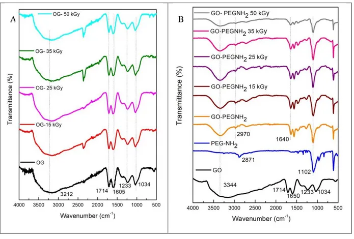
The FT-IR results indicated the presence of oxygenated functional groups on the surface of GO. According to Zhao et al. [14] the main bands corresponding to the hydroxyl groups (~3000 cm-1) carbonyl esters (~1600 cm-1), carboxylic acids (~1700 cm-1), carboxyl C-O (~1300 cm-1) and epoxy (~1080 cm-1).
For GO-PEG-NH2, bands at 1640 cm-1 of carbonyls from amides were observed. This result
demonstrates that PEG-NH2 has been successfully grafted onto the surface of GO by the amidation
reaction.
Figure 1 shows that after gamma irradiation, the samples of GO and GO-PEG-NH2 spectra
Figure 1: FT-IR spectra of (A) GO, GO gamma irradiated with 15, 25, 35 and 50 kGy and (B) GO,
PEG-NH2, GO-PEG-NH2, GO-PEG-NH2 irradiated with 15, 25, 35 and 50 kGy.
3.1.2
Raman spectroscopy
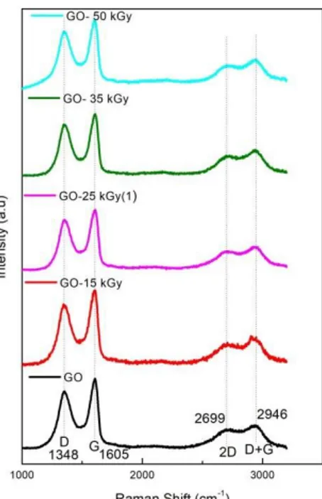
Raman spectroscopy is a technique used to analyze the existence of defects and the extent of functionalization [15]. In this sense, it is presented as a complementary analysis to FT-IR in the evaluation of GO and GO-PEG-NH2 surfaces. For both GO samples the spectra (Figure 2) shows D
band at (~1,348 cm-1), the G band (~1600 cm-1), 2D (~2699 cm-1), and D+G call at (~2,946 cm-1), in agreement with the studies carried out by Zhao et al. [14]. The D functions are associated with the material disorder, while the G functions contribute to the graphitic organization [16].
Figure 2: Raman spectra of GO, GO gamma irradiated with 15, 25, 35 and 50 kGy.
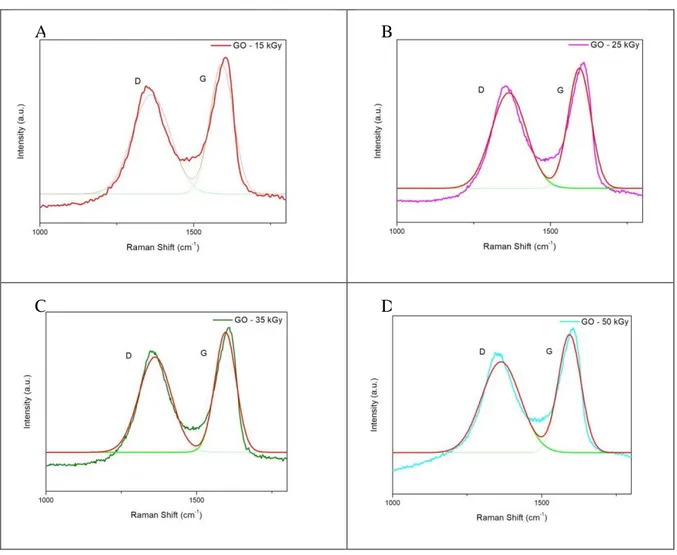
Figure 3 shows D and G bands of the GO, with their respective bands deconvoluted by Gaussian function.
Figure 3: Raman spectra of GO and curves deconvoluted.
D and G bands of irradiated samples were deconvoluted using the Gaussian function as shown in Figure 4.
Figure 4: Raman spectra of samples GO irradiated and curves deconvoluted, (A) GO-15 kGy, (B) GO-25 kGy, (C) GO-35 kGy, (D) GO-50 kGy.
The intensity (ID1 / IG) and half height (ωd1 / ωg) ratios of all samples were calculated as shown
in Table 1. The (ID1 / IG) ration indicates the degree of the material’s organization, while (ωd1 / ωg)
ration corresponds to amount of structural defects [17] formed as a consequence of oxygenated groups incorporated on their surface. With these data, it was possible to relate the contribution of D and G bands.
A B
Table 1: Values of (ωd1 / ωg) and (ID1 / IG).
In Table 1, the results indicate that there is no difference in the GO when irradiated even at 50 kGy.
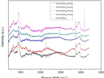
On GO-PEG-NH2 Raman spectra (Figure 5) the following bands are observed D band at 1351
cm-1, the G band at 1600 cm-1, the 2D at 2688 cm-1 and the so-called D + G at 2933 cm-1 for all samples [14]. These results corroborated with FT-IR showing that the irradiation does not affect GO and GO-PEG-NH2 structures.
Figure 5: Raman spectra of samples GO-PEG-NH2, GO-PEG-NH2 irradiated with 15, 25, 35 and
4. CONCLUSION
The methods for the synthesis of GO and GO-PEG-NH2 proved to be effective since there was
confirmation of the surface oxidation of materials and functionalization with the PEG-NH2 group.
The analyzes performed by FT-IR and Raman spectroscopy indicated that the radiation doses used for sterilization of the nanocomposites did not cause modifications on their surface, neither on the functional groups.
ACKNOWLEDGMENT
The authors would like to thank CAPES for scholarship granted to Raynara M. S. Jacovone and CNPq for scholarship granted to Jaqueline J. S. Soares.
REFERENCES
[1] MEHL H.; MATOS F. C.; NEIVA E. G.; DOMINGUES S. H.; ZARBIN A. J. G. Efeito da variação de parâmetros reacionais na preparação de grafeno via oxidação e redução do grafite.
Química Nova, v.37, n°10, pp.1639-1645, (2014).
[2] GULZAR A.; YANG P.; HEI F.; XU J.; YANG D.; XU L.; JAN M. O. Bioapplications of graphene constructed functional nanomaterials, Chemico-Biological Interactions, v.262, pp.69-89, (2017).
[3] NISHIDA E.; TAKITA H.; KANAYAMA I.; TSUJI M.; AKASAKA T.; SUGAYA T.; SAKAGAMI R.; KAWANAMI M. Graphene oxide coating facilitates the bioactivity of scaffold material for tissue engineering. Japanese Journal of Applied Physics, v.53, 13 May. (2014).
[4] ZHANG Y.; NAYAK T. R.; HONG H.; CAI W. Graphene: a versatile nanoplatforms for biomedical applications. Nanoscale, v.4, pp.3833-3842, (2012).
[5] KRISHNA K. V.; MÉNARD M.; VERMA S.; BIANCO A. Graphene-based nanomaterials for nanobiotechnology and biomedical applications. Nanomedicine, v.8, pp.1669–1688, (2013).
[6] XU Z.;WANG S.; LI Y.; WANG M.; HUANG P. Shi. Covalent functionalization of graphene oxide with biocompatible poly (ethylene glycol) for delivery of paclitaxel. Applied materials e
interfaces, v.6, pp.17268-17276, (2014).
[7] FENG L.; LIU Z.; Graphene in biomedicine: opportunities and challenges. Nanomedicine, v.6, pp. 317-324, (2011).
[8] CLELAND M. L.; Industrial Applications of Electron Accelerators, Ion beam applications. IBA
Technology Group 151, New York. (2005).
[9] HUMMERS W. S.; OFFERMAN R. E.; Preparation of graphitic oxide. J. Am. Chem. Soc., v.80, pp.1339–1339, (1958).
[10] MUTTER M.; Soluble polymers in organic synthesis: I. Preparation of polymer reagents using polyethylene glycol with terminal amino groups as polymeric component. Tetrahedron
Letters, Germany, n.31, pp.2839-2842 (1978).
[11] YANG K.; FENG L.; HONG H.; CAI W.; LIU Z.; Preparation and functionalization of graphene nanocomposites for biomedical applications. Nature Protocols, v.8, n.12, (2013).
[12] COLLINS C. J. Reactions of primary aliphatic amines with Nitrous acid. Advan. Chem.
Phys, v.4, (1970).
[13] AWASTHI G.; KUMAR A.; SANGUI A.; SINGH S. S. Biochemical Laboratory Manual,
International E-Publication, pp. 30-31, (2013).
[14] ZHAO J.; LIU L.; LI F.; Graphene Oxide: Physics and Applications. London: Springer, 161 p. (2015).
[15] GEORGAKILAS V. Functionalization of graphene. Wiley-VCH, p.426, (2014).
[16] KING A. A. K.; DAVIES B. R.; NOORBEHESHT N.; NEWMAN P.; CHURCH T. L.; HARRIS A. T.; RAZAL J. M.; MINETT A. I. Characterization of Graphene oxide and its derivatives. Scientific Reports, (2016).
[17] CANÇADO L. G.; JORIO A.; FERREIRA E. H. M.; STAVALE F.; ACHETE C. A.; CAPAZ R. B.; MOUTINHO M. V. O.; LOMBARDO A.; KULMALA T.; FERRARI A. C.
Quantifying defects in graphene via Raman spectroscopy at different excitation energies,