Evaluation of
Arenaria montana
L. hydroethanolic extract as a
chemopreventive food ingredient: A case study focusing a dairy product
(yogurt)
Franciely S. Oliveira
a,b,1, Andreia Ribeiro
a,b,1, Lillian Barros
b, Ricardo C. Calhelha
b, João C.M. Barreira
b,
Bogdan D. Junior
c, Rui M.V. Abreu
b, Maria Filomena Barreiro
a,⇑, Isabel C.F.R. Ferreira
b,⇑aLaboratory of Separation and Reaction Engineering - Laboratory of Catalysis and Materials (LSRE-LCM), Polytechnic Institute of Bragança, Campus de Santa Apolónia, 1134, 5301-857 Bragança, Portugal
bMountain Research Centre (CIMO), ESA, Polytechnic Institute of Bragança, Campus Santa Apolónia, 1172, 5300-253 Bragança, Portugal cUTFPR, Universidade Federal Tecnológica do Paraná, Campus Campo Mourão Via Rosalina Maria dos Santos,1233, Campo Mourão, Paraná, Brazil
a r t i c l e
i n f o
Article history:
Received 1 January 2017
Received in revised form 30 August 2017 Accepted 14 September 2017
Available online 20 September 2017
Keywords: Arenaria montana
VEGFR-2 phosphorylation inhibition Antiangiogenic potential
Functionalized yogurts
a b s t r a c t
Natural ingredients are valuable options to be exploited in the design of innovative food formulations with health benefits. Therefore, it was evaluated the potential use ofArenaria montanaL. hydroethanolic extract (rich in apigenin derivatives) as a chemopreventive agent in functional foods. Apigenin is recog-nized as inhibiting VEGFR-2, which is the key receptor involved in angiogenesis. The obtained extract was also able to inhibit the VEGFR-2 phosphorylation through an enzymatic assay (IC50=63mg/mL). Thereafter, free and microencapsulated forms were incorporated in yogurt. The obtained products main-tained the nutritional value along the tested 3 days of storage, as also free sugars and fatty acids profiles, in comparison with the control samples. Nevertheless, the VEGFR-2 phosphorylation inhibition was not exhibited as intended. Even this behavior for the microencapsulated forms can be attributed to the pro-tecting effect of the alginate matrix, further studies are required in order to better understand the shown performance.
Ó2017 Elsevier Ltd. All rights reserved.
1. Introduction
The growth of new blood vessels from pre-existing ones is called angiogenesis, which is a fundamental process for embryonic
development as well as normal tissue homeostasis (Patan, 2000).
Under normal physiological conditions, there is a balance between stimulatory and inhibitory factors that regulates the process of
angiogenesis (Fan, Yeh, Leung, Yue, & Wong, 2006). However, in
pathological cases, like tumor progression, the stimulation becomes excessive, resulting in the uncontrolled growth of new vessels, which facilitate tumor development and metastasis, through the production of abnormally large amounts of angiogen-esis factors, such as VEGF (vascular endothelial growth factor) (Saghiri, Asatourian, Orangi, Sorenson, & Sheibani, 2015). The VEGF is involved in the vascular permeability, as well as in the induction of endothelial cell proliferation, migration and survival, mainly
through their membrane receptor tyrosine kinase VEGFR-2 (also
known as KDR) (Soares et al., 2013).
It is recognized that diet contributes to more than 40% of the
chronic angiogenic diseases (Losso, 2007). Nevertheless, foods are
nowadays designed not only for nutritional purposes, but also to prevent diseases and to improve physical and mental well-being (Bigliardi & Galati, 2013). Therefore, several studies have been con-ducted in order to identify functional foods active against excessive
angiogenesis (Losso, 2007).
Plants are recognized as a source of bioactive compounds, for example phenolic compounds with reported antitumor properties (Stagos et al., 2012).Arenaria montanaL., an herbaceous plant from
the mountainous regions of southwestern Europe (Timité et al.,
2011), contains apigenin derivatives, specifically apigenin
6-C-hexoside-8-C-hexoside, apigenin 6-C-hexoside-8-C-pentoside,
apigenin 200-O-pentosyl-6-C-hexoside, apigenin-6-C-glucoside,
apigenin 200-O-acetylpentosyl-6-C-hexoside and apigenin 200
-O-feruloylhexosyl-6-C-hexoside (Pereira et al., 2014). Apigenin has
received increasing attention due to its bioactive properties
such as anti-inflammatory (Wang & Huang, 2013), anti-mutagenic
(Patel, Modi, Chiosis, & Taldone, 2011) and, particularly,
http://dx.doi.org/10.1016/j.jff.2017.09.027
1756-4646/Ó2017 Elsevier Ltd. All rights reserved.
⇑Corresponding authors.
E-mail addresses: barreiro@ipb.pt (M.F. Barreiro), iferreira@ipb.pt (I.C.F.R. Ferreira).
1 Both authors contributed equally.
Contents lists available atScienceDirect
Journal of Functional Foods
antiangiogenic and anticancer properties (Choudhury et al., 2013; He et al., 2015; Johnson & Mejia, 2013). This flavone has shown the capacity to inhibit the cellular proliferation of several cancer
cell lines such as hepatocellular (Kim, Jeon, & Nam, 2011),
pancre-atic (He et al., 2015; Johnson & Mejia, 2013), colorectal (Banerjee
& Mandal, 2015), multiple myeloma (Zhao et al., 2011) and
leuke-mia (Budhraja et al., 2012), prevent the growth of new blood vessels
(metastasis) (Osada, Imaoka, & Funae, 2004), change the
microenvi-ronment of the cancer cells growth and reduce the cancer cells
glucose uptake (Lefort & Blay, 2013).
Although appreciated and consumed worldwide, yogurt is a product with limited amounts of bioactive compounds. This limita-tion can be overcome, by enrichment with natural extracts from
plants or fruits, as proposed by some authors (Caleja et al., 2016;
Martins et al., 2014). However, these extracts, which contain phe-nolic compounds and other bioactive molecules, most often are
unstable under environmental and processing conditions,e.g.light,
moisture, temperature and food matrix effects (Dias, Ferreira, &
Barreiro, 2015). Also, they might become inactive after being
metabolized (Heleno, Martins, Queiroz, & Ferreira, 2015). In this
context, microencapsulation, a process through which the bioac-tive compound is entrapped or covered by a material, allows
pro-tection from these adverse conditions (Dias, Ferreira et al., 2015).
In the present study, the ability ofA. montanahydroethanolic
extract (rich in apigenin derivatives) to inhibit the phosphorylation of VEGRF-2 (enzyme involved in the angiogenesis process) was evaluated. Thereafter, the extract was microencapsulated using an atomization/coagulation technique with alginate as the encap-sulating material. The obtained microspheres were characterized by optical microscopy (to inspect morphology and size) and the encapsulation estimated by HPLC-DAD. As a subsequent applica-tion step, the produced microspheres, as well as the free extract, were incorporated into yogurt samples in order to evaluate their role in functional dairy products with chemopreventive (antiangio-genic) potential.
2. Materials and methods
2.1. Standards and reagents
Sodium alginate was provided from Fluka Chemie (Steinheim, Switzerland) and calcium chloride di-hydrate was obtained from PanReac AppliChem SAU (Barcelona, Spain). For chromatographic analysis, HPLC-grade acetonitrile was acquired from Fisher Scien-tific (Lisbon, Portugal). Ethanol, used as solvent, was acquired from Pronalab (Lisbon, Portugal). The fatty acids methyl ester (FAME) reference standard mixture (standard 47885-U) was purchased from Sigma (St. Louis, MO, USA), as well as the sugar standards. Apigenin was obtained from Extrasynthesis (Genay Cedex, France) and sorafenib from Molekula (Newcastle, United Kingdom). Water was treated in a Milli-Q water purification system (TGI Pure Water Systems, Greenville, SC, USA). All other chemicals and solvents were of analytical grade and purchased from common sources.
2.2. Plant species and preparation of the extract
A. montanaflowers and leafy stems (roughly the upper 15 cm of the dense clumps produced in spring) are commonly gathered to be used in traditional preparations. Samples were collected in full blossom along paths through oak trees. Voucher specimens were deposited at the Herbarium of the Escola Superior Agrária de
Bra-gança (BRESA) according withPereira et al. (2014). A sample for
analysis was prepared by putting together the material from differ-ent specimens, thereafter lyophilized (FreeZone 4.5, Labconco,
Kansas City, MO, USA), reduced to a fine dried powder (20 mesh) and mixed to obtain homogeneity.
The hydroethanolic extract was prepared by stirring the sample
(1 g; 150 rpm) with 30 mL of an ethanol:water mixture (80:20,v/
v), at room temperature, during 1 h, and subsequently filtered thought a Whatman paper filter No. 4. The solid residue was re-extracted under the same conditions and the obtained supernatant mixed with the one previously obtained. The ethanol of the com-bined extracts was removed by evaporation under reduced pres-sure (Büchi R-210, Flawil, Switzerland), while the water was eliminated by lyophilisation.
2.3. VEGFR-2 enzymatic inhibition assay
TheA. montanahydroethanolic extract was assessed for VEGFR-2 inhibition activity using the Z’-LYTE-Tyr1 Peptide assay kit (Invit-rogen, Cat. PV3190), according to the procedure recommended by the manufacturer. Briefly, assays were performed with a total of
20mL in 384-well plates using the fluorescence resonance energy
transfer technology. Each well plate was filled with 2.5mL of the
tested extract in different concentrations, 5mL of VEGFR-2 solution
and 2.5mL of ATP solution, and incubated during 1 h at room
tem-perature. The development reagents (5mL) were then added to
each well, followed by a stop reagent after a second incubation of 1 h. The fluorescence was read at 445 nm and 520 nm
(excita-tion 400 nm) using a Biotek FLX800 micro-plate. Gen5TMSoftware
was used for data analysis. Sorafenib was used as positive control (Guimarães et al., 2016).
The results were expressed as IC50values (mg/mL), which
repre-sent the concentration of the sample needed to inhibit 50% of the VEGFR-2 phosphorylation.
2.4. Microencapsulation of the hydroethanolic extract and characterization of the microspheres
2.4.1. Microencapsulation
The microspheres were produced by using an
atomization/co-agulation technique previously described byMartins et al., 2014;
Dias et al., 2015andRibeiro et al., 2016. Briefly, the encapsulating material (calcium alginate) was formed by using sodium alginate
and a calcium chloride (CaCl2) as the coagulation agent (source
of Ca2+ ions). The atomization solution was prepared by firstly
mix 50 mg of theA. montanaextract with 10 mL of water. After
complete dissolution, 400 mg of sodium alginate were added and the preparation kept under stirring during 2 h. To produce the microspheres, a NISCO VarJ30 (Zurich, Switzerland) system, consti-tuted by a pressure controller, gas source (nitrogen) and a syringe pump, was used. The previously prepared alginate solution con-taining the hydroethanolic extract was atomized using the follow-ing conditions: feed rate of 0.2 mL/min and a nitrogen pressure of
0.1 bar. The atomized droplets upon in contact with the CaCl2
solu-tion (250 mL at concentrasolu-tion of 4% (w/v)) coagulate promptly fix-ing the microspheres shape. For total consolidation, they remained in contact with the coagulation solution during 4 h under stirring at 200 rpm at room temperature. Finally, microspheres were recovered by filtration under reduced pressure, washed twice with water (100 mL), lyophilized and stored in the dark.
2.4.2. Characterization
The microspheres formation was monitored during the coagula-tion step through Optical microscopy (OM) (initial and final times). The apparatus was a Nikon Eclipse 50i equipped with a camera (Nikon Digital Sight, Tokyo, Japan) for image acquisition. The encapsulation efficiency (EE) was evaluated by HPLC-DAD by quantifying the non-encapsulated apigenin derivatives, as
and the two washing solutions were analysed and the obtained responses summed. The encapsulation efficiency was then calcu-lated according to the following expression:
EE¼ ½ðMe-t Me-neÞ=ðMe-tÞ 100
in which Me-trepresents the theoretical amount of extract, i.e. the
amount of extract used in the microencapsulation process, and
Me-necorresponds to the non-encapsulated extract.
2.5. Functionalization of yogurt with free and microencapsulated extracts
2.5.1. Preparation of the yogurt samples
A natural yogurt with 5% fat was selected to test the
incorpora-tion of the free and microencapsulated A. montana extracts. Six
yogurt portions, with 125 g each, were prepared: (i) two samples
of the base yogurt (used as the control sample, i.e. withoutA.
mon-tanaextract); (ii) two samples of yogurt with the free extract (16
mg each); (iii) two samples of yogurt with the microencapsulated extract (70 mg of lyophilized microspheres each). All the prepared samples contain an equivalent amount of extract, either in its free or encapsulated form (the same equivalent ratio of mg extract/g
yogurt). The used amount of extract was twice the IC50 value
(obtained in the VEGFR-2 enzymatic inhibition assay), in accor-dance with the recommended daily intake of apigenin derivatives
(4–24 mg) (Brasil, 2013). According with our previous results
(Pereira et al., 2014), theA. montanahydroethanolic extract
con-tains 48% (w/w) of apigenin derivatives (Supplementary table).
All the subsequent analyses were performed with lyophilized yogurt samples obtained immediately after incorporation (right after the addition of the free or microencapsulated extracts) (t0), and after 3 days (t3). The t0 and t3 yogurt samples were
condi-tioned at 4°C until analysis.
2.5.2. Nutritional value of the yogurt samples
For the assessment of the nutritional composition of the yogurt samples, moisture, protein, fat, carbohydrates and ash were
deter-mined following AOAC procedures (AOAC, 2005). The crude protein
content (estimated as N6.38) was determined using the Kjeldahl
method; the fat was determined by extracting a known weight of powdered sample with petroleum ether; the ash content was
determined by incineration at 600 ± 15°C; and the total
carbohy-drates obtained by difference. Total energy was calculated as
Energy (kcal) = 4(protein weight (g) + carbohydrate weight
(g)) + 9(lipid weight (g)).
Free sugars were determined from defatted samples by HPLC coupled with a refraction index (RI) detector. The compounds were identified by chromatographic comparison with injected authentic standards, and quantification performed by the internal standard (melezitose) method. Sugars content was expressed in g/100 g of the yogurt.
Fatty acids were analysed by gas-chromatography coupled with a flame ionization detector (GC-FID). The identification was made by comparison with the relative retention times of fatty acid methyl esters of standards. The results were expressed as relative percentages.
2.5.3. VEGFR-2 phosphorylation inhibition
The lyophilized yogurt samples (1 g) were extracted with 30 mL
of ethanol: water (80:20 v/v) at room temperature during 1 h
under stirring. The obtained extract was filtered through a What-man paper filter No. 4 and the remaining solid residue subjected to an additional extraction. The combined extracts were evapo-rated under reduced pressure in a rotatory evaporator until com-plete removal of ethanol. Finally, the evaporated extract was dissolved in water at a concentration of 8 mg/mL for the evaluation
of VEGFR-2 phosphorylation inhibition according with the
methodology described in Section2.3.
2.6. Statistical analysis
All the assays were carried out in triplicate and the results expressed as mean values ± standard deviation (SD), and maintain-ing the decimal places allowed by the magnitude of the standard deviation. An ANOVA with type III sums of squares was performed using the general linear model (GLM) procedure. The dependent variables were analysed using a 2-way ANOVA, with the factors ‘‘functionalizing type” (FT) and ‘‘storage time” (ST). When a
statis-tically significant interaction (FAST) was detected, the two
fac-tors were evaluated simultaneously by the estimated marginal means (EMM) plots for all the levels of each single factor. Alterna-tively, if no statistical significant interaction was verified, the means were compared using Tukey’s honest significant difference (HSD) multiple comparison test to evaluate the FA effect, or by a t-student test to assess the effect of ST.
All statistical tests were performed at a 5% significance level using IMB’s SPSS (Statistics for Windows, version 22.0 (IBM Corp., Armonk, NY, USA).
3. Results and discussion
3.1. VEGFR-2 phosphorylation inhibition by the A. montana extract
In this work, theA. montanahydroethanolic extract was
evalu-ated for its ability to interact with the VEGFR-2 kinase domain, using an enzymatic fluorescence resonance energy transfer (FRET)
based assay. The results are presented inFig. 1. The hydroethanolic
extract showed ability to inhibit the enzymatic phosphorylation,
and the obtained IC50value was 63 ± 3mg/mL. It was also possible
to observe that the percentage of VEGFR-2 phosphorylation inhibi-tion increased as the extract concentrainhibi-tion increases. The sorafenib
(positive control) showed a lower IC50 value, i.e. a high rate of
VEGFR-2 phosphorylation inhibition. This synthetic drug was approved by the Food and Drug Administration (FDA) and has been commonly used for the treatment of pathological angiogenesis once it binds to receptors on the surface of the endothelial cells inhibiting, namely, the VEGFR-2. However, it reveals several toxic-ities for human organism. Diarrhea, fatigue, abdominal pain, hep-atic toxicity, arterial and venous thrombotic events, gastric cancer and AVC are some of the side effects ascribed to sorafenib (Gotink & Verheul, 2010).
It should be also highlighted that A. montanahydroethanolic
extract gave a lower IC50value (higher VEGFR-2 phosphorylation
inhibition) than the methanolic extract (269 ± 9mg/mL) and
infu-sion (301 ± 13mg/mL) obtained from Roman chamomile
(Chamae-melum nobileL.) (Guimarães et al., 2016).
3.2. Microencapsulation of the A. montana extract with alginate
TheA. montanahydroethanolic extract was microencapsulated with alginate using the atomization/coagulation process and fol-lowing previously optimized conditions, as described in some
for-mer works of the authors (Dias, Barros et al., 2015; Martins et al.,
2014; Ribeiro et al., 2016).
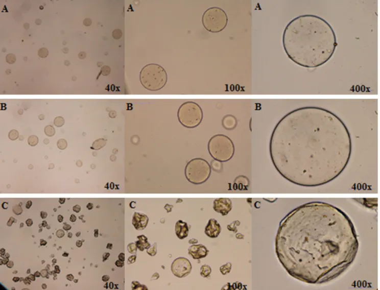
The produced microspheres were analysed by OM immediately
after the atomization process (Fig. 2A) and after 4 h under stirring
in a coagulation solution of calcium chloride (Fig. 2B). After
solution the number of microspheres with spherical morphology increased. Moreover, the microspheres were perfectly individual-ized (no agglomerates were detected). The size of the
microparti-cles was estimated by OM as ranging between 14 and 88mm,
size slightly larger than the one obtained in previous works of
the authors Martins et al. (2014), Dias, Barros et al. (2015) and
Ribeiro et al. (2016). The lyophilized microparticles can be
observed inFig. 2C, showing a shape similar to the one observed
for the hydrated forms obtained after the encapsulation process (round and pear-like form), although they showed a roughened surface due to the water removal, a consequence of the drying pro-cess. Additionally, the small brown spots observed in hydrated and lyophilized microspheres were associated with the presence of the extract inside the microparticles, in conformity with the work
development byRibeiro et al. (2016)andCaleja et al. (2016).
The encapsulation efficiency (EE), estimated through an indirect method based on the quantification of the non-encapsulated
api-genin derivatives by HPLC-DAD, was determined as 100% (the
quantitative analysis of the coagulation and the two wash solu-tions revealed that the apigenin derivatives were present only in residual concentrations or absent).
3.3. Yogurts functionalized with free and microencapsulated A. montana extracts
3.3.1. Nutritional value
The yogurt samples were analysed for their nutritional compo-sition regarding ash, fat, protein and carbohydrates (including indi-vidual quantification of galactose and lactose) contents, as well as 0
10 20 30 40 50 60 70
0.1 1 10 100
%
Phosphorylation
inhibition
Extract concentration [µg/mL]
Fig. 1.Percentage of VEGFR-2 phosphorylation inhibition as a function of theA. montanaextract concentration.
total energy. The studied ‘‘functionalized type” (FT) considered absent (no added extract), free and microencapsulated forms. The ‘‘storage time” (ST) considered 0 (t0) and 3 days (t3).
Before the study of each individual factor, it is necessary to
analyse the possible interaction (FTST) between the various
fac-tors under study. That is, check whether the effect of a given factor over the different levels of the second follows a specific trend. Thereby, for all the evaluated parameters, it was aimed to assess the ST effects regardless of having pure or functionalized yogurts, as well as evaluating the influence of FT, independently of the number of days kept in storage.
Table 1shows the obtained results for macronutrients composi-tion, individual sugars (galactose and lactose) and energetic value. It can be verified that the interaction was significant in the case of fat, protein and ash. Interestingly, these parameters were also not significantly affected by each individual factor in most cases, except protein, along ST, and fat, according to FT. Protein content was higher at initial ST (t0), while fat content was higher in the functionalized yogurts (both free and microencapsulated forms), although these differences cannot be unambiguously interpreted, due to the significant interaction between factors. For all the remaining parameters, the lack of a significant interaction allowed
performing multiple comparisons for each individual factor, but their individual effects were only significant (p-value < 0.05) in the case of lactose (for ST), which proved to be significantly higher at t0, when compared to the value obtained at t3. All other param-eters remained approximately invariable for both factors in study (ST and FT).
The results obtained are in agreement with other reports on
yogurts (Caleja, Barros et al., 2016; Serafeimidou, Zlatanos,
Kritikos, & Tourianis, 2013). According with Serafeimidou et al. (2013), the yogurts prepared with cow and sheep milk had similar amounts of moisture and proteins, but lower ash content in com-parison with the cow yogurt samples studied in the present work. However, ash values were in accordance with the ones reported by
Caleja, Barros et al. (2016)for yogurts fortified with chamomile
(Matricaria recutitaL.) and fennel (Foeniculum vulgareMill.)
aque-ous extracts. Furthermore, the same composition in galactose and lactose was described.
In the case of fatty acids (Table 2) the interaction between the
factors was significant for all the analysed molecules. Thus, it is not possible to indicate unequivocal changes induced by each one of the factors. Nevertheless, the performed multiple compar-isons indicated some general trends. The increase in ST induced a
Table 1
Nutritional value of the control yogurts and of those containing the extract (free or microencapsulated) at storage times of 0 and 3 days (mean ± SD). Moisture (g/
100 g) Fat (g/100 g) Protein (g/100 g) Carbohydrates (g/100 g) Ash (g/100 g) Energy (kcal/100 g) Galactose Lactose Total
Storage time (ST) 0 days 88 ± 1 2.6 ± 0.2 4.0 ± 0.1 0.5 ± 0.1 3.9 ± 0.4 4 ± 1 0.6 ± 0.1 57 ± 2 3 days 89 ± 1 2.7 ± 0.2 3.8 ± 0.1 0.6 ± 0.1 3.4 ± 0.4 4 ± 1 0.6 ± 0.1 57 ± 3
p-value (n = 27) t-Student test 0.054 0.065 <0.001 0.284 <0.001 0.108 0.062 0.394 Functionalization type
(FT)
Absent 88 ± 1 2.4 ± 0.1 3.9 ± 0.2 0.6 ± 0.1 3.7 ± 0.4 4 ± 1 0.6 ± 0.1 56 ± 2 Free extract 89 ± 1 2.9 ± 0.1 3.9 ± 0.1 0.5 ± 0.1 3.7 ± 0.4 4 ± 1 0.5 ± 0.1 58 ± 3 Microencapsulated
extract
89 ± 1 2.7 ± 0.1 3.9 ± 0.1 0.6 ± 0.1 3.6 ± 0.5 4 ± 1 0.6 ± 0.1 57 ± 2
p-value (n = 18) Tukey’s HSD test 0.352 <0.001 0.762 0.205 0.801 0.308 0.051 0.090 STFT (n = 54) p-value 0.756 0.007 <0.001 0.214 0.506 0.506 0.007 0.819
Table 2
Main fatty acids and total saturated, monounsaturated and polyunsaturated fatty acids (relative percentage) in the control yogurts and in those containing the extract (free or microencapsulated) at storage times of 0 and 3 days (mean ± SD).
C4:0 C6:0 C10:0 C12:0 C14:0 C16:0 C18:0 C18:1 C18:2 SFA MUFA PUFA Storage time(ST) 0 days 2 ± 1 2.4 ± 0.4 3.3 ± 0.2 3.7 ± 0.1 11 ± 1 31 ± 1 11 ± 1 24 ± 1 3.1 ± 0.2 69 ± 1 27 ± 1 4.6 ± 0.2
3 days 3 ± 1 2.8 ± 0.2 3.2 ± 0.2 3.5 ± 0.2 11 ± 1 30 ± 1 11 ± 1 24 ± 1 3.2 ± 0.2 68 ± 1 27 ± 1 4.7 ± 0.4
p-value (n = 27) t-Student test <0.001 <0.001 0.059 <0.001 0.062 0.002 0.485 0.473 0.702 0.216 0.345 0.160 Functionalization
type (FT)
Absent 2.6 ± 0.4 2.7 ± 0.2 3.2 ± 0.1 3.5 ± 0.2 11 ± 1 30 ± 1 12 ± 1 24 ± 1 3.2 ± 0.2 68 ± 1 27 ± 1 4.8 ± 0.3 Free extract 2.3 ± 0.5 2.3 ± 0.4 3.5 ± 0.1 3.8 ± 0.1 11 ± 1 31 ± 1 11 ± 1 24 ± 1 3.1 ± 0.2 69 ± 1 27 ± 1 4.5 ± 0.3 Microencapsulated
extract 3.6 ± 0.2 2.8 ± 0.2 3.1 ± 0.2 3.6 ± 0.1 11 ± 1 30 ± 1 11 ± 1 24 ± 1 3.1 ± 0.2 69 ± 1 27 ± 1 4.6 ± 0.3
p-value (n = 18) Tukey’s HSD test <0.001 <0.001 <0.001 0.001 0.055 0.061 <0.001 0.080 0.085 0.064 0.236 0.056 STFT (n = 54) p-value <0.001 <0.001 0.004 <0.001 <0.001 <0.001 <0.001 <0.001 <0.001 <0.001 <0.001 <0.001 Butiric acid (C4:0), caproic acid (C6:0); capric acid (C10:0); lauric acid (C12:0); myristic acid (C14:0); palmitic acid (C16:0); stearic acid (C18:0); oleic acid (C18:1); linoleic acid (C18:2); SFA - Saturated fatty acids; MUFA - Monounsaturated fatty acids; PUFA - Polyunsaturated fatty acids. In addition to the tabled fatty acids, twenty more were also detected in minor amounts (each one <2%).
Table 3
Percentage of VEGFR-2 phosphorylation inhibition of the yogurt samples functionalized withA. montanaextracts (free and microencapsulated) relative to the control (yogurt without extracts).
Storage time (days) VEGFR-2 phosphorylation inhibition
1mg/mL 10mg/mL 100mg/mL
Free extract 0 0.99 ± 0.01 1.80 ± 0.03 5.10 ± 0.03
3 2.44 ± 0.06 3.12 ± 0.05 0.85 ± 0.05 Microencapsulated extract 0 1.31 ± 0.06 1.84 ± 0.07 4.13 ± 0.03
slight increase in C4:0, C6:0 and C12:0 percentages, and minor reductions in C12:0 and C16:0 percentages. The remaining fatty acids proportions remained nearly constant. In what concerns FT effect, yogurts incorporated with the microencapsulated extract showed higher values of C4:0 and C6:0, while those incorporated with the free extract showed higher percentages of C10:0 and C12:0. Finally, the control yogurts (yogurts absent of extract) showed slightly higher percentages of C18:0. All the other fatty acids, kept their overall percentages roughly constant, either in response to ST and FT factors, which indicates that the fatty acids profile suffer only slight changes. The general proportions (SFA > -MUFA > PUFA) are in general agreement with previous works (Serafeimidou et al., 2013).
3.3.2. Evaluation of the VEGFR-2 phosphorylation inhibition
The yogurt functionalized with the free and microencapsulated extracts were evaluated for their potential VEGFR-2 phosphoryla-tion inhibiphosphoryla-tion. In general, yogurt samples added with the extract, in both free and microencapsulated forms, revealed very low
inhi-bition ability (low inhiinhi-bition percentages) (Table 3).
The absence, or scarcity, of VEGFR-2 phosphorylation inhibition observed for the yogurt samples prepared with the free extract was not foreseen based on its ascribed potential and the used amount
(2IC50), which was also chosen to guarantee the recommended
daily intake of apigenin derivatives. Due to this observation, a sam-ple yogurt 20-fold more concentrate was prepared. The main objective was to evaluate the influence of the extract concentration on the VEGFR-2 phosphorylation activity of the prepared yogurt. However, the obtained results (data not shown) revealed, once again, very low inhibition percentages pointing out for the extract degradation and/or bioactivity loss. In fact, proteins, the predomi-nant constituents of the yogurt matrix, present ability to conjugate with phenolic compounds (major constituents of the
hydroethano-lic extract) leading to bioavailability loss (Ozdal, Capanoglu, &
Altay, 2013).
When the microencapsulated forms were used, the determined VEGFR-2 phosphorylation inhibition was also absent, or scarce. In fact, the extract was, in this case, protected inside the alginate microspheres, material reported as stable in acid pH conditions (Cui, Goh, Kim, Choi, & Lee, 2000), thus effective to protect it in
contact with the yogurt, an acidic food matrix (Caleja, Barros
et al., 2016). This behavior was also reported byRibeiro et al. (2016)in cottage cheese functionalized with rosemary extracts microencapsulated in alginate microspheres. This procedure is, in principle, effective to avoid the loss of bioavailability during the shelf life of the product, thus until its ingestion and release in slight alkaline medium (intestinal conditions) due to the disruption of the alginate microparticles.
4. Conclusions
A. montanahydroethanolic extract, an extract rich in apigenin derivatives (weight content of 48%, as reported in a previous work
of the group (Pereira et al., 2014), revealed VEGFR-2
phosphoryla-tion inhibiphosphoryla-tion ability. The extract was successfully encapsulated in an alginate matrix, being incorporated in yogurts presenting nutri-tional properties similar to the ones incorporated with free extract or without extract (control sample). However, after incorporation of the free extract in yogurts, the antiangiogenic activity was not effective since only minor antiangiogenic ability was detected, independently of the used concentration. For the microencapsu-lated forms, and based on the successful applied microencapsula-tion process, the extract protecmicroencapsula-tion in acidic medium was expected, thus supporting the absence of the antiangiogenic activ-ity of the produced yogurts. Accordingly, more studies are
neces-sary to understand the stability of the bioactive compounds of the extract, namely their interactions with the food matrix con-stituents and behavior (for both free and microencapsulated forms) under simulated gastrointestinal conditions in order to develop functional foods with effective chemopreventive effects against pathological angiogenesis.
Acknowledgements
The authors are grateful to the Foundation for Science and Tech-nology (FCT, Portugal) and FEDER for CIMO (UID/AGR/00690/2013) financial support. To POCI-01-0145-FEDER-006984 (LA LSRE-LCM) funded by ERDF through POCI-COMPETE2020 and FCT. To NORTE-01-0145-FEDER-000006, funded by NORTE 2020, under PT2020 through ERDF. L. Barros, R.C. Calhelha and J.C.M. Barreira acknowledge the FCT for their post-doctoral grants (SFRH/BPD/107855/2015, SFRH/BPD/68344/2010 and SFRH/BPD/72802/2010, respectively).
The authors also thank Ana Maria Carvalho for providingArenaria
montanaL. samples.
Appendix A. Supplementary material
Supplementary data associated with this article can be found, in
the online version, athttp://dx.doi.org/10.1016/j.jff.2017.09.027.
References
AOAC (2005). Official methods of analysis of AOAC international. In W. Horwitz, & G. Latimer (Eds.) (18th ed.) Gaithersburg, MD: AOAC International.
Banerjee, K., & Mandal, M. (2015). Oxidative stress triggered by naturally occurring flavone apigenin results in senescence and chemotherapeutic effect in human colorectal cancer cells.Redox Biology, 5, 153–162.
Bigliardi, B., & Galati, F. (2013). Innovation trends in the food industry. The case of functional foods.Trends in Food Science & Technology, 31, 118–129.
Brasil (2013). Agência Nacional de Vigilância Sanitária (ANVISA). Consulta Pública n
°14, de 14 de maio. Diário Oficial da União (D.O.U) de 15 de maio. Relator:
Dirceu Brás Aparecido Barbano.
Budhraja, A., Gao, N., Zhang, Z., Son, Y., Cheng, S., Wang, X., ... Shi, X. (2012). Apigenin induces apoptosis in human leukemia cells and exhibits anti-leukemic activity in vivo.Molecular Cancer Therapeutics, 11, 132–142.
Caleja, C., Barros, L., Antonio, A. L., Carocho, M., Oliveira, M. B. P. P., & Ferreira, I. C. F. R. (2016). Fortification of yogurts with different antioxidant preservatives: A comparative study between natural and synthetic additives.Food Chemistry, 210, 262–268.
Caleja, C., Ribeiro, A., Barros, L., Barreira, J. C. M., Antonio, A. L., Oliveira, M. B. P. P., ... Ferreira, I. C. F. R. (2016). Cottage cheeses functionalized with fennel and chamomile extracts: Comparative performance between free and microencapsulated forms.Food Chemistry, 199, 720–726.
Choudhury, D., Ganguli, A., Dastidar, D. G., Acharya, B. R., Das, A., & Chakrabarti, G. (2013). Apigenin shows synergistic anticancer activity with curcumin by binding at different sites of tubulin.Biochimie, 95, 1297–1309.
Cui, J., Goh, J., Kim, P., Choi, S., & Lee, B. (2000). Survival and stability of bifidobacteria loaded in alginate poly-l-lysine microparticles. International Journal of Pharmaceutics, 210, 51–59.
Dias, I., Barros, L., Fernandes, I., Ruphuy, G., Oliveira, M. B. P. P., Santos-Buelga, C., ... Ferreira, I. C. F. R. (2015). A nutraceuticals formulations based onFragaria vesca L. vegetative parts: characterization of the bioactives and application in k-carrageenan gelatina.Journal of Functional Foods, 16, 243–255.
Dias, M. I., Ferreira, I. C. F. R., & Barreiro, M. F. (2015). Microencapsulation of bioactives for food applications. Food & Function. http://dx.doi.org/10.1039/ c4fo01175a.
Fan, T. P., Yeh, J. C., Leung, K. W., Yue, P. Y., & Wong, R. N. (2006). Angiogenesis: From plants to blood vessels.Trends Pharmacological Sciences, 27, 297–309.
Gotink, K. J., & Verheul, H. M. W. (2010). Anti-angiogenic tyrosine kinase inhibitors: What is their mechanism of action?Angiogenesis, 13, 1–14.
Guimarães, R., Calhelha, R. C., Froufe, H., Abreu, R., Carvalho, A. M., Queiroz, M. J. R. P., & Ferreira, I. C. F. R. (2016). Wild Roman chamomile extracts and phenolic compounds: Enzymatic assays and molecular modelling studies with VEGFR-2 tyrosine kinase.Food and Function, 7, 79–83.
He, J., Ning, C., Wang, Y., Ma, T., Huang, H., Ge, Y., ... Jiang, Y. (2015). Natural plant flavonoid apigenin directly disrupts Hsp90/Cdc37 complex and inhibits pancreatic cancer cell growth and migration.Journal of Functional Foods, 18, 10–21.
Heleno, S., Martins, A., Queiroz, M. J. R. P., & Ferreira, I. C. F. R. (2015). Bioactivity of phenolic acids: Metabolites versus parent compounds: A review. Food Chemistry, 173, 501–513.
on human pancreatic cancer cells,in vitro.Food and Chemical Toxicology, 60, 83–91.
Kim, B. R., Jeon, Y. K., & Nam, M. J. (2011). A mechanism of apigenin-induced apoptosis is potentially related to anti-angiogenesis and anti-migration in human hepatocellular carcinoma cells. Food and Chemical Toxicology, 49, 1626–1632.
Lefort, É. C., & Blay, J. (2013). Apigenin and its impact on gastrointestinal cancers. Molecular Nutrition & Food Research, 57, 126–144.
Losso, J. N. (2007). In: J. N. Losso, F. Shahidi & D. Bagchi (Eds.),Screening functional foods as inhibitors of angiogenesis biomarkers in anti-angiogenic functional and medicinal foods. CRC Press.
Martins, A., Barros, L., Carvalho, A., Santos-Buelga, C., Fernandes, I., Barreiro, F., & Ferreira, I. C. (2014). Phenolic extracts of Rubus ulmifoliusschott flowers: Characterization, microencapsulation and incorporation into yogurts as nutraceutical sources.Food Function, 5, 1091–1100.
Osada, M., Imaoka, S., & Funae, Y. (2004). Apigenin suppresses the expression of VEGF, an important factor for angiogenesis, in endothelial cells via degradation of HIF-1aprotein.FEBS Letters, 575, 59–63.
Ozdal, T., Capanoglu, E., & Altay, F. (2013). A review on protein-phenolic interactions and associated changes.Food Research International, 51, 954–970.
Patan, S. (2000). Vasculogenesis and angiogenesis as mechanisms of vascular network formation, growth and remodeling.Journal of Neuro-Oncology, 50, 1–15.
Patel, H. J., Modi, S., Chiosis, G., & Taldone, T. (2011). Advances in the discovery and development of heat-shock protein 90 inhibitors for cancer treatment.Expert Opinion on Drug Discovery, 6, 559–587.
Pereira, E., Barros, L., Calhelha, R. C., Dueñas, M., Carvalho, A. M., Santos-Buelga, C., & Ferreira, I. C. F. R. (2014). Bioactivity and phytochemical characterization of Arenaria montanaL.Food and Function, 5, 1848–1855.
Ribeiro, A., Caleja, C., Barros, L., Santos-Buelga, C., Barreiro, M. F., & Ferreira, I. C. F. R. (2016). Rosemary extracts in functional foods: Extraction, chemical characterization and incorporation of free and microencapsulated forms in cottage cheese.Food and Function, 7, 2185–2196.
Saghiri, M. A., Asatourian, A., Orangi, J., Sorenson, C. M., & Sheibani, N. (2015). Functional role of inorganic trace elements in angiogenesis - Part II: Cr, Si, Zn, Cu, and S.Critical Reviews in Oncology/Hematology, 96, 143–155.
Serafeimidou, A., Zlatanos, S., Kritikos, G., & Tourianis, A. (2013). Change of fatty acid profile, including conjugated linoleic acid (CLA) content, during refrigerated storage of yogurt made of cow and sheep milk.Journal of Food Composition and Analysis, 31, 24–30.
Soares, P., Costa, R., Froufe, H. J. C., Calhelha, R. C., Peixoto, D., Ferreira, I. C. F. R., ... Queiroz, M. R. P. (2013). 1-Aryl-3-[4-(thieno[3,2-d]pyrimidin-4-yloxy)phenyl] ureas as VEGFR-2 Tyrosine Kinase Inhibitors: Synthesis, biological evaluation, and molecular modelling studies.BioMed Research International.http://dx.doi. org/10.1155/2013/154856.
Stagos, D., Amoutzias, G. D., Matakos, A., Spyrou, A., Tsatsakis, A. M., & Kouretas, D. (2012). Chemoprevention of liver cancer by plant polyphenols. Food and Chemical Toxicology, 50, 2155–2170.
Timité, G., Mitaine-Offer, A., Miyamoto, T., Tanaka, C., Mirjolet, J., Duchamp, O., & Lacaille-Dubois, M. (2011). Unusual oleanane-type saponins from Arenaria montana.Phytochemistry, 72, 503–507.
Wang, Y., & Huang, K. (2013).In vitroanti-inflammatory effect of apigenin in the Helicobacter pylori-infected gastric adenocarcinoma cells.Food and Chemical Toxicology, 53, 376–383.