Genome-wide analysis of the Glycerol-3-Phosphate Acyltransferase (GPAT)
gene family reveals the evolution and diversification of plant GPATs
Edgar Waschburger
5, Franceli Rodrigues Kulcheski
4, Nicole Moreira Veto
1, Rogerio Margis
1,2,3, Marcia
Margis-Pinheiro
1and Andreia Carina Turchetto-Zolet
11
Programa de Pós-Graduação em Genética e Biologia Molecular, Departamento de Genética, Universidade
Federal do Rio Grande do Sul (UFRGS), Porto Alegre, RS, Brazil.
2
Centro de Biotecnologia e Programa de Pós-Graduação em Biologia Celular e Molecular, Universidade
Federal do Rio Grande do Sul (UFRGS), Porto Alegre, RS, Brazil.
3
Departamento de Biofísica, Universidade Federal do Rio Grande do Sul (UFRGS), Porto Alegre, RS,
Brazil.
4
Departamento de Biologia Celular, Embriologia e Genética, Universidade Federal de Santa Catarina
(UFSC), Florianópolis, SC, Brazil.
5
Graduação em Biotecnologia, Departamento de Biologia Molecular e Biotecnologia, Universidade Federal
do Rio Grande do Sul (UFRGS), Porto Alegre, RS, Brazil.
Abstract
sn-Glycerol-3-phosphate 1-O-acyltransferase (GPAT) is an important enzyme that catalyzes the transfer of an acyl group from acyl-CoA or acyl-ACP to the sn-1 or sn-2 position of sn-glycerol-3-phosphate (G3P) to generate lysophosphatidic acids (LPAs). The functional studies of GPAT in plants demonstrated its importance in controlling storage and membrane lipid. Identifying genes encoding GPAT in a variety of plant species is crucial to understand their involvement in different metabolic pathways and physiological functions. Here, we performed genome-wide and evolutionary analyses of GPATs in plants. GPAT genes were identified in all algae and plants studied. The phylogen-etic analysis showed that these genes group into three main clades. While clades I (GPAT9) and II (soluble GPAT) include GPATs from algae and plants, clade III (GPAT1-8) includes GPATs specific from plants that are involved in the biosynthesis of cutin or suberin. Gene organization and the expression pattern of GPATs in plants corroborate with clade formation in the phylogeny, suggesting that the evolutionary patterns is reflected in their functionality. Overall, our results provide important insights into the evolution of the plant GPATs and allowed us to explore the evolutionary mechanism underlying the functional diversification among these genes.
Keywords: Plant lipids, GPAT enzymes, phylogeny, evolution, gene expression.
Received: March 21, 2017; Accepted: August 1, 2017.
Introduction
Lipids from plants are composed of several types of fatty acids and their derivatives, such as lipid polyesters, glycerolipids and sterols. They are involved in a wide range of metabolic reactions, playing important physiological roles in plant development, as major components of cellular membranes, storage, extracellular protective layers and sig-naling molecules (Chenet al., 2011a). A complex network of genes and proteins is involved and controls the biosyn-thesis of different lipids. sn-Glycerol-3-phosphate 1-O-acyltransferase (GPAT; Enzyme Commission [EC]
2.3.1.15) is an important enzyme in glycerolipid biosyn-thesis, which is involved in different metabolic pathways and physiological functions. GPAT catalyzes the first step in the synthesis of almost all membrane phospholipids. GPAT transfers an acyl group from acyl-CoA or acyl-ACP at the sn-1 or -2 position of a glycerol 3-phosphate generat-ing lysophosphatidic acids (LPAs) (Zheng et al., 2003; Takeuchi and Reue, 2009). LPA is a substrate for the pro-duction of several important glycerolipid intermediates, such as storage lipids, extracellular lipid polyesters and membrane lipids (Li-Beissonet al., 2013).
Other enzymes involved in triacylglycerol (TAG) biosynthesis have also been studied. Diacylglycerol acyl-transferase (DGAT; EC 3.2.1.20) was demonstrated to be crucial for enhancing the control of seed oil content through bioengineering (Liuet al., 2012). These enzymes have also Send correspondence to Andreia Carina Turchetto Zolet.
Departa-mento de Genética, Universidade Federal do Rio Grande do Sul, Av. Bento Gonçalves 9500, Prédio 43312, 91501-970 Porto Alegre, RS, Brazil. E-mail: carina.turchetto@ufrgs.br.
been widely studied in relation to their evolutionary history (Turchetto-Zoletet al., 2011, 2016). Evolutionary studies were also performed for lysophosphatidic acid acyltrans-ferase (LPAAT, EC 2.3.1.51) (Körbeset al., 2016), that uses lysophosphatidic acid (LPA) to yield phosphatidic acid (PA), and for phospholipid:diacylglycerol acyltrans-ferase (PDAT; EC 2.3.1.158) (Panet al., 2015). Identifying all acyltransferases genes involved in plant glycerolipid biosynthesis, such as GPAT, is crucial for the understand-ing the involvement of these genes in different metabolic pathways and physiological functions. Besides, this knowl-edge can contribute to the development of engineered plant oils containing desired nutritional or industrial properties.
GPATs were first characterized biochemically over 60 years ago from animal and plant tissues (Weisset al., 1939; Kornberg and Pricer, 1987). The reaction involving GPAT activity has already been characterized in bacteria (Zhang and Rock, 2008), fungi (Zheng and Zou, 2001), ani-mals (Gimeno and Cao, 2008; Wendelet al., 2009), and plants (Murata and Tasaka, 1997; Chenet al., 2011a; Yang
et al., 2012). Different GPATs were characterized in plants and their activity was observed in three distinct plant subcellular compartments, i.e., plastid, endoplasmic reticu-lum (ER) and mitochondria (Giddaet al., 2009). The mito-chondrial and ER GPATs are membrane-bound forms with acyl-CoA and acyl-ACP as natural acyl donors, while the plastidial GPAT is a soluble form and uses acyl-ACP as its natural acyl substrate (Zhenget al., 2003). Comparative analysis of GPATs from evolutionarily diverse organisms has revealed that these enzymes contain at least four highly conserved amino acid sequence motifs that are essential for both acyltransferase activity and the glycerol-3-phosphate substrate binding (Lewinet al., 1999).
Studies demonstrated the presence of 10 GPAT genes in the model plant Arabidopsis thaliana genome, named GPAT1-9 and Soluble GPAT (plastidial form). GPAT9 plays an essential role in plant membrane and storage lipid biosynthesis (Gidda et al., 2009; Chen et al., 2011a; Shockeyet al., 2015). The plastidial form of GPATs (also known as ATS1 in Arabidopsis) is involved in thede novo
biosynthesis of glycerolipids within chloroplasts (Ohlrogge and Browse, 1995). Its fatty acid substrates are synthesized within the chloroplast, yielding 16:0-ACP, 18:0-ACP and 18:1-ACP, which can be used by either the soluble GPATs or hydrolyzed by acyl-ACP thioesterases (Sánchez-García
et al., 2010). Certain evidence suggests that acyl substrate preference (i.e., saturated vs. unsaturated acyl-ACPs) of the plastidial soluble GPAT may partially control the chilling tolerance in plants, by mediating the fatty acid composition at the sn-1 position of phosphatidylglycerol (PG), and, thus, affecting membrane fluidity of the plant aerial tissue (Nishidaet al., 1987, 1993). Finally, the remaining eight GPATs (GPAT1-8) (Zheng et al., 2003; Beisson et al., 2007; Giddaet al., 2009; Yanget al., 2012) are not required for membrane or storage lipid biosynthesis, but may affect
the composition and quantity of cutin or suberin in
Arabidopsis thaliana(Beisson et al., 2012; Yang et al., 2012); Brassica napus (Chen et al., 2011b, 2014) and
Oryza sativa(Menet al., 2017).
Previous studies revealed that: (i) there are multiple copies of GPAT genes in plant genomes, (ii) different GPAT gene paralogs can encode enzymes with different glycerolipid synthesizing ability, and (3) GPATs may be involved in many different metabolic and physiologic path-ways. All these findings shed new light on glycerolipid biosynthetic pathway in plants and emphasize the need for a deeper understanding of the complexity of plant GPATs. In this study, we performed a genome-wide comparative analysis, including a phylogenetic approach, gene structure comparison and gene expression analyses to provide fur-ther insights into the present-day diversity and ortholog/pa-ralog relationship of plant GPATs.
Materials and Methods
Identification of GPAT genes and their homologs in plants
To identify GPAT genes and their homologs, we first performed a literature survey to find GPAT genes that have already been characterized in the model plantA. thaliana. Then, we retrieved theA. thalianaGPAT sequences using BLAST and keyword searches in the Phytozome database (http://www.phytozome.net/). TheA. thalianaGPATs se-quences (soluble GPAT [AT1G32200], GPAT1
[AT1G06520], GPAT2 [AT1G02390], GPAT3
[AT2G38110], GPAT4 [AT4G01950], GPAT5
[AT3G11430], GPAT6 [AT1G01610], GPAT7
[AT5G06090], GPAT8 [AT4G00400] and GPAT9 [AT5G60620]) were used as queries to perform BLASTp and TBLASTx searches in the Phytozome database. BLAST searches were conducted against 39 plant species genomes and proteomes available in Phytozome, including algae (Chlamydomonas reinhardtii, Volvox carteri,
Coccomyxa subellipsoidea C-169, Micromonas pusilla
CCMP1545, Micromonas pusilla sp and Ostreococcus lucimarinus); the lycophyteSelaginella moellendorffii, the mosses Physcomitrella patensand Sphagnum fallax; the single living representative of the sister lineage to all other extant flowering plantsAmborella trichopoda; monocots (Brachypodium distachyon, Oryza sativa,Panicum hallii, Setaria italica, Setaria viridis,Sorghum bicolor andZea mays); and eudicots (Aquilegia coerulea, Mimulus guttatus,Manihot esculenta,Ricinus communis,Populus
trichocarpa, Medicago truncatula, Phaseolus vulgaris,
Glycine max, Cucumis sativus, Arabidopsis lyrata,
Arabidopsis thaliana,Eutrema salsugineum,Capsella
ru-bella, Capsella grandiflora, Brassica rapa, Gossypium
raimondii, Theobroma cacao, Citrus sinensis, Citrus
clementina, Eucalyptus grandis, Solanum tuberosum,
amino acid sequences corresponding to each GPAT or pu-tative GPAT were downloaded from the Phytozome data-base. All taxa were indicated by three-letter acronyms in which the first letter is the first letter of the genus and the next two letters are the first two letters of the species name (e.g. Osa corresponds toO. sativa). The sequences were identified in all analyses using the acronym followed by the protein accession number (e.g., Osa_LOC_Os01g44069 corresponds toOryza sativa). The names for the previously reportedA. thalianaGPATs were added before the acro-nym, and the accession number (e.g., GPAT1-Ath_AT1G06520 corresponds toA. thaliana GPAT1). A detailed description of the sequences used in this study, in-cluding their corresponding accession numbers, protein length, presence of protein domain and intron numbers is provided in Supplementary Table S1.
Sequence alignment and phylogenetic analyses
The nucleotide and protein sequences were aligned using MUSCLE (Edgar, 2004) implemented in Molecular Evolutionary Genetics Analysis - MEGA version 7.0 (Ku-maret al., 2016). The multiple alignments were manually inspected and edited and only unambiguously aligned posi-tions were included in the final analysis. The phylogenetic relationships were reconstructed following nucleotide and protein sequence alignments using a Bayesian method car-ried out in BEAST1.8.4 (Drummondet al., 2012). ProTest 2.4 (Abascalet al., 2005) was used to select the best model of protein evolution. The JTT+I+G model was the best model indicated by ProtTest for the protein sequences dataset. The best model for nucleotide evolution was se-lected in jModelTest (Posada, 2008), and the best fit model was GTR+I+G. The Birth-death processes was selected as a tree prior to Bayesian analysis, and was run for 60,000,000 generations with Markov chain Monte Carlo (MCMC) al-gorithms for both amino acid and nucleotide sequences.
Tracer 1.6 (Rambaut et al., 2014;
http://beast.bio.ed.ac.uk/Tracer) was used to verify the con-vergence of the Markov chains and the adequate effective sample sizes (> 200). The trees were visualized and edited using FigTree v1.4.3 (http://tree.bio.ed.ac.uk/software/fig-tree).
Gene structure analysis
In order to determine the intron/exon distribution in the GPAT genes of plants and understand the rules and pos-sible consequences of gene structure and organization on protein functionality and evolutionary changes among spe-cies (Wanget al., 2013), a comparative analysis of exon/in-tron organization was performed from genomic DNA sequences deposited in the Piece2.0 databases (Wanget al., 2016). Basically, we submitted a query sequence set (in multi-FASTA format) consisting of genomic and CDS for GPATs and putative GPATs from seven representative species (A. thaliana,G. max,O. sativa,B. distachyon, S.
moellendorffii,P. patensandV. carteri) to GSDraw and re-trieved the gene structures with conserved protein motifs and phylogenetic trees. We also performed searches for each GPAT in all species deposited in the Piece database and retrieved gene structure organization and intron phase for all these species. The online Gene Structure Display Server (Guoet al., 2007; http://gsds.cbi.pku.edu.cn) was also used to analyze the intron/exon distribution and intron phase patterns along with the phylogenetic tree for the seven species cited above.
Detection of transmembrane domains and conserved motifs
Potential transmembrane domains in GPAT protein sequences were predicted using the TMHMM-2.0 program (Kroghet al., 2001) provided by the CBS Prediction Ser-vers (http://www.cbs.dtu.dk/services/TMHMM-2.0/) and in PROTTER (Omasitset al., 2014) in representative spe-cies (A. thaliana,O. sativa,S. moellendorffii,P. patensand
V. carteri). Potential functional motifs of GPAT proteins were identified using the multiple expectation maximiza-tion for motif elicitamaximiza-tion (MEME) utility program (http://meme.sdsc.edu) (Baileyet al., 2006). The sequence
logo was constructed with WebLogo
(http://weblogo.berkeley.edu/logo.cgi) (Crooks et al., 2004).
Gene expression prediction
Microarray data available at the
GENEVESTIGATOR web site
(https://www.genevestigator.com) (Hruzet al., 2008) were used to determine tissue specificity and intensity of expres-sion of GPAT and putative GPAT genes ofA. thaliana,G. max,O. sativaandZ. mays. The Hierarchical Clustering tool implemented in GENEVESTIGATOR was used to perform this analysis. The highest expression values were considered for genes with more than one probe set. The ex-pression data were gene-wise normalized and hierarchi-cally clustered based on Pearson coefficients. The percent expression potential of GPAT and putative GPAT genes in different anatomical regions and developmental stages was represented in heat maps.
Results
Genome-wide identification of GPAT homologs sequences in plants
Currently, there are 10 genes annotated as GPATs in
the A. thaliana genome. These 10 genes are named as:
the presence of GPAT homologs genes in 39 species (six al-gae and 33 land plants) (Table 1, Tables S1 and S2). Candi-date GPAT genes were found in all examined plant geno-mes. Interestingly, the BLAST searches againstA. thaliana
returned 11 putative GPAT sequences, among them 10 are known GPATs (soluble GPAT, GPAT9 and GPAT1-8). One of these, AT3G11325, is annotated as a member of the phospholipid/glycerol acyltransferase protein family in the Table 1- Taxonomy data, number of GPATs per species, and clade distribution based on the phylogeny.
Clade
Family Species Acronymon Number of genes I (GPAT9) II (Soluble GPAT) III (GPAT1-8)
Funariaceae Physcomitrella patens Ppa 9 1 1 7
Sphagnaceae Sphagnum fallax Sfa 12 2 1 9
Selaginellaceae Selaginella moellendorffii Smo 12 1 1 10 Amborellaceae Amborella trichopoda Atr 7 0 1 6
Poaceae Brachypodium distachyon Bdi 18 1 1 16
Poaceae Oryza sativa Osa 18 1 1 16
Poaceae Panicum hallii Pha 18 1 1 16
Poaceae Setaria italica Sit 20 1 1 18
Poaceae Setaria viridis Svi 19 1 1 17
Poaceae Sorghum bicolor Sbi 16 1 1 14
Poaceae Zea mays Zma 17 2 1 14
Ranunculaceae Aquilegia coerulea Aco 15 2 1 12
Phrymaceae Mimulus guttatus Mgu 13 1 1 11
Solanaceae Solanum lycopersicum Sly 10 1 1 8
Solanaceae Solanum tuberosum Stu 13 1 0 12
Myrtaceae Eucalyptus grandis Egr 12 1 2 10
Euphorbiaceae Manihot esculenta Mês 11 1 0 10
Salicaceae Populus trichocarpa Ptr 10 0 1 9
Euphorbiaceae Ricinus communis Rco 10 1 1 8
Rutaceae Citrus sinensis Csi 9 1 0 8
Rutaceae Citrus clementina Ccl 10 1 1 8
Malvaceae Gossypium raimondii Gra 17 2 3 12
Malvaceae Theobroma cacao Tca 12 1 1 10
Brassicaceae Arabidopsis lyrata Aly 10 1 1 8
Brassicaceae Arabidopsis thaliana Ath 11 1 1 9
Brassicaceae Brassica rapa Bra 17 2 3 12
Brassicaceae Capsella grandiflora Cgr 11 1 1 9
Brassicaceae Capsella rubella Cru 11 1 1 9
Brassicaceae Eutrema salsugineum Esa 10 1 1 8
Cucurbitaceae Cucumis sativus Csa 8 1 1 6
Fabaceae Glycine max Gma 28 3 2 25
Fabaceae Medicago truncatula Mtr 12 2 1 9
Fabaceae Phaseolus vulgaris Pvu 12 2 1 9
Chlamydomonadaceae Chlamydomonas reinhardtii Cre 2 1 1 0
Volvocaceae Volvox carteri Vca 2 1 1 0
Coccomyxaceae Coccomyxa subellipsoidea Csu 2 1 1 0
Mamiellaceae Micromonas pusilla Mpu 2 1 1 0
Mamiellaceae Micromonas sp. Msp 2 1 1 0
Phytozome database and is more similar to Arabidopsis GPAT5 and GPAT7 (80% and 76.2%, respectively).
GPAT genes were ubiquitously found in all algae and land plants studied. In total, we retrieved 450 sequences from 39 species (Table 1). The algae species have two puta-tive GPATs genes, except forO. lucimarinusthat presented three. The mossesP. patensandS. fallaxpresented nine and 12 putative GPAT genes, respectively. The lycophyteS. moellendorffiipresented 12, whileA. trichopodahas seven putative GPAT genes. Among the monocot species, B. distachyon,O. sativaand P. halliipresented 18 putative GPAT genes, whileS. italica,S. viridis,S. bicolorandZ. mayspresented 20, 19, 16 and 17 putative GPAT genes, re-spectively. In the eudicot species, the number of genes ranged from eight (C. sativus) to 28 (G. max).C. sinensis
presented nine and C. clementina, A. lyrata, E. salsugi-neum, S. lycopersicum,P. trichocarpa, R. comunis, pre-sented 10 putative GPAT genes.M. esculenta,A. thaliana,
C. grandifloraandC. rubellapresented 11 putative GPAT
genes.E. grandis,T. cacao,M. truncatulaandP. vulgaris
presented 12, whileM. guttatusandS. tuberosumpresented 13 putative GPAT genes.A. coeruleapresented 15 putative GPAT genes.G. raimondiiandB. rapapresented 17 puta-tive GPAT genes. To verify the reliability of the BLAST re-sults, the 450 protein sequences retrieved were subjected to InterPro and Pfam analyses (Table S1), and most of them were classified into the acyltransferase family (Pfam: PF01553). This family contains acyltransferases involved in phospholipid biosynthesis and proteins of unknown function.
Phylogenetic relationships of the GPATs in plants
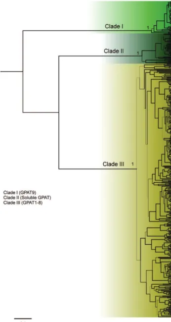
To investigate the evolutionary relationships among the plant GPATs, we reconstructed phylogenetic trees us-ing the protein sequences of putative GPATs identified by homology searches in 39 species. In Figure 1, a compact view of the tree based on protein sequences is shown (the entire, expanded view, including species names and acces-sion numbers, can be found in Figures 2–5). The phylogen-etic analysis of GPAT amino acid sequences resulted in a well-resolved tree, revealing the formation of three main clades (Figure 1). The first one (named clade I) includes GPAT9 sequences, the second (clade II) includes the solu-ble GPAT sequences, and the third (clade III) includes GPAT1-8 and GPAT-like proteins. The algal GPAT se-quences are placed in the GPAT9 and soluble GPAT cla-des, suggesting that these GPATs are the most ancient forms. No algae GPATs were placed within the GPAT1-8 clade (clade III), indicating that these GPATs are plant spe-cific and evolved in land plants to provide pathways for functions not present in other organisms.
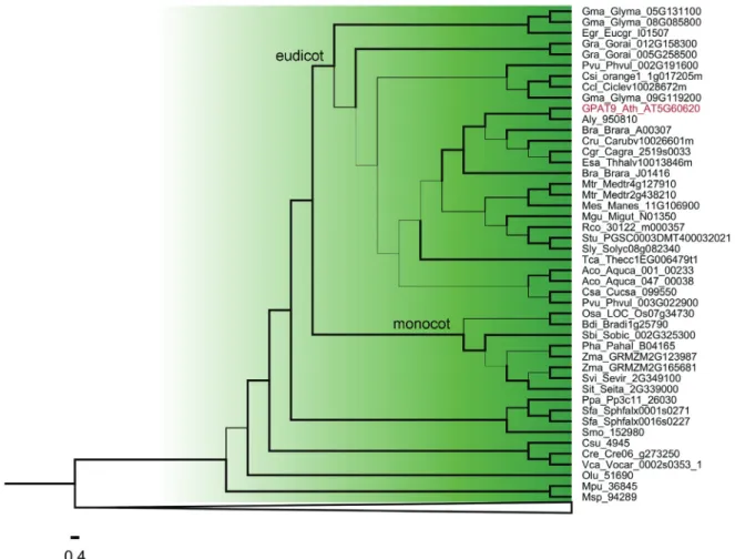
Within clade I (GPAT9) (Figure 2) and clade II (solu-ble GPAT) (Figure 3), the algal GPAT9 and algal solu(solu-ble GPAT are phylogenetically divergent from the land plant GPAT9 and land plant soluble GPAT. Among the land
plants, GPATs from basal plants (moss and lycophyte), monocots and eudicots species diverged from each other and formed distinct clusters. Most of the species studied present only one sequence of GPAT9 and soluble GPAT, except forG. max,G. raimondii,B. rapa,M. truncatula,E. grandisandZ. mays,these possibly presenting gene dupli-cation events.
Figure 3- Phylogenetic relationships among GPAT genes belonging to Clade II from Figure 1. Thicker lines present posterior probability > 0.9. The com-plete list of species is presented in Table S1.
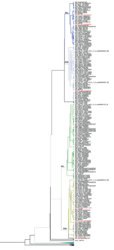
includes GPAT1-3. Within subclade IIIe we observed a separate group of sequences that included only monocot species related to the well characterized GPAT3 fromO. sativa. The GPATs fromP. patensandS. fallax(moss) and
S. moellendorffii (lycophyte), two basal lineages of land plants, are phylogenetically more related with GPAT4-8 (subclade IIIa) and GPAT6 (subclade IIIb), implying that GPAT4-6-8 are the most ancient forms of GPATs exclu-Figure 4- Phylogenetic relationships among GPAT genes belonging to
Clade III (subclades IIIa, IIIb, IIIc and IIId) from Figure 1. Thicker lines present posterior probability > 0.9.
sive of land plants (GPAT1-8). Within subclade IIIa, most of the species presented only one sequence. The species that presented more than one sequence are G. max, M. gutattus,B. rapa,A. lyrata,A. thaliana(well characterized GPAT 4 and GPAT8) andE. salsugineum.This indicates that duplication events that originated GPAT 4 and GPAT8 were independent, lineage specific events. Subclade IIIb (GPAT6) is closely related with subclade IIIa suggesting that GPAT4, GPAT8 and GPAT6 have a common ancestral gene and diverged from duplication events. GPAT5 and GPAT7 within subclade IIId are also likely resulted from independent and lineage-specific duplication events. GPAT1, GPAT2 and GPAT3 (subclade IIIe) are closely re-lated and may have originated by duplication events in vas-cular plants. TheA. thalianaAT3G11325 gene retrieved in BLAST searches and annotated as Phospholipid/glycerol acyltransferase family protein in the Phytozome database is placed in subclade IIId, close to GPAT5 and GPAT7. This sequence also presents an acyltransferase domain.
Comparative analysis of gene structure and organization of GPATs
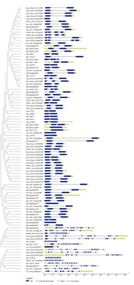
To explore possible mechanisms underlying gene structure and organization of GPAT genes during evolu-tion, we compared the exon–intron organization pattern of GPAT genes from plant and algae species (Table S1 and Figure 6). The length (in base pairs) of exons and introns were counted manually by aligning the cDNA sequences to their corresponding genomic DNA sequences. These anal-yses revealed that the number of introns per gene ranged from zero to 14. Most of the putative GPAT sequences re-trieved by BLAST searches (280) have only one intron. The number of introns and the gene organization were fairly conserved within the GPAT clades. The number of introns in algal GPAT9 genes ranged from zero to seven, while most of the GPAT9 genes from land plants have 11 introns, suggesting a possible gain of introns in land plant GPAT9 genes. The same pattern was observed for soluble GPAT. The gene structure analysis for GPAT1-8 showed that most of the species have one intron, with some exceptions, such asA. thalianaGPAT 4 and 8 that have three introns (Figure 6). Although several plant genes carry introns, a significant portion of plant genes lack introns. Genes that are not inter-rupted by introns are called intronless genes or single-exon genes. Since intronless genes are very important in under-standing evolutionary patterns of related genes and geno-mes, we verified the intronless for GPAT genes. Twenty nine out of 450 sequences included in this study are intron-less genes. Most of them belong to clade III (GPAT1-8). For GPAT9 genes, only the algalO. lucimarinusandM. pusillaGPAT9 genes are intronless.
In addition, intron phases across all GPATs of repre-sentative species (Figure 6) were investigated. The analysis showed that the intron phase pattern is quite variable across plant GPATs. Phase 0 was majority across genes with only
one intron, that is the case of most GPAT1-8s. The intron phase 2,0,2,0,0,1,2,0,2,2,2 is strikingly conserved across putative GPAT9 genes that grouped into the clade I to-gether with GPAT9 from A. thaliana. For the soluble GPAT the intron phase pattern is 1,0,0,2,0,0,2,2,0,0,0.
Evaluation of GPAT protein properties
After the examination of gene structure, we continued our analysis with a focus on the protein properties of 450 putative GPATs, including protein length, presence of pu-tative transmembrane domains, and conserved motifs. Overall, the length of the GPAT amino acid sequences ranged from 237 to 621 residues (see Table S1 for details). Conserved motifs in the representative proteins from plant and algae species are depicted in Figure 7. Analysis of the amino acid sequences of the 10 members of GPATs in plants revealed that all have a plsC acyltransferase domain in the C-terminal region. A second domain in the N-ter-minal region that is homologs to conserved motifs of the HAD-like hydrolase superfamily is found in some GPATs (GPAT4-8). The C-terminal acyltransferase domain of the GPAT family possesses the classic H(X)4D motif of PlsC class acyltransferases (Figures 7). Predictions of trans-membrane (TrM) structures showed that at least one region of GPAT1-9 proteins contained a highly probable TrM se-quence (Table S3, Figure S1), while no TrM was identified for plastid GAPT, indicating that GPAT1-9 proteins are as-sociated with membrane systems and that plastid GPAT is a soluble form.
Expression profiling of GPAT genes in model monocot and eudicot plants
The available plant expression data from GENEVESTIGATOR was used to obtain information about potential functional roles of each GPAT. We ana-lyzed the temporal and spatial expression patterns of the GPAT genes in plant tissues, using public microarray ex-pression data of the eudicotsA. thaliana(Table 2, Figures S2 and S3) andG. max(Table 2, Figures S4 and S5) and the monocotsO. sativa (Table 2, Figures S6 and S7) andZ. mays(Table 2, Figures S8 and S9). We found probes for nine GPATs inA. thaliana (GPAT1-6, GPAT8, GPAT9 and soluble GPATs). ForG. max, eight out of 28 genes identified in our BLAST searches presented available probes (Glyma.01G014200, Glyma.09G207900,
Glyma.02G249300, Glyma.14G028300,
Glyma.07G069700, Glyma.03G078600,
Glyma.01G113200, Glyma.02G010600). For the monocots, we found 17 available probes for 17 putative
GPATs for Z. mays (GRMZM2G165681,
GRMZM2G123987, GRMZM2G065203,
GRMZM2G177150, GRMZM2G147917,
GRMZM2G064590, GRMZM2G124042,
GRMZM2G166176, GRMZM2G083195,
Waschburger
et
al.
Species Gene (Clade) Anatomical parts Development stages
Arabidopsis thaliana
GPAT1 - AT1G06520 (IIIe) inflorescence, flower, stame, stigma, ovary, petal, suspensor, re-plum
GPAT2 - AT1G02390 (IIIe) Lateral root cap protoplast, root epidermis and lateral root cap protoplast, senescent leaf
mature siliques
GPAT3 - AT2G38110 (IIIe) lateral root cap protoplast, root epidermis and lateral root cap pro-toplast, root hair cell propro-toplast, guard cell propro-toplast, guard cell
-GPAT4 - AT4G01950 (IIIa) guard cell protoplast, root endodermis and quiescent center cell, root culture, seedling culture, cotyledon and leaf pavement cell, guard cell, seedling, cotyledon, pedicel
germinated seed, seedling, young rosette, developed rosette, bolt-ing, developed flower, flowers and siliques
GPAT5 - AT3G11430 (IIId) root endodermis and quiescent center cell, root stele cell
-GPAT6 - AT1G01610 (IIIb) flower stamen, stigma, pela, sepal
-GPAT7 - AT5G06090 (IIId)
-GPAT8 - AT4G00400 (IIIa) guard cell protoplast, root endodermis and quiescent center cell, root culture, seedling culture, cotyledon and leaf pavement cell, trichome and leaf petiole epidermis cell, cotyledon and leaf guard cell, shoot vascular tissue and bundle sheath cell, guard cell, seedling, cotyledon
germinated seed, seedling, young rosette, developed rosette, bolt-ing, developed flower, flowers and siliques
GPAT9 - AT5G60620 (I) embryo, suspensor, endosperm, micropylar endosperm peripheral endosperm chalazal endosperm, cotyledon and leaf pavement cell
senescence
Soluble GPAT - AT1G32200 (II) cotyledon, shoot apex, pedicel, shoot, leaf primordia, axillary shoot germinated seed, seedling, young rosette, developed rosette, bolt-ing, developed flower
Glycine max Glyma.01G014200 (II) shoot, trifoliolate leaf, inner integument, shoot apical meristem lowers and siliques
Glyma.09G207900 (II) syncytium, paraveinal mesophyll cell palisade parenchyma cell, seedling, shoot apical meristem, axillary meristem
inflorescence, embryo, suspensor, inner integument
fruit formation
Glyma.02G249300 (IIId) - flowering
Glyma.14G028300 (IIIe) leaf flowering
Glyma.07G069700 (IIIa) seedling, shoot apical meristem, axillary meristem, inflores-cence, suspensor, pod, testa, shoot
-Glyma.03G078600 (IIIb) pod
-Glyma.01G113200 (IIIb) root hair
-Glyma.02G010600 (IIIe) seedling, pod
-Oryza sativa LOC_Os01g44069/OS01G0631400 (IIIe) pistil, stigma, ovary
-LOC_Os10g27330/OS10G0413400 (IIIc) inflorescence
-LOC_Os03g52570/OS03G0735900 (IIIc) inflorescence germination
analysis
and
evolution
of
plant
GPAts
365
Species Gene (Clade) Anatomical parts Development stages
LOC_Os05g38350/OS05G0457800 (IIId) -
-LOC_Os11g45400/OS11G0679700 (IIIe) seedling, leaf, inflorescence, anther, pistil
-LOC_Os02g02340/OS02G0114400 (IIIa) root seedling, tillering stage
LOC_Os05g20100/OS05G0280500 (IIIe) root
-LOC_Os08g03700/OS08G0131300 coleoptile
-LOC_Os01g19390/OS01G0299300 (IIIe) -
-LOC_Os12g37600/OS12G0563000 (IIIe) coleoptile
-LOC_Os03g61720/OS03G0832800 (IIIe) seedling, leaf germination
LOC_Os01g14900 (IIIe) -
-LOC_Os05g37600/OS05G0448300 (IIIb) -
-LOC_Os10g41070 (IIIe) pollen
-LOC_Os01g22560/OS01G0329000 (IIIe) sperm cell, leaf
-LOC_Os07g34730/OS07G0531600 (I) sperm cell, flag leaf, collar
-Zea mays GRMZM2G165681 (I) elongation zone, placento-chalazal region, brace root, spikelet, ova-ry, central starchy endosperm, conducting zone
-GRMZM2G123987 (I) spikelet, central starchy endosperm, pericarp, ovary
-GRMZM2G065203 (IIIe) style(silk), adult leaf, sheath, husk leaf primordium, foliar leaf primordium
-GRMZM2G177150 (IIIe) husk leaf primordium
-GRMZM2G147917 (IIIc) meyocite
-GRMZM2G064590 (IIIc) tassel, shoot, husk leaf primordium *not high enough quantities
-GRMZM2G124042 (IIIc) shoot
-GRMZM2G166176 (IIId) embryo sac, adult leaf, maturation zone
-GRMZM2G083195 (IIIb) husk leaf primordium, foliar leaf blade inflorescence formation
GRMZM2G059637 (IIId) root, cortex, adult leaf, root tip, maturation zone
-GRMZM2G072298 (IIIe) shoot
-GRMZM2G156729 (IIIe) -
-GRMZM2G070304 (IIIe) meyocite, pistil
-GRMZM2G033767 (IIIe) sheath
-GRMZM2G020320 (IIIa) adult leaf, root tip
-GRMZM2G131378 root tip
GRMZM2G156729, GRMZM2G070304,
GRMZM2G033767, GRMZM2G020320,
GRMZM2G131378, GRMZM2G159890) and 17 probes
for O. sativa (LOC_Os01g44069, LOC_Os10g27330,
LOC_Os03g52570, LOC_Os01g63580,
LOC_Os05g38350, LOC_Os11g45400,
LOC_Os02g02340, LOC_Os05g20100,
LOC_Os08g03700, LOC_Os01g19390,
LOC_Os12g37600, LOC_Os03g61720,
LOC_Os01g14900, LOC_Os05g37600,
LOC_Os10g41070, LOC_Os01g22560,
LOC_Os07g34730). We analyzed 105 anatomical parts and 10 developmental stages fromA. thaliana, 68 anatomi-cal parts and five developmental stages fromG. max, 85 an-atomical parts and 7 developmental stages from Z. may, and38 anatomical parts and 9 developmental stages from
O. sativa.
In silicoanalyses of the expression profiles showed that all plant GPAT genes present some expression level in developmental stages and anatomical parts. However, dif-ferent expression patterns across difdif-ferent tissues and plant developmental stages were found across different GPATs within each species (Table 2). For example, theA. thaliana
GPAT1 and GPAT6 genes are more expressed in inflores-cence parts, while GPAT2 and GPAT3 are more expressed in root parts. TheA. thaliana GPAT4 and GPAT8 genes presented high expression in guard cell protoplast, root endodermis and quiescent center cell, root culture, seedling culture, cotyledon and leaf pavement cell, guard cell, seed-ling, cotyledon, pedicel. The GPAT9 gene fromA. thaliana
is more expressed in embryo, suspensor, endosperm, micropylar endosperm, peripheral endosperm, chalazal en-dosperm, cotyledon and leaf pavement cell; while soluble GPAT is more expressed in cotyledon, shoot apex, pedicel, shoot, leaf primordia and axillary shoot. The G. max
Glyma.01G113200 gene included in subclade IIIb (GPAT6) is more expressed in radicle, maturation zone and root hair, while Glyma.14G028300 (included in subclade IIIe and closely related with GPAT1 fromA. thaliana) and Glyma.02G249300 (included in subclade IIId – GPAT5
and 7) are more expressed in flower cluster (raceme), flower, androecium, stamen, anther and pollen.O. sativa
LOC_Os01g63580 included in the GPAT6 group (subclade IIIb) is more expressed in inflorescence, panicle, spikelet, coleoptile, anther and pistil.O. sativaLOC_Os01g44069 included in the GPAT1 group (subclade IIIe) is highly ex-pressed in stigma.O. sativa LOC_Os11g45400 grouped into subclade IIId is more expressed in seedling, leaf, inflo-rescence, anther and pistil.Z. maysGRMZM2G070304 in-cluded in subclade IIIe (GPAT1-3) is highly expressed in spikelet cell, floret cell, stamen cell, anther cell and meyo-cite.
Discussion
phylogenetic tree (Figure 1) and named clade I, clade II and clade III. Clade III is the most diversified clade and were further subdivided into five subclades (IIIa, IIIb, IIIc, IIId and IIIe).
Clade I includes GPAT9 homologs from six algae species and 31 plant species. Most species studied possess only one GPAT9 gene, and we were not able to find GPAT9 homologs inA. trichopodaandP. trichocarpa. Our phylogenetic analysis showed that GPAT9 is a very diver-gent clade. It has already been reported that A. thaliana
GPAT9 is more closely related to the mammalian ER-localized GPAT3 and GPAT4 compared to other members of the A. thalianaGPAT family (GPAT1–8), suggesting that the divergence of the GPAT9 gene from the GPAT1–8 of this species occurred prior to the evolutionary split be-tween plants and mammals (Giddaet al., 2009) and that they have experienced different patterns of evolution. GPAT9 has been demonstrated to be involved in TAG biosynthesis and to be present in several algal species that also produce an abundance of TAGs (Khozin-Goldberg and Cohen, 2011; Iskandarovet al., 2016). Heterologous ex-pression of a GPAT9 homolog from the oleaginous green microalgaLobosphaera incisainC. reinhardtiiincreased TAG content by up to 50% (Iskandarovet al., 2016). In the oilseed plantR. comunis, GPAT9 (30122.m000357) pres-ents higher expression compared to other GPATs in endo-sperm tissue, suggesting that it is likely important in castor oil synthesis (Brownet al., 2012). Our expression analysis showed thatA. thalianaGPAT9 presents higher levels of expression in embryo, suspensor, endosperm, micropylar endosperm, peripheral endosperm, chalazal endosperm, cotyledon and leaf pavement cell. Studies withA. thaliana
showed that reduced GPAT9 expression impacts the amount and composition of TAGs in seeds (Shockeyet al., 2015). Another study demonstrated that GPAT9 exhibits sn-1 acyltransferase activity with high specificity for acyl-CoA, thus confirming its role in seed TAG biosynthesis, and provides comprehensive evidence in support of its role in the production of both polar and non-polar lipids in leaves, as well as lipid droplets in pollen (Singeret al., 2016). The exon/intron structures (11 introns and 12 exons) and intron phase patterns (2,0,2,0,0,1,2,0,2,2,2) are con-served in almost all GPAT9 genes of land plants, which di-verge form algae GPAT9 that present seven introns/six exons and intron phase patterns (2,2,2,2,0,1,1). These dif-ferences suggest that the structure of the land plant GPAT9 gene was established and retained after the divergence of land plants from algae. It also indicates an intron gain throughout Embryophyta evolution. In addition, all protein sequences grouped into clade I and classified as GPAT9 present at least one putative transmembrane domain (TMD), indicating that they are membrane proteins.
The soluble, plastid-localized GPAT homologs from six algae species and 30 plant species are grouped into clade II. Most species studied possess only one gene and we
were not able to find homologs in S. tuberosum, M.
esculentaandC. sinensis. Soluble plastid GPAT was the
first GPAT to be identified in plants (Murata and Tasaka, 1997). This enzyme is essential for chloroplasts glyce-rolipid synthesis that are primarily converted into galac-tolipids, which serve as major structural and functional components of photosynthetic membranes (Dörmann and Benning, 2002). Analyses of gene expression in A. thaliana,G. maxandZ. maysshowed that soluble GPAT is predominantly expressed in green tissues, this being cor-roborated by the fact that this protein is involved in chlo-roplast lipid biosynthesis. A similar pattern was also observed for soluble GPAT of Helianthus annuus
(HaPLSB) (Payá-Milanset al., 2015). These authors dem-onstrated that HaPLSB expression increased during cotyle-don development, which was consistent with the elevated rate ofde novo chloroplast membrane lipid biosynthesis during the early stages of plant growth, and was maintained at high levels in mature leaves. The transmembrane domain prediction demonstrated that all sequences grouped into clade II have no TMD, confirming their soluble form for all species studied, as already was shown for A. thaliana
(Nishidaet al., 1993; Kim, 2004; Chandra-Shekaraet al., 2007) and H. annuus (Payá-Milans et al., 2015). The exon/intron structures (11 introns and 12 exons) and intron phase patterns (1,0,0,2,0,0,2,2,0,0,0) are conserved among land plants within clade I. The soluble GPAT from algae presents only one intron, this indicating an intron gain throughout Embryophyta evolution also for this gene.
The remaining GPATs (GPAT1-8) were included in the clade III and are present only in Embryophyta lineages. It was shown forA. thaliana that members of GPAT1-8 clearly affect the composition and quantity of cutin or suberin (Beissonet al., 2007, 2012; Yanget al., 2012). None of these GPATs seem to be required for the synthesis of membrane or storage lipids. These results demonstrated
in vivothat a GPAT enzyme can catalyze the transfer of
cutin formation in leaves (Liet al., 2007), and GPAT6 re-quired for cutin synthesis in flowers (Yanget al., 2012). GPAT5 and GPAT7 also resulted from an independent and lineage-specific duplication event that appears to have oc-curred after monocot/eudicot divergence. GPAT5 has been shown to be involved in suberin synthesis in roots and seeds (Beisson et al., 2007). GPAT1 is closely related with GPAT2 and GPAT3. These GPATs were shown to be lo-cated in the mitochondria. Most of the GPAT1-8 members present a conservation of intron/exon structure (one in-tron/2 exons). However, these GPATs presented a variable gene expression pattern, indicating that they can be differ-entially regulated depending on plant tissue.
In conclusion, our study provides a comprehensive genomic analysis of GPAT genes in plants, covering phylo-genetic, gene structure, protein properties, and gene expres-sion analysis. These results can improve our understanding of the evolutionary history of GPAT genes in plants and shed light on their function. Phylogenetic analysis indicates that plant GPATs can be grouped into three distinct clades, which is further supported by their conservation and varia-tion in gene structure, protein properties, motif occurrences and gene expression patterns. Our study, together with pre-vious studies, suggests that the presence of several genes encoding GPATs in land plants may be related to their ad-aptation to a terrestrial environment. Current knowledge re-garding the functions of plant GPATs is limited to few species. To obtain a more thorough understanding of the function of GPATs in plants, the functional characteriza-tion of GPATs in more species will be necessary.
Acknowledgments
This work was financially supported by Conselho Nacional de Desenvolvimento Científico e Tecnológico (CNPq; grant number: 306202/2016-6) and Coordenação de Aperfeiçoamento de Pessoal de Nível Superior (CAPES).
References
Abascal F, Zardoya R and Posada D (2005) ProtTest: Selection of best-fit models of protein evolution. Bioinformatics 21:2104-2105.
Bailey TL, Williams N, Misleh C and Li WW (2006) MEME: Dis-covering and analyzing DNA and protein sequence motifs. Nucleic Acids Res 34:W369-W373.
Beisson F, Li Y, Bonaventure G, Pollard M and Ohlrogge JB (2007) The acyltransferase GPAT5 is required for the syn-thesis of suberin in seed coat and root of Arabidopsis. Plant Cell 19:351-368.
Beisson F, Li-Beisson Y and Pollard M (2012) Solving the puz-zles of cutin and suberin polymer biosynthesis. Curr Opin Plant Biol 15:329-337.
Brown AP, Kroon JTM, Swarbreck D, Febrer M, Larson TR, Gra-ham IA, Caccamo M and Slabas AR (2012) Tissue-specific whole transcriptome sequencing in castor, directed at
under-standing triacylglycerol lipid biosynthetic pathways. PLoS One 7:e30100.
Chandra-Shekara AC, Venugopal SC, Barman SR, Kachroo A and Kachroo P (2007) Plastidial fatty acid levels regulate re-sistance gene-dependent defense signaling in Arabidopsis. Proc Natl Acad Sci U S A 104:7277-82.
Chen X, Snyder CL, Truksa M, Shah S and Weselake RJ (2011a) sn-Glycerol-3-phosphate acyltransferases in plants. Plant Signal Behav 6:1695-1699.
Chen X, Truksa M, Snyder CL, El-Mezawy A, Shah S and Weselake RJ (2011b) Three homologous genes encoding sn-glycerol-3-phosphate acyltransferase 4 exhibit different expression patterns and functional divergence inBrassica napus. Plant Physiol 155:851-865.
Chen X, Chen G, Truksa M, Snyder CL, Shah S and Weselake RJ (2014) Glycerol-3-phosphate acyltransferase 4 is essential for the normal development of reproductive organs and the embryo inBrassica napus. J Exp Bot 65:4201-4215. Crooks GE, Hon G, Chandonia JM and Brenner SE (2004)
WebLogo: A sequence logo generator. Genome Res 14:1188-1190.
Dörmann P and Benning C (2002) Galactolipids rule in seed plants. Trends Plant Sci 7:112-118.
Drummond AJ, Suchard MA, Xie D and Rambaut A (2012) Bayesian phylogenetics with BEAUti and the BEAST 1.7. Mol Biol Evol 29:1969-1973.
Edgar RC (2004) MUSCLE: Multiple sequence alignment with high accuracy and high throughput. Nucleic Acids Res 32:1792-1797.
Gidda SK, Shockey JM, Rothstein SJ, Dyer JM and Mullen RT (2009)Arabidopsis thalianaGPAT8 and GPAT9 are local-ized to the ER and possess distinct ER retrieval signals: Functional divergence of the dilysine ER retrieval motif in plant cells. Plant Physiol Biochem 47:867-879.
Gimeno RE and Cao J (2008) Mammalian glycerol-3-phosphate acyltransferases: new genes for an old activity. J Lipid Res 49:2079-2088.
Guo AY, Zhu QH, Chen X and Luo JC (2007) GSDS: a gene structure display server. Yi Chuan 29:1023-1026.
Hruz T, Laule O, Szabo G, Wessendorp F, Bleuler S, Oertle L, Widmayer P, Gruissem W and Zimmermann P (2008) Gene-vestigator v3: A reference expression database for the meta-analysis of transcriptomes. Adv Bioinformatics 2008:420747.
Iskandarov U, Sitnik S, Shtaida N, Didi-Cohen S, Leu S, Khozin-Goldberg I, Cohen Z and Boussiba S (2016) Cloning and characterization of a GPAT-like gene from the microalga Lobosphaera incisa(Trebouxiophyceae): overexpression in Chlamydomonas reinhardtii enhances TAG production. J Appl Phycol 28:907-919.
Khozin-Goldberg I and Cohen Z (2011) Unraveling algal lipid metabolism: Recent advances in gene identification. Bio-chimie 93:91-100.
Kim HU (2004) Plastid lysophosphatidyl acyltransferase Is essen-tial for embryo development in Arabidopsis. Plant Physiol 134:1206-1216.
Kolattukudy PE (2001) Polyesters in higher plants. Adv Biochem Eng Biotechnol 71:1-49.
lyso-phosphatidic acid acyltransferase (LPAAT) gene family. Mol Phylogenet Evol 96:55-69.
Kornberg A and Pricer WE (1987) Enzymatic synthesis of the coenzyme A derivatives of long chain fatty acids. J. Biol. Chem. 1953:3293-43.
Krogh A, Larsson B, von Heijne G and Sonnhammer ELL (2001) Predicting transmembrane protein topology with a hidden Markov model: Application to complete genomes. J Mol Biol 305:567-580.
Kumar S, Stecher G and Tamura K (2016) MEGA7: Molecular Evolutionary Genetics Analysis version 7.0 for bigger data-sets. Mol Biol Evol 33:1870-4.
Lewin TM, Wang P and Coleman RA (1999) Analysis of amino acid motifs diagnostic for the sn-glycerol-3-phosphate acyl-transferase reaction. Biochemistry 38:5764-5771.
Li-Beisson Y, Shorrosh B, Beisson F, Andersson MX, Arondel V, Bates PD, Baud S, Bird D, DeBono A, Durrett TP,et al. (2013) Acyl-lipid metabolism. Arabidopsis Book 11:e0161. Li Y, Beisson F, Koo AJ, Molina I, Pollard M and Ohlrogge J
(2007) Identification of acyltransferases required for cutin biosynthesis and production of cutin with suberin-like monomers. Proc Natl Acad Sci U S A 104:18339-18344. Liu Q, Siloto RMP, Lehner R, Stone SJ and Weselake RJ (2012)
Acyl-CoA:diacylglycerol acyltransferase: Molecular biol-ogy, biochemistry and biotechnology. Prog Lipid Res 51:350-377.
Men X, Shi J, Liang W, Zhang Q, Lian G, Quan S, Zhu L, Luo Z, Chen M and Zhang D (2017) Glycerol-3-Phosphate Acyl-transferase 3 (OsGPAT3) is required for anther development and male fertility in rice. J Exp Bot 12:erw445.
Murata N and Tasaka Y (1997) Glycerol-3-phosphate acyltrans-ferase in plants. Biochim Biophys Acta - Lipids Lipid Metab 1348:10-16.
Nishida I, Frentzen M, Ishizaki O and Murata N (1987) Purifica-tion of isomeric forms of acyl-[acyl-carrier-protein]:glyc-erol-3-phosphate acyltransferase from greening squash cot-yledons. Plant Cell Physiol 28:1071-1079.
Nishida I, Tasaka Y, Shiraishi H and Murata N (1993) The gene and the RNA for the precursor to the plastid-located glyc-erol-3-phosphate acyltransferase of Arabidopsis thaliana. Plant Mol Biol 21:267-277.
Ohlrogge J and Browse J (1995) Lipid biosynthesis. Plant Cell 7:957-970.
Omasits U, Ahrens CH, Müller S and Wollscheid B (2014) Prot-ter: Interactive protein feature visualization and integration with experimental proteomic data. Bioinformatics 30:884-886.
Pan X, Peng FY and Weselake RJ (2015) Genome-wide analysis of PHOSPHOLIPID:DIACYLGLYCEROL ACYL-TRANSFERASE (PDAT) genes in plants reveals the eudi-cot-wide PDAT gene expansion and altered selective pres-sures acting on the core eudicot PDAT paralogs. Plant Physiol 167:887-904.
Payá-Milans M, Venegas-Calerón M, Salas JJ, Garcés R and Martínez-Force E (2015) Cloning, heterologous expression and biochemical characterization of plastidial sn-glycerol-3-phosphate acyltransferase from Helianthus annuus. Phytochemistry 111:27-36.
Posada D (2008) jModelTest: Phylogenetic model averaging. Mol Biol Evol 25:1253-1256.
Rambaut A, Suchard MA, Xie D and Drummond AJ (2014) Tracer v1.6, Available from http://tree.bio.ed.ac.uk/soft-ware/tracer/.
Ranathunge K, Schreiber L and Franke R (2011) Suberin research in the genomics era-New interest for an old polymer. Plant Sci 180:339-413.
Rensing SA, Lang D, Zimmer AD, Terry A, Salamov A, Shapiro H, Nishiyama T, Perroud P-F, Lindquist EA,et al.(2008) The Physcomitrellagenome reveals evolutionary insights into the conquest of land by plants. Sci New Ser 319:64-69. Sánchez-García A, Moreno-Pérez AJ, Muro-Pastor AM, Salas JJ,
Garcés R and Martínez-Force E (2010) Acyl-ACP thio-esterases from castor (Ricinus communisL.): An enzymatic system appropriate for high rates of oil synthesis and accu-mulation. Phytochemistry 71:860-869.
Schreiber L (2010) Transport barriers made of cutin, suberin and associated waxes. Trends Plant Sci 15:546-553.
Shockey J, Regmi A, Cotton K, Adhikari N, Browse J, Bates PD, Wallace J and Buckler ES (2015) Identification of Arabidopsis GPAT9 (At5g60620) as an essential gene in-volved in triacyglycerol biosynthesis. Plant Physiol 170:163-179.
Singer SD, Chen G, Mietkiewska E, Tomasi P, Jayawardhane K, Dyer JM and Weselake RJ (2016) Arabidopsis GPAT9 con-tributes to synthesis of intracellular glycerolipids but not surface lipids. J Exp Bot 67:4627-4638.
Takeuchi K and Reue K (2009) Biochemistry, physiology, and ge-netics of GPAT, AGPAT, and lipin enzymes in triglyceride synthesis. Am J Physiol Endocrinol Metab 296:E1195-E1209.
Turchetto-Zolet AC, Maraschin FS, de Morais GL, Cagliari A, Andrade CM, Margis-Pinheiro M and Margis R (2011) Evo-lutionary view of acyl-CoA diacylglycerol acyltransferase (DGAT), a key enzyme in neutral lipid biosynthesis. BMC Evol Biol 11:263-277.
Turchetto-Zolet AC, Christoff AP , Kulcheski FR, Loss-Morais G, Margis R and Margis-Pinheiro M (2016) Diversity and evolution of plant diacylglycerol acyltransferase (DGATs) unveiled by phylogenetic, gene structure and expression analyses. Genet Mol Biol 39:524-538.
Wang Y, Xu L, Thilmony R, You FM, Gu YQ and Coleman-Derr D (2016) PIECE 2.0: An update for the plant gene structure comparison and evolution database. Nucleic Acids Res 45:1015-1020.
Wang Y, You FM, Lazo GR, Luo MC, Thilmony R, Gordon S, Kianian SF and Gu YQ (2013) PIECE: A database for plant gene structure comparison and evolution. Nucleic Acids Res 41:D1159-D1166.
Weiss BS, Kennedy PE and Kiyasu YJ (1939) The enzymatic synthesis of triglycerides. J Biol Chem 235:40-44.
Wendel AA, Lewin TM and Coleman RA (2009) Glycerol-3-phosphate acyltransferases: Rate limiting enzymes of tria-cylglycerol biosynthesis. Biochim Biophys Acta - Mol Cell Biol Lipids 1791:501-506.
Zhang Y-M and Rock CO (2008) Thematic review series: Gly-cerolipids. Acyltransferases in bacterial glycerophospho-lipid synthesis. J Lipid Res 49:1867-1874.
Zheng Z and Zou J (2001) The initial step of the glycerolipid path-way: Identification of glycerol 3-phosphate/dihydro-xyacetone phosphate dual substrate acyltransferases in Saccharomyces cerevisiae. J Biol Chem 276:41710-41716.
Zheng Z, Xia Q, Dauk M, Shen W, Selvaraj G and Zou J (2003) Arabidopsis AtGPAT1, a member of the membrane-bound glycerol-3-phosphate acyltransferase gene family, is essen-tial for tapetum differentiation and male fertility. Plant Cell 15:1872-1887.
Internet resources
GENEVESTIGATOR tools - https://www.genevestigator.com (January 1, 2017)
FigTree software - http://tree.bio.ed.ac.uk/software/figtree/ (March 1, 2017)
The plant genomics resouces (Phytozome) https://phytozome.jgi.doe.gov/pz/portal.html (October 1, 2016)
PIECE database - http://wheat.pw.usda.gov/piece/GSDraw.php (December 1, 2016)
TMHMM-2.0 software- http://www.cbs.dtu.dk/services/ (De-cember 1, 2016)
SMART software - http://smart.embl-heidelberg.de/ (December 1, 2016)
PROTTER software - http://wlab.ethz.ch/protter/start/ (Decem-ber 1, 2016)
Supplementary material
The following online material is available for this article: Table S1- Information on the GPAT sequences retrieved in this study.
Table S2 - Information about similarity with the respective Arabidopsis gene
Table S3 - Predictions of transmembrane domains of plant GPAT proteins.
Figure S1 - Properties of GPAT protein sequences of repre-sentative species.
Figure S2 - Microarray data analysis of GPATs in anatomi-cal parts ofArabidopsis thaliana.
Figure S3 - Microarray data analysis of GPATs in develop-mental stages ofArabidopsis thaliana.
Figure S4 - Microarray data analysis GPATs in anatomical parts ofGlycine max.
Figure S5 - Microarray data analysis from of GPATs in de-velopmental stages ofGlycine max.
Figure S6 - Microarray data analysis of GPATs in anatomi-cal parts ofOryza sativa.
Figure S7 - Microarray data analysis of GPATs in develop-mental stages ofOryza sativa.
Figure S8 -Microarray data analysis of GPATs in anatomi-cal parts ofZea mays.
Figure S9 - Microarray data analysis of GPATs in develop-mental stages ofZea mays.
Associate Editor: Loreta B. Freitas


