Development of a multiplex PCR tool for screening of
pathogens in teleost farmed fish
Micaela Ferreira Pinto
Development of a multiplex PCR tool for screening
of pathogens in teleost farmed fish
Micaela Ferreira Pinto
Dissertação para obtenção do Grau de Mestre em Biotecnologia dos
Recursos Marinhos
Dissertação de Mestrado realizada sob a orientação da Doutora Clélia
Afonso e co-orientação da Especialista Teresa Baptista
ii
Title: Development of a multiplex PCR tool for screening of pathogens in teleost farmed fish
Título: Desenvolvimento de uma ferramenta de multiplex PCR para rastreio de agentes patogénicos em peixes teleósteos de aquacultura
Copyright © Micaela Ferreira Pinto
A Escola Superior de Turismo e Tecnologia do Mar e o Instituto Politécnico de Leiria têm o direito, perpétuo e sem limites geográficos, de arquivar e publicar este trabalho de projeto através de exemplares impressos reproduzidos em papel ou de forma digital, ou por qualquer outro meio conhecido ou que venha a ser inventado, e de a divulgar através de repositórios científicos e de admitir a sua cópia e distribuição com objetivos educacionais ou de investigação, não comerciais, desde que seja dado crédito ao autor e editor.
iii
Faça as coisas o mais simples que puder, porém não se restrinja às mais
simples.
iv
v Agradecimentos
Nesta etapa final, não poderia deixar de mostrar a minha gratidão a todas as pessoas que de alguma forma contribuíram, direta ou indiretamente, para a elaboração desta dissertação.
Em primeiro lugar tenho de agradecer à minha família, principalmente aos meus pais por todo o apoio incondicional e porque sem eles este sonho não seria possível.
Às minhas duas orientadoras Doutora Clélia Afonso e Especialista Teresa Baptista agradeço todo o acompanhamento e disponibilidade demonstrados ao longo desta etapa.
Ao professor Américo Rodrigues pela sua disponibilidade e contribuição ao longo do estudo.
Ao Tiago Gomes por toda a ajuda e por estar sempre lá para mim.
Não poderia deixar também de agradecer à Adriana Januário pela paciência e encorajamento que me deu ao longo deste ano.
Deixo também um agradecimento a todas as pessoas que de alguma forma contribuíram nesta etapa e que não foram anteriormente mencionadas.
vi
vii Resumo
A aquacultura desempenha um papel cada vez mais importante na produção de alimentos em todo o mundo (Martins et al., 2015). Espécies dos géneros Aeromonas,
Vibrio, Edwardsiella e Streptococcus são patógenos que infetam peixes (Zhang et al.,
2014), causando elevadas perdas económicas em aquacultura.
A corvina (Argyrosomus regius, Asso, 1801) é um dos maiores peixes da família Sciaenidae a nível mundial, e uma espécie de elevado valor comercial no Sudoeste da Europa, sendo neste momento objeto de grande interesse em todo o Mediterrâneo visando a sua produção comercial (Amoedo, 2011). Devido à sua elevada fertilidade, ampla distribuição e boa aceitação por parte dos consumidores torna-se um bom candidato à produção em aquacultura podendo atingir preços de mercado médio-altos (6 euros/kg, INE 2010).
Como ainda não foram reportadas manifestações patológicas relevantes nesta espécie, o principal método de prevenção é o controlo da densidade nos tanques, sendo necessário pesquisar eventuais patógenos que afetem a corvina em diferentes estádios do seu ciclo e mesmo em condições de stress.
O objetivo deste estudo foi o desenvolvimento de uma ferramenta de multiplex-PCR para a deteção precoce de Edwardsiella tarda, Photobacterium damselae subsp.
piscicida, Vibrio alginolyticus, Vibrio anguillarum e Vibrio harveyi em peixes de
aquacultura, incluindo a corvina, que permita de forma prática e eficiente a deteção destes patógenos.
Concluiu-se que é possível através de ferramentas de multiplex-PCR, a deteção dos patogénicos Edwardsiella tarda, Vibrio alginolyticus, Vibrio anguillarum e Vibrio
harveyi até um limite mínimo de 0,4 ng de DNA/µl para V. anguillarum; 0.5 ng/µl para E. tarda; 1.5 ng/µl para V. harveyi e 5.6 ng/µl para V. alginolyticus, podendo, no futuro, estas
técnicas ter aplicação prática cada vez mais extensa.
Palavras-chave: Argyrosomus regius, Aquacultura, diagnóstico de doenças, multiplex-PCR.
viii
ix Abstract
Aquaculture plays an increasingly important role in food production worldwide (Martins et al., 2015). Species of the four genera Aeromonas, Vibrio, Edwardsiella and
Streptococcus are major pathogens that infect fish (Zhang et al., 2014), causing high
economic loss in aquaculture.
The meagre (Argyrosomus regius, Asso, 1801) is one of the largest fish from the Sciaenidae family worldwide, and a species of high commercial value in southwestern Europe. This species shows great interest for commercial production throughout the Mediterranean (Amoedo, 2011). Due to its high fertility, wide distribution and good acceptance by consumers, becomes a good candidate for aquaculture production that could reach medium-high market prices (6 euros/kg, INE 2010).
As have not yet been reported relevant pathological manifestations in this species, the primary method of prevention is to control the crop density, which requires research into possible pathogens affecting the meagre in different stages of the life cycle and even in stress conditions.
The aim of this study was to develop a multiplex-PCR tool for early detection of
Edwardsiella tarda, Photobacterium damselae subsp. piscicida, Vibrio alginolyticus, Vibrio anguillarum and Vibrio harveyi in farmed fish, including meagre, enabling practical and
efficient detection of these pathogens.
In conclusion, it is possible through multiplex-PCR tools the detection of
Edwardsiella tarda, Vibrio alginolyticus, Vibrio anguillarum and Vibrio harveyi as low as
0.4 ng/µl for V. anguillarum; 0.5 ng/µl for E. tarda; 1.5 ng/µl for V. harveyi and 5.6 ng/µl for
V. alginolyticus, may, in the future, practical application of these techniques increasingly
extensive.
x
xi Table of Contents
Resumo……… vii
Abstract……… ix
List of Figures……….. xiii
List of Tables………... xv
List of Abbreviations……….. xvii
Chapter I. General Introduction………... 19
1. Fish Diseases on Aquaculture………....21
2. Traditional and Molecular Diagnosis……….21
3. Major bacterial pathogens on Aquaculture………..….... 22
4. PCR diagnosis……….…. 25
5. m-PCR……….….. 28
5.1 State of the art………... 28
5.2 Tool Development……….….. 29
6. Test organism………... 30
7. Aims of the study……….…. 31
Chapter II. Development of a m-PCR tool for detection of four bacterial pathogens on Aquaculture……… 32
1. Introduction……….. 34
2. Materials and Methods……….. 34
2.1. Bacterial strains and culture conditions……….. 34
2.2. DNA extraction from bacterial pure culture and confirmation………. 35
2.3. Primers used in this study……… 35
2.4. m-PCR analysis……….. 36
2.5. Specificity and sensitivity of m-PCR assay………… 36
2.6. Experimental fish infection……… 36
2.7. Tool validation and Spiking on tissues……….. . 38
3. Results……….. 38
xii
3.2. Specificity and sensitivity of m-PCR assay………...… 40
3.3. Experimentally fish infection……… 42
3.4. Tool validation……….... 42
4. Discussion and Conclusion………...………44
Chapter III. Concluding Remarks and Future Perspectives………... 48
xiii List of Figures
Chapter II. Development of a m-PCR tool for detection of four bacterial pathogens on Aquaculture
Figure 2.1 – m-PCR at different annealing temperatures. M – 250 bp ladder; Lane 1 – 49ºC; Lane 2 – 51ºC; Lane 3 – 53°C; Lane 4 – 55ºC……….……… 39
Figure 2.2 – m-PCR at different annealing temperatures and different concentrations of MgCl2. Lane 1 – 250 bp ladder; Lane 2 - 49ºC, 2 mM MgCl2; Lane 3 – 49ºC, 4 mM MgCl2; Lane 4 – 49ºC, 6 mM MgCl2; Lane 5 – 49ºC, 8 mM MgCl2; Lane 6 - 51ºC, 2 mM MgCl2; Lane 7 - 51ºC, 4 mM MgCl2; Lane 8 - 51ºC, 6 mM MgCl2; Lane 9 - 51ºC, 8 mM MgCl2; Lane 10 - 53ºC, 2 mM MgCl2; Lane 11 - 53ºC, 4 mM MgCl2; Lane 12 - 53ºC, 6 mM MgCl2; Lane 13 - 53ºC, 8 mM MgCl2……….…..…….. 40
Figure 2.3 - Amplification of assay primers in monoplex reaction. M - 250 bp ladder; Lane 1 - E. tarda (426 bp); Lane 2 - Phdp (267 bp); Lane 3 - V. alginolyticus (773 bp); Lane 4 – V. anguillarum (519 bp); Lane 5 – V. harveyi (606 bp)………... 41
Figure 2.4 – m-PCR sensitivity test. Lane 1 – 250 bp ladder; Lane 2 – mixture of different DNA (64.6 ng/µl for E. tarda; 89.5 ng/µl for V. alginolyticus; 110.8 ng/µl for V.
anguillarum; 24 ng/µl for V. harveyi); Lane 3 – mixture of different DNA (32.3 ng/µl for E. tarda; 44.8 ng/µl for V. alginolyticus; 55.4 ng/µl for V. anguillarum; 12 ng/µl for V. harveyi);
Lane 4 – mixture of different DNA (16.2 ng/µl for E. tarda; 22.4 ng/µl for V. alginolyticus; 27.7 ng/µl for V. anguillarum; 6 ng/µl for V. harveyi); Lane 5 - mixture of different DNA (8.1 ng/µl for E. tarda; 11.2 ng/µl for V. alginolyticus; 13.9 ng/µl for V. anguillarum; 3 ng/µl for
V. harveyi); Lane 6 - mixture of different DNA (4 ng/µl for E. tarda; 5.6 ng/µl for V. alginolyticus; 6.9 ng/µl for V. anguillarum; 1.5 ng/µl for V. harveyi); Lane 7 - mixture of
different DNA (2 ng/µl for E. tarda; 2.8 ng/µl for V. alginolyticus; 3.5 ng/µl for V.
anguillarum; 0.8 ng/µl for V. harveyi); Lane 8 - mixture of different DNA (1 ng/µl for E. tarda; 1.4 ng/µl for V. alginolyticus; 1.7 ng/µl for V. anguillarum; 0.4 ng/µl for V. harveyi);
Lane 9 - mixture of different DNA (0.5 ng/µl for E. tarda; 0.7 ng/µl for V. alginolyticus; 0.9 ng/µl for V. anguillarum; 0.2 ng/µl for V. harveyi); Lane 10 - mixture of different DNA (0.3 ng/µl for E. tarda; 0.3 ng/µl for V. alginolyticus; 0.4 ng/µl for V. anguillarum; 0.1 ng/µl for V.
xiv
Figure 2.5 – m-PCR applied to spiked spleen, liver and kidney from an apparently healthy meagre (A. regius). M – 250 bp ladder; Lane 1 – Spleen tissue spiked with mixture of different test pathogens; Lane 2 – Liver tissue spiked with mixture of different test pathogens; Lane 3 – Kidney tissue spiked with mixture of different test pathogens; Lane 4 – Blood sample from infected symptomatic fish infected with E. tarda; Lane 5 – control of the reaction, without DNA………..…….. 43
Figure 2.6 – m-PCR applied to tissue and blood samples from A. regius. M – 250 bp ladder; Lane 1 – Tissue from control fish; Lane 2 – Tissue from an infected fish with E.
tarda; Lane 3 – Tissue from an infected fish with V. alginolyticus; Lane 4 – Tissue from an
infected fish with V. anguillarum; Lane 5 - Tissue from an infected fish with V. harveyi; Lane 6 - Blood sample from infected symptomatic fish infected with E. tarda; Lane 7 – Control of the reaction, without DNA……….………. 44
xv List of Tables
Chapter II. Development of a m-PCR tool for detection of four bacterial pathogens on Aquaculture
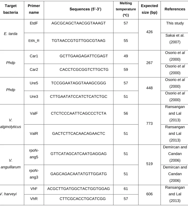
Table I - Characteristics of the primers used in this study and their references……… 36
xvi
xvii List of Abbreviations
FAO – Food and Agriculture Organization HBSS – Hank’s Balanced Salt Solution m-PCR – multiplex-PCR
NCBI – National Center for Biotechnology Information PBS – Phosphate Buffered Saline
PCR – Polimerase Chain Reaction
Phdp – Photobacterium damselae subsp. piscicida TSB – Tryptone Soya Broth
xviii
19
Chapter I.
General Introduction
Chapter I. General Introduction
20
Chapter I. General Introduction
21 1. Fish Diseases on Aquaculture
It may be assumed that fish are continually bathed in an aqueous suspension of microorganisms and most of the members of the normal microflora of water can be bacterial fish pathogen candidates (Chang et al., 2012). The appearance and development of a fish disease is the result of the interaction among pathogen, host and environment (Toranzo et al., 2005). Fish diseases, especially those caused by Gram-negative bacteria, are a serious problem in aquaculture (Castro et al., 2014). Regarding the infectious diseases caused by bacteria in marine fish, although pathogenic species have been described in the majority of the existing taxonomic groups, only a relatively small number are responsible of important economic losses in cultured fish worldwide (Toranzo et al., 2005). Disease outbreaks have direct effects on fish production, causing severe economic losses in the aquaculture sector (Martins et al., 2015). The possibility of outbreaks is increased if fish are stressed, as what happens with inappropriate water temperature, low dissolved oxygen, high nitrite levels, and high culture densities (Perera
et al., 1997; Shoemaker et al., 2000). Healthy looking fish without any clinical signs or
lesions can carry some pathogens and create a serious risk for spread of contagious diseases in fish populations (Onuk et al., 2010).
2. Traditional and Molecular diagnosis
Traditionally disease diagnosis is obtained by culturing bacteria on agar plates followed by phenotypic and serological characterization of the pathogen, or by histological examination (Pazos et al., 1996). Biochemical tests, DNA homology, and protease variability techniques have also been used (Chen et al., 1995), but these techniques have some disadvantages, such as the need for initial isolation of the pathogen and insufficient sensitivity to detect low levels of pathogen, which can be overcome by molecular techniques such as polymerase chain reaction (PCR) used to increase sensitivity and specificity of pathogen detection (Altinok, 2011). Molecular methods have slowly established a place in the diagnosis of disease in aquaculture (Chatterjee and Haldar, 2012). Blood testing is preferable to tissue analyses because it does not require the sacrifice of the sampled fish and is suitable for monitoring fish on farms (Gonzalez et al., 2003).
Chapter I. General Introduction
22
3. Major bacterial pathogens on Aquaculture
In the last decade, edwardsiellosis, caused by Edwardsiella tarda, a Gram-negative, motile, rodshaped, member of the family Enterobacteriaceae has become an important bacterial pathogen in aquaculture (Castro et al., 2012). Affecting commercial fish species worldwide (Castro et al., 2011; Zhang et al., 2014) including flatfish, this bacteria also has been reported on environment contaminated by animals or man (Castro et al., 2014), being a possible source of zoonoses.
Members of the species Photobacterium damselae are frequently associated with disease outbreaks, and have been described as emergent fish pathogens in aquaculture systems (Martins et al., 2015). Photobacteriosis or Pasteurelosis is a septicemia caused by the halophilic, Gram-negative bacteria, Photobacterium damselae subsp. piscicida, a member of Vibrionaceae family which shares the same specie with Photobacterium
damselae subsp. damselae (Osorio et al., 2000). It is commonly called
“pseudotuberculosis” due to the fact that, in the chronic form, the diseased fish showed whitish tubercles in the internal organs which consist of bacterial colonies, necrotic phagocytes and granuloma in several internal organs of infected fish accumulations (Toranzo et al., 1991; Noya et al., 1995; Margariños et al., 1996). This disease is considered one of the most dangerous bacterial diseases in aquaculture worldwide due to its wide host range, high mortality rate, and ubiquitous distribution (Barnes et al., 2005; Andreoni and Magnani, 2014). The disease was first described in wild populations of white perch (Morone americanus) and striped bass (Morone saxatilis) in 1963, when a massive epizootic occurred in Chesapeake Bay (USA) (Snieszko et al., 1964). However, its taxonomic position remained controversial until DNA–DNA hybridization studies (Gauthier
et al., 1995) provided evidence for its definitive reclassification in the genus Photobacterium, as Photobacterium damselae subspecies piscicida (Phdp) closely related
to the subspecies damselae (Pdd) (Amagliani et al., 2009). Phenotypic homogeneity of
Photobacterium damselae subsp. piscicida allows us to distinguish it from other
subspecies - Photobacterium damselae subsp. damselae - by significant biochemical and physiological characteristics such as motility, nitrate reduction, the formation of gas from glucose and the production of hemolysin, urease, and amylase (Magariños et al., 1996; Romalde, 2002). The disease is important in Europe, where since 1990 there have been recorded several photobacteriosis outbreaks in different countries including Spain (Toranzo et al., 1991), France (Baudin-Laurencin et al., 1991), Italy (Ceschia et al., 1991), Greece (Bakopoulos et al., 1995) and Portugal (Baptista et al., 1996). Photobacteriosis
Chapter I. General Introduction
23
seems to be more prevalent during the summer months (Frerichs and Roberts, 1989; Magariños et al., 1996) at higher water temperatures (greater than 23°C) and salinities (20-30) (Hawke et al., 1987; Magariños et al., 1996) and when water quality is low (Magariños et al., 1996). Although the optimum growth temperature of this microorganism is between 22.5°C and 30°C, Phdp can grow between 15°C and 32.5ºC (Magariños et al., 1992; Magariños et al.,1996). Moreover, it has been demonstrated that although Phdp cells may exist in a dormant state, they are capable of resuming rapid resuscitation and division when nutrient conditions are suitable (Magariños et al., 1994).
Vibriosis is one of the most important and the oldest recognized fish disease in marine aquaculture worldwide (Gonzalez et al., 2003). Vibrio spp. are Gram-negative and halophilic bacteria widely spread in sea- and brackish water worldwide (Messelhäusser et
al., 2010). The main feature of this bacterial group is their capacity to cause serious
alimentary intoxication associated with the consumption of raw or undercooked contaminated fish or shellfish posing a considerable public health threat as agents of sporadic and epidemic human infections, therefore representing an important microbial group in the field of food safety and quality (Espiñeira et al., 2010). Bacterial interaction or colonization with challenged organisms is a very complex process (Chatterjee and Haldar, 2012). During certain periods of the year, pathogenic Vibrio withstand unfavourable environmental conditions within aquaculture settings and when favourable environmental conditions are re-established, Vibrio are once again able to cause disease in wild animals (Chatterjee and Haldar, 2012). Ben-Haim et al. in 2003 advanced the hypothesis that aquaculture settings serve as foci or reservoirs for pathogenic Vibrio strains (Naylor et al., 2000; Chatterjee and Haldar, 2012). Vibrio harveyi, Vibrio anguillarum, Vibrio
alginolyticus, Vibrio ordalii and Vibrio vulnificus are considered opportunistic pathogens of
fish (Ghittino et al., 2003; Dalmasso et al., 2009; Zhang et al., 2014). Major Vibrio species
viz. V. harveyi, V. parahaemolyticus, V. alginolyticus, V. anguillarum, V. vulnificus, and V. splendidus are usually associated with shrimp diseases (Chatterjee and Haldar, 2012). V. harveyi is associated with luminescent vibriosis in shrimps e.g., Litopenaeus vannamei
and Penaeus monodon, and it is the most important etiological agent for mass mortality in
P. monodon (Lavilla-Pitogo et al., 1998; Lavilla-Pitogo and De la Pena, 1998; Austin et al., 2003; Guzmán et al., 2010; Chatterjee and Haldar, 2012). Internal symptoms of disease in fish caused by strains of Vibrio include intestinal necrosis, anaemia, ascitic fluid, petechial haemorrhages in the muscle wall, liquid in the air bladder, haemorrhaging and/or bloody exudate in the peritoneum, swollen intestine, haemorrhaging in or on internal organs, and pale mottled liver (Austin and Austin, 1999). External symptoms include sluggish behaviour, twirling, spiral or erratic movement, lethargy, darkened
Chapter I. General Introduction
24
pigment, eye damage/exophthalmia, haemorrhaging in the mouth, gill damage, white and/or dark nodules on the gills and/or skin, fin rot, haemorrhaging at the base of the fins, distended abdomen, haemorrhaging on the surfaces and muscles, ulcers, and haemorrhaging around the vent (Thompson et al., 2004a).
Vibrio harveyi, and Vibrio anguillarum are the most frequently isolated marine Vibrio species (Arias et al., 1999; Pujalte et al., 1999; Pujalte et al., 2003; Frans et al.,
2011; Chatterjee and Haldar, 2012), having been associated with large-scale losses of larval and juvenile penaeids and also causing several opportunistic diseases to fishes (Hispano et al., 1997; Company et al., 1999; Diggles et al., 2000; Alcaide et al., 2001; Liu
et al., 2003; Zorrilla et al., 2003; Chatterjee and Haldar, 2012). Due to the plasticity of Vibrio genomes, with frequent horizontal gene transfer events, species boundaries are
very narrow in the marine environment (Fraser et al., 2007; Chatterjee and Haldar, 2012). Hence, the identification of Vibrio-related species isolated from the marine environment is sometimes difficult (Chatterjee and Haldar, 2012). Vibrio harveyi is one of the most commonly isolated marine Vibrio species, and can easily be found both as free-living or associated to the intestinal microbiota of marine animals (Ramesh et al., 1990; Makemson and Hermosa Jr, 1999; Pujalte et al., 2003). Moreover, V. harveyi is the dominant heterotrophic species in western Mediterranean seawater and marine bivalves during the warm season (Ortigosa et al., 1994; Arias et al., 1999; Pujalte et al., 1999; Pujalte et al., 2003). Although not included among the main classical fish pathogens, V. harveyi has been related to several opportunistic infections of ornamental or edible cultured fish in the last decade (Kraxberger-Beatty et al., 1990; Saeed, 1995; Hispano et al., 1997; Company
et al., 1999; Pujalte et al., 2003), and recent reports confirm the virulence of some strains
for gilthead sea bream, silver mullet, salmon and seahorse (Balebona et al., 1998; Álvarez
et al., 1998; Zhang and Austin, 2000; Alcaide et al., 2001; Pujalte et al., 2003).
Vibrio anguillarum is a Gram-negative bacterium that causes haemorrhagic
septicaemia in fish, a disease that leads to great economic losses in fish farming worldwide (Hong et al., 2007). The causative agent, Vibrio anguillarum, was initially isolated by Canestrini (1893) (Gonzalez et al., 2003) and since the first identification, vibriosis has been described in anadromous and catadromous species (Toranzo and Barja, 1990; Austin and Austin, 1999; Pedersen et al., 1999a; Gonzalez et al., 2003) and is reported to produce disease in more than 48 fish species (Austin and Austin, 1999; Gonzalez et al., 2003). Among the 23 different O-serogroups described for Vibrio
anguillarum, only serogroups O1, O2 and O3 are important as the causative agent of
mortalities in farmed and feral fishes, with the remaining serogroups considered to comprise mainly environmental strains isolated from water and sediment (Sørensen and
Chapter I. General Introduction
25
Larsen, 1986; Pedersen et al., 1999b; Gonzalez et al., 2003). Microflora associated with healthy and diseased sea bass (Dicentrarchus labrax) and sea bream (Sparus aurata) larvae were also investigated (Grisez, 1997; Grisez et al., 1997; Pedersen et al., 1999a;
Pedersen et al., 1999b; Gonzalez et al., 2003) and was demonstrated that V. anguillarum constituted a significant part of the intestinal microflora of these larvae during feeding with rotifers and that V. anguillarum was dominant during outbreaks of disease, causing high mortalities among the larvae (Pedersen et al., 1999b).
These bacterial pathogens are important etiological agents that hamper aquaculture production sharing common morphological characteristics and cause similar clinical signs in diseased fish, making the rapid diagnosis of multiple and secondary infections through culture difficult (Zhang et al., 2014).
4. PCR diagnosis
The rapid development of molecular biological techniques offers significant advantages for workers involved in fish disease diagnosis (Chatterjee and Haldar, 2012), so in recent years, the number of publications describing new molecular techniques or methods has increased significantly (Hirono et al., 1996; Brasher et al., 1998; Romalde et
al., 1999; Cerdà-Cuéllar et al., 2000, Botella et al., 2002; Conejero and Hedreyda, 2003;
Avendaño-Herrera et al., 2004; Thompson et al., 2005; Bramha Chari and Dubey, 2006; Bauer and Rørvik, 2007; Beaz-Hidalgo et al., 2008; Amagliani et al., 2009; Espiñeira et
al., 2010; Altinok, 2011; Chang et al., 2012; Ransangan and Lal, 2013; Castro et al., 2014;
Zhang et al., 2014). Individual PCR assays have been developed for detection and identification of the fish pathogens (Altinok et al., 2008). There has been much interest in the development of specific PCR protocols, many of them based on the amplification of 16S rRNA genes, for detecting a variety of Gram negative and Gram positive bacterial fish pathogens in fish samples and complex substrates (Brown et al., 1994; Cunningham, 2002; Romalde and Toranzo, 2002; Beaz-Hidalgo et al., 2008). However, a large number of individual PCR reactions would be necessary if single primer sets were used to screen a large number of clinical samples, resulting in a relatively costly and time-consuming process (Altinok, 2011).
Currently, fish photobacteriosis diagnosis is carried out through standard microbiological methods, which are time-consuming and laborious, relying on pathogen culture and isolation steps and the complete protocol always includes biochemical and serological confirmation, leading to an extension of the time needed for the final diagnosis (Amagliani et al., 2009). The diagnosis of bacterial infection in aquatic animal has been
Chapter I. General Introduction
26
based on the microbiological analysis using bacteriological culture, morphological characteristic and biochemical tests. Biochemical tests often lead to misinterpretation of results because of strain metabolic variability (Thyssen et al., 1998; Botella, et al., 2002; Amagliani et al., 2009). Additional drawbacks are related to the slow growth of this species and its inhibition by other fast-growing bacteria present in the same samples (Romalde, 2002; Amagliani et al., 2009) and to the viable, but non-culturable form that Phdp can also assume (Magariños et al., 1994). Several authors have reported that 16S rRNA gene does not provide a sufficient phylogenetic resolution at the species level for
Vibrio or Photobacterium species (Osorio and Klose, 2000; Thompson et al., 2005;
Martins et al., 2015). According to previous studies (Osorio et al., 1999; Osorio and Klose, 2000) the subspecies Photobacterium damselae subsp. damselae and Photobacterium.
damselae subsp. piscicida have 100% homology between sequences of 16S rRNA gene
and only 91% homology between sequences of toxR gene (Martins et al., 2015).
An array of phenotypic and genomic techniques has become available for the identification of Vibrio species in the last three decades (Vandamme et al., 1996; Rademaker et al., 1998; Savelkoul et al., 1999; Olive and Bean, 1999; Rademaker et al., 2000; Dijkshoorn et al., 2001; Gurtler and Mayall, 2001; Van Belkum et al., 2001; Chatterjee and Haldar, 2012).
V. alginolyticus has been reported to easily out number other Vibrio species in
environmental samples (Oliver and Kaper, 2001; Ransangan and Lal, 2013), which may cause the detection of other bacteria difficult (Ransangan and Lal, 2013). Differentiation of these bacteria using phenotypic characterization and 16S rRNA sequencing is also difficult because of high genome homology among Vibrio species (Thompson et al., 2005; Ransangan and Lal, 2013). Recently, several genes of Vibrio anguillarum, such as hemolysin, angE, rpoN, and 16S rRNA genes, were cloned using PCR (Wiik et al., 1995; Hirono et al., 1996; Gonzalez et al., 2003; Liu et al., 2004; Demircan and Candan, 2006). Sigma factor σ54 is responsible for regulating the genes providing coordination between carbon and nitrogen fixation in bacteria (Demircan and Candan, 2006). This factor is also necessary for decarboxylic acid transportation, toluene and xylene catabolism, hydrogenase biosynthesis, and the translation of gene coding for flagella production and nitrogen fixation (Merrick, 1993; Demircan and Candan, 2006). O’Toole et al. (1997) were the first group to sequence the 2218 bp rpoN gene; then Gonzalez et al. (2003) amplified the 519 bp portion of this gene to identify Vibrio anguillarum in fish blood and other tissues (Demircan and Candan, 2006). However, extensive database comparisons demonstrate that differences in the 16S gene sequence between V. anguillarum and closely related
Chapter I. General Introduction
27
species are insufficient to warrant the design of species-specific PCR primers using that gene as a target (Gonzalez et al., 2003).
Among the currently available V. anguillarum gene sequences in the EMBL database, Gonzalez et al. (2003) selected rpoN gene coding for the cellular sigma factor σ54 as a PCR target. As a housekeeping gene, rpoN is expected to be present in all V.
anguillarum isolates (Gonzalez et al., 2003). Moreover, regions of high sequence
variability are found in this gene compared to the homologous genes in other Vibrio species (Gonzalez et al., 2003).
Considering the damages that these bacteria can bring about to fish and human, a rapid, simple, simultaneous and low cost detection method is necessary (Ransangan and Lal, 2013). Compared to traditional methods, these molecular techniques can avoid problems that are inherent in investigating organisms for which no culture medium, cell lines (for viruses) or detection method is available (Lievens et al., 2011).
Although this bacterium has been reclassified as Listonella anguillarum based on 5S rRNA gene sequence analysis, it is still commonly referred to as Vibrio anguillarum (MacDonell and Colwell, 1984; Hong et al., 2007). Recently, putative virulence genes of V.
anguillarum were identified by random genome sequencing (Rodkhum et al., 2006a; Hong et al., 2007). Di Lorenzo et al. (2003) have also determined the complete sequence of the
virulence plasmid pJM1 from V. anguillarum, which affects marine fish (Hong et al., 2007). The phylogenetic relationships between V. anguillarum and other Vibrio species have been reported, based on comparisons of the DNA sequences of PCR amplicons generated using specific primers that target the recA and 16S rRNA genes (Dorsch et al., 1992; Kita-Tsukamoto et al., 1993; Urakawa et al., 1997; Thompson et al., 2004b; Hong et
al., 2007). However, these genes are not useful in discriminating between closely related
strains, due to the very high degrees of sequence identity among these strains (Hong et
al., 2007). Gonzalez et al. (2003) have demonstrated that the annealing temperature is
very important in detecting the PCR product using specific primers for the rpoN gene (Hong et al., 2007). The expected band also appeared at the normal annealing temperature with Vibrio ordalii, which is known to be a very difficult strain to differentiate from V. anguillarum (Hong et al., 2007). A multiplex PCR has been reported for the specific detection of V. anguillarum using primers that target five hemolysin genes (Rodkhum et al., 2006b; Hong et al., 2007). This method also fails to discriminate V.
ordalii from V. anguillarum reliably, and, consequently, there remains a need for specific
Chapter I. General Introduction
28 5. m-PCR
When multiple bacterial pathogens are likely to occur, as in the aquatic environment, amplification of multiple target genes in a single reaction mixture is possible with the multiplex PCR (m-PCR) method (Brasher et al., 1998, Del Cero et al., 2002, Panicker et al., 2004; Panangala et al., 2007), thus reducing cost, time and effort without compromising the test utility (Panangala et al., 2007). Although simultaneous detection of several pathogens with a multiplex PCR (mPCR) has been widely applied to the detection of multiple viruses and bacteria in clinical specimens, this approach has not been widely used in the detection of fish pathogens (Osorio et al., 2000; Del Cerro et al., 2002; Mata et al., 2004; Altinok et al., 2008; Castro et al., 2014), and reports of applications of these techniques on a routine basis in diagnostic laboratories are few (Chatterjee and Haldar, 2012).
5.1 State of the art
Several attempts to develop methods for the rapid and accurate diagnosis of edwardsiellosis have been made, including PCR-based methods (Castro et al., 2014). Of these, a PCR protocol employing the gene etfD (which encodes the upstream region of the fimbrial gene) reported by Sakai et al. (2007) was shown to be the most rapid and sensitive method for the accurate detection of E. tarda in infected fish (Castro et al., 2014).
The plasmid content has proved to be very different depending on the geographical origin of subsp. piscicida strains (Magariños et al., 1992, Magariños et al., 1996), but Osorio et al. (2000), proved that, regardless of geographical origin and source of isolation, strains of Photobacterium damselae subsp. piscicida show one band with a molecular weight of 267 bp corresponding to ureC gene.
In 2006, Bramha Chari and Dubey developed PCR-based identification methods for V. harveyi targeting a partial 16S rRNA gene (Bramha Chari and Dubey, 2006; Chatterjee and Haldar, 2012). Fukui and Sawabe modified the method by developing a one step colony PCR targeting the same 16S rRNA gene to identify pathogenic V. harveyi from aquaculture settings (Fukui and Sawabe, 2007; Chatterjee and Haldar, 2012). Similarly, Conejero and Hedreyda in 2003 targeted the toxR gene for identification of V.
harveyi from aquaculture systems (Conejero and Hedreyda, 2003; Chatterjee and Haldar,
2012). However, the most precise method to identify V. harveyi along with V. campbellii and V. parahaemolyticus was developed by Haldar et al. in 2010, using multiplex PCR; this method was so accurate that the individual detection limit of all three target species
Chapter I. General Introduction
29
ranged from 10 to 100 cells per PCR tube, using primer concentrations of 0.25 to 0.5 μmol/l (Haldar et al., 2010; Chatterjee and Haldar, 2012).
The main drawback of molecular methodology is that it does not discriminate between DNA from alive and dead microorganisms (Espiñeira et al., 2010). Other methodological approaches are focused on the detection of mRNA, because this molecule is less stable than DNA and therefore will not be detected in a sample unless there are viable microorganisms that synthesize it during the enrichment phase (Birmingham et al., 2008; Espiñeira et al., 2010).
5.2 Tool Development
The ability to determine bacterial pathogens using multiplex PCR method was reported dependent to the target genes (Ransangan and Lal, 2013). However, success of this method depends on the selection of target gene, which should be species-specific, widely distributed and also stable in the genome (Chatterjee and Haldar, 2012). Fortunately, both housekeeping and virulent genes can equally serve as good targets for multiplex PCR amplification (Ransangan and Lal, 2013). However, Bauer and Rørvik (2007) also showed that single gene (ToxR) can be used as the target PCR amplification of similar bacterial species (Ransangan and Lal, 2013). Nevertheless, virulent genes could serve a better target for multiplex PCR amplification because of their divergence and because they are highly conserved among Vibrionaceae (Osorio and Klose, 2000; Ransangan and Lal, 2013).
The 16S rRNA gene (about 1,500 bp in length) consists of highly conserved regions and is present in almost all bacteria which may reveal deep-branching (e.g., classes, phyla) relationships, while variable regions may be demonstrated to be useful in discriminating species within the same genus (Chatterjee and Haldar, 2012). This feature has prompted researchers to use 16S rRNA both as a phylogenetic marker and as an identification tool (Wiik et al., 1995; Chatterjee and Haldar, 2012). It has been demonstrated that different selective media are not quite selective or species-specific (Chatterjee and Haldar, 2012). Detection of different marine bacteria on selective media and subsequent colony hybridization with species-specific probes (probe is a fragment of DNA or RNA of variable length, used in DNA or RNA samples to detect the presence of nucleotide sequences), based on variable target regions of the 16S rRNA and other specific genes have been evaluated as an alternative fast screening tool for identification of marine bacteria (Martínez-Picado et al., 1996; Cerdà-Cuéllar et al., 2000; Cerdà-Cuéllar and Blanch, 2002; Tanaka et al., 2002; Sloan et al., 2003; Chatterjee and Haldar, 2012).
Chapter I. General Introduction
30
However, there is nearly 100% 16S rRNA gene homology among many closely related bacterial species, viz. V. scophthalmi and V. ichthyoenteri, thus there is a significant possibility of cross-hybridization and misidentification of closely related species (Cerdà-Cuéllar et al., 2000; Chatterjee and Haldar, 2012).
It is not easy to incorporate more than six primer sets because of the cross-reaction in m-PCR, and the challenges inherent in size discrimination among PCR products by conventional electrophoresis (Warsen et al., 2004; Chang et al., 2012).
6. Test organism
The species belonging to the Scianidae family and selected for this present experiment is Argyrosomus regius known as Meagre. It is a good candidate for the diversification on commercial aquaculture in Mediterranean and Eastern Atlantic for its good flesh, easy management and high growth rate (Jiménez et al., 2005; El-Shebly et al., 2007; Roo et al., 2010; Velazco-Vargas et al., 2013).
Meagre (Argyrosomus regius) aquaculture has recently developed, starting in the mid-1990s in Southern France, and is much less advanced than for developed farm fish species such as sea bass, sea bream or turbot (Martínez-Llorens et al., 2011). The production of meagre (A. regius) began in the second half of the 90s, following an agreement between Italian and French producers, which resulted in the first commercial production in 1997 in France (Amoedo, 2011), spreading, in the years following, to other Mediterranean countries and increasing its production rapidly.
Being a eurihaline species has an easy adaptation to different environments, including growth in earthen ponds with brackish water, also tolerating the imprisonment as demonstrated by its presence in large aquariums and achieving high growth rates and good food conversion levels (Calderón et al., 1997; Shepherd et al., 2002; Amoedo, 2011). The organoleptic characteristics of aquaculture meagre are considered very good, characterized by a high protein content and low lipid content (1.5 to 4%) compared to other species of fish, tolerating long periods of cold (1ºC with ice cover) on storage conditions (with a shelf life about 9 days of refrigeration), characteristics that give the fish farming meagre the category of a product of excellence (Poli et al., 2003; Amoedo, 2011). This species does not have significant pathological manifestations, being parasites like
Amyloodinium ocellatum, Gyrodactylus spp. and the bacterium V. anguillarum its major
pathogens, having already been developed treatment for those, so the control the crop density is the primary method of prevention (Amoedo, 2011). Meagre could be interesting for aquaculture: high flesh quality and flavour, high commercial value over 2 kg, rapid
Chapter I. General Introduction
31
growth between 16 and 20°C, high tolerance to salinity, excellent biological characteristics, because they withstand captivity perfectly, with high growth and good feed conversion ratio (Calderón et al., 1997; Martínez-Llorens et al., 2011). Juveniles (Age 1) eat small demersal fish and crustaceans (mysids and shrimp) and when they reach 30 to 40 cm, they feed on pelagic fish and cephalopods (Calderón et al., 1997; Martínez-Llorens
et al., 2011). Meagre production has been increasing in recent years with a significant
production in 2006, as a result of the achievement of reproduction in captivity (Martínez-Llorens et al., 2011). According to Apromar (2008), meagre production in that year was around 845 tm with which a new growth is observed compared with 2005, which is indicative of the establishment and the importance of its production (Martínez-Llorens et
al., 2011).
The sciaenid meagre (Argyrosomus regius) is found in the Mediterranean and Black Sea and along the Atlantic coasts of Europe and the west coast of Africa (Poli et al., 2003). Meagre lives in inshore and shelf waters, close to the bottom or near the surface (depth range 15–200 m); it also enters estuaries and coastal lagoons (Chao, 1986; Griffiths and Heemstra, 1995; Poli et al., 2003). The fish can reach over 50 kg in the wild, the largest size recorded being 182 cm total length and 103 kg of body weight (Quéro and Vayne, 1987; Poli et al., 2003). Its flesh quality is much appreciated (regius for royal quality of flesh) (Poli et al., 2003). In 2003 only a few attempts have been made at farming this species in Europe, between these, only one French farm (Les Poissons du Soleil), with hatchery, nursery and on growing facilities, has succeeded in the artificial reproduction and rearing of meagre and just two French and two Italian marine farms have intensively grown fry up to market sizes (Poli et al., 2003).
7. Aims of the study
The aim of the present study was to develop a multiplex-PCR tool for detection of
Edwardsiella tarda, Photobacterium damselae subsp. piscicida, Vibrio harveyi, Vibrio alginolyticus and Vibrio anguillarum in tissue and blood samples of diseased fish.
32
Chapter II.
Development of an m-PCR tool for detection of four
bacterial pathogens on Aquaculture
Chapter II. Development of an m-PCR tool for detection of four bacterial pathogens on Aquaculture
33
Chapter II. Development of an m-PCR tool for detection of four bacterial pathogens on Aquaculture
34 1. Introduction
Aquaculture is an emerging industrial sector which requires continued research with scientific and technical developments, and innovation (Toranzo et al., 2005). Bacterial diseases are one major problem causing fish mortality and economic loss in aquaculture (Ransangan and Lal, 2013). Fast detection of these pathogens is important for management and control of disease outbreaks (Martins et al., 2015).
Considering the damages that some bacteria can cause to humans and fish, it is necessary a fast, simple, simultaneous (detection of different pathogens in one single reaction at the same time) and low cost detection method being its development the primary objective of this study.
We have selected as development targets of a multiplex PCR tool the following pathogens: Vibrio alginolyticus, Vibrio anguillarum, Vibrio harveyi and still Edwardsiella
tarda and Photobacterium damselae subsp. piscicida. Vibrio species under optimum
temperature and salinity conditions arise in high amounts in aquatic organisms (Espiñeira
et al., 2010) and E. tarda has recently become an emerging bacterial pathogen in
aquaculture (Castro et al., 2014), affecting a wide range of cultivated species. It is associated with life-threatening sepsis and infections in various animals, including humans (Castro et al., 2014). Members of Photobacterium damselae species have been described also as emerging pathogens of fish in aquaculture systems (Martin et al., 2015), causing sepsis (Osorio et al., 2000). Pathogenicity, frequency of relapses and their severity, determined the choice of these species. The development of this molecular tool is also important because it covers pathogens that affect a wide range of cultivated organisms such as bivalve mollusks, crustaceans and fish, decreasing costs for companies that already have different types of cultures or it may be a mean to encourage aquaculture companies to diversify their production.
2. Materials and Methods
2.1. Bacterial strains and culture conditions
In total, five bacterial species were used in this study: Edwardsiella tarda (ACC 36.1), Photobacterium damselae subsp. piscicida (AQP 17.1), Vibrio harveyi (DSM 19023), Vibrio anguillarum (AQV 55.1) and Vibrio alginolyticus (CECT 521). All bacteria were cultured on Tryptone Soya Broth (TSB; Himedia), supplemented with 1.5% of sodium chloride (NaCl; Panreac), except for E. tarda that was with only 1%, and growth curves were established.
Chapter II. Development of an m-PCR tool for detection of four bacterial pathogens on Aquaculture
35
2.2. DNA extraction from bacterial pure culture and confirmation
Bacterial DNA extraction was performed with NZY Tissue gDNA isolation kit (nzytech, genes & enzymes) for 1 ml of pure culture.
For confirmation, the PCR reactions (20 µL) were set with 10 µL of Master Mix (NZYTaq 2x Colourless Master Mix), 1 µL MgCl2,1 µL of each primer, 5.5 µL of sterile distilled water and 1.5 µL of template DNA. The PCR reaction was initiated with a denaturation of 5 minutes at 95ºC, followed by 45 cycles of denaturation for 30 seconds at 95°C, annealing 30 seconds at 51°C and extension for 1 minute at 72°C. The final extension step consisted of 10 minutes at 72°C. After addition of the dye RED safe amplified products were separated on agarose gel at 1.5% (w/v) with 1x TAE buffer and visualized in UV transilluminator. The horizontal electrophoresis ran for 60 minutes at a voltage of 70 V.
Bacterial DNA extracted was quantified with a Nanodrop 2000 Spectrophotometer (Thermo scientific) in ng/µl.
2.3. PCR primers used in this study
Primers used in this study are listed in Table I with respective target bacteria, name, forward and reverse sequences, melting temperatures (ºC), expected size band (bp) and reference of the author. EtdF primer was design with the help tool Primer-Blast from NCBI (http://www.ncbi.nlm.nih.gov/tools/primer-blast/) (Ye et al., 2012).
Chapter II. Development of an m-PCR tool for detection of four bacterial pathogens on Aquaculture
36
Table I - Characteristics of the primers used in this study and their references. Target bacteria Primer name Sequences (5’-3’) Melting temperature (ºC) Expected size (bp) References E. tarda EtdF AGCGCAGCTAACGGTAAAGT 57 426 This study EtfA_R TGTAACCGTGTTGGCGTAAG 55 Sakai et al. (2007) Phdp Car1 GCTTGAAGAGATTCGAGT 49 267 Osorio et al (2000)
Car2 CACCTCGCGGTCTTGCTG 59 Osorio et al
(2000) Phdp Ure5 TCCGGAATAGGTAAAGCGGG 57 448 Osorio et al (2000)
Ure3 CTTGAATATCCATCTCATCTGC 51 Osorio et al (2000) V. alginolyticus ValF CTCTCCCAATTCAGCCCTCTA 56 773 Ransangan and Lal (2013) ValR GACTCTTCACAACAGAACTC 51 Ransangan and Lal (2013) V. anguillarum rpoN-ang5 GTTCATAGCATCAATGAGGAG 51 519 Demircan and Candan (2006) rpoN-ang3 GAGCAGACAATATGTTGGATG 51 Demircan and Candan (2006) V. harveyi VhF ACGCTTGATGGCTACTGGTGGAG 61 606 Ransangan and Lal (2013) VhR CTTCGCACCTGCATCGG 57
Chapter II. Development of an m-PCR tool for detection of four bacterial pathogens on Aquaculture
37 2.4. m-PCR analysis
Multiplex-PCR reactions were tested using NZYTaq polymerase, variable annealing temperatures (49ºC, 51ºC, 53ºC and 55ºC) and 0.125 µM of each primer concentrations. Amplification conditions were: 5 min. at 95ºC, 45 cycles (95ºC, 30s; 50±10ºC, 30s; 72ºC, 1 min.) and finally at 72ºC for 7 min.
Was also performed an m-PCR reaction with variable concentrations of MgCl2 (2, 4, 6 and 8 mM) maintaining annealing temperatures and conditions mentioned above.
All reaction products were analyzed by electrophoresis in agarose gel (1,5%), using a 250 bp ladder (Alfa Aesar) as a molecular weight marker.
2.5. Specificity and sensitivity of m-PCR assay
The specificity of the primers was tested using purified bacterial DNA of the respective strains. For a multiplex-PCR conditions (25 µL) we added 12.5 µL of Master Mix (NZYTaq 2x Colourless Master Mix), 1.25 µL MgCl2, 1 µL of each primer, 4.45 µL of sterile distilled water and 2 µL of mixture of different template DNA (with double quantity of
E. tarda purified DNA). Amplification conditions were: 5 min. at 95ºC, 45 cycles (95ºC,
30s; 51ºC, 30s; 72ºC, 1 min.) and finally at 72ºC for 7 min.
Sensitivity was tested with eight successive dilutions of a mixture of the four bacterial pathogens purified DNA from a known concentration of 64.6 µg/µL for E. tarda; 89.5 µg/µL for V. alginolyticus; 110.8 µg/µL for V. anguillarum and 24 µg/µL for V. harveyi. Multiplex – PCR reaction (20 µL) added 10 µL of Master Mix (NZYTaq 2x Colourless Master Mix), 1 µL MgCl2,0.25 µL of each primer (except for E. tarda that was 0.5 µL of each forward and reverse primers), 3.5 µL of sterile distilled water and 3 µL of mixture of different template DNA. Amplifications conditions were the same used for specificity testing.
2.6. Experimental fish infection
After bacteria growth curves were established, the applicability of the mPCR protocol in infected fish was determined through the inoculation of batches of three meagre (average 30-36 g) with 0.1ml of a suspension of each bacteria at a concentration of 106 CFUml-1. Three batches of fish were inoculated for each bacterium separately. As the negative control, three batches of fish were inoculated with sterile Hank’s Balanced Salt Solution (HBSS) and maintained under the same conditions as the experimentally infected fish. The fish were maintained at equal density of 6 kgm-3 with continuous
Chapter II. Development of an m-PCR tool for detection of four bacterial pathogens on Aquaculture
38
aeration and a water temperature of 22±1ºC. One week post-infection, the kidney, liver, spleen and blood samples were collected from all fish and kept at -80ºC. Classical microbiology analysis was also performed to the liver, kidney and spleen samples.
2.7. Tool validation and spiking on tissues
When the tool was validated and the conditions set, the total DNA from tissues and blood collected were extracted with DNeasy Blood & Tissue Kit (QIAGEN) and m-PCR was performed in these conditions (20 µL) added 10 µL of Master Mix (NZYTaq 2x Colourless Master Mix), 2 µL MgCl2,0.5 µL of each primer, 1 µL of sterile distilled water and 3 µL of tissue total DNA. Amplification conditions were: 5 min. at 95ºC, 45 cycles (95ºC, 30s; 51ºC, 30s; 72ºC, 1 min.) and finally at 72ºC for 7 min.
To make it possible to predict the behavior of this tool in opportunistic infection situation in which they occur one or more pathogens, pathogens were cultured, as already mentioned above. After homogenization with 100 µl of Phosphate Buffered Saline (PBS) by repeated pipetting of each sample (0.1 to 0.2g) of kidney, liver and spleen of a healthy meagre - previously analyzed with classical microbiology - the samples were spiked with 100 µl of the mixture of the five different pathogens with a concentration of 340 CFUml-1 for E. tarda (exponential phase), 410 CFUml-1 for V. alginolyticus (exponential phase, almost in stationary phase), 810 CFUml-1 for V. anguillarum (exponential phase), 740 CFUml-1 for V. harveyi (exponential phase) and let them incubate at 25ºC during 1h. Finished incubation, total DNA was extracted with DNeasy Blood & Tissue Kit (QIAGEN) and m-PCR was performed along electrophoresis.
3. Results
3.1. m-PCR analysis
Different annealing temperatures (49ºC, 51ºC, 53ºC and 55ºC) were tested and were obtained four expected size bands (E. tarda – 426 bp; V. anguillarum – 519 bp; V.
harveyi – 606 bp; V. alginolyticus – 773 bp) at 49ºC, 51ºC and 53ºC and only three bands
at 55ºC (E. tarda – 426 bp; V. anguillarum – 519 bp; V. harveyi – 606 bp). For Phdp was no amplification, so the expected band of 267 bp did not show up. We concluded that the temperature of 51°C is better to distinguish the bands of four different pathogens: E. tarda,
Chapter II. Development of an m-PCR tool for detection of four bacterial pathogens on Aquaculture
39
Figure 2.1 – m-PCR at different annealing temperatures. M – 250 bp ladder; Lane 1 – 49ºC; Lane 2 – 51ºC; Lane 3 – 53°C; Lane 4 – 55ºC.
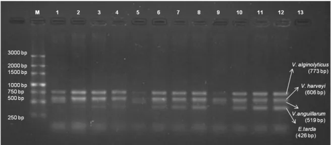
Different concentrations of MgCl2 (2 mM, 4 mM, 6 mM and 8 mM) were also tested along the different annealing temperatures mentioned above and the best results obtained were four expected size bands (E. tarda – 426 bp; V. anguillarum – 519 bp; V. harveyi – 606 bp; V. alginolyticus – 773 bp). It follows that the temperature of 51°C along with between 4 and 6 mM of MgCl2 is better to distinguish the bands of the four different pathogens: E. tarda, V. anguillarum, V. harveyi and V. alginolyticus (Figure 2.2).
Chapter II. Development of an m-PCR tool for detection of four bacterial pathogens on Aquaculture
40
Figure 2.2 – m-PCR at different annealing temperatures and different concentrations of MgCl2. Lane 1 – 250 bp ladder; Lane 2 - 49ºC, 2 mM MgCl2; Lane 3 – 49ºC, 4 mM MgCl2; Lane 4 – 49ºC, 6 mM MgCl2; Lane 5 – 49ºC, 8 mM MgCl2; Lane 6 - 51ºC, 2 mM MgCl2; Lane 7 - 51ºC, 4 mM MgCl2; Lane 8 - 51ºC, 6 mM MgCl2; Lane 9 - 51ºC, 8 mM MgCl2; Lane 10 - 53ºC, 2 mM MgCl2; Lane 11 - 53ºC, 4 mM MgCl2; Lane 12 - 53ºC, 6 mM MgCl2; Lane 13 - 53ºC, 8 mM MgCl2.
3.2. Specificity and sensitivity of m-PCR assay
The specificity of the primers was tested using purified bacterial DNA of the respective strains, yielding bands at expected sizes (E. tarda – 426 bp; V. anguillarum – 519 bp; V. harveyi – 606 bp; V. alginolyticus – 773 bp), which can be identified on agarose gels with clearness and without overlapping sizes, except for Pdhp, that doesn´t show the expected band of 267 bp but a non-specific band over 1500 bp (Figure 2.3).
Chapter II. Development of an m-PCR tool for detection of four bacterial pathogens on Aquaculture
41
Figure 2.3 - Amplification of assay primers in monoplex reaction. M - 250 bp ladder; Lane 1 - E. tarda (369 bp); Lane 2 - Phdp (expected size 267 bp); Lane 3 - V. alginolyticus (773 bp); Lane 4 – V. anguillarum (519 bp); Lane 5 – V. harveyi (606 bp).
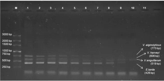
The detection limit of the multiplex PCR amplification was as low as 0.4 ng/µl for V.
anguillarum; 0.5 ng/µl for E. tarda; 1.5 ng/µl for V. harveyi and 5.6 ng/µl for V. alginolyticus
Chapter II. Development of an m-PCR tool for detection of four bacterial pathogens on Aquaculture
42
Figure 2.4 – m-PCR sensitivity test. Lane 1 – 250 bp ladder; Lane 2 – mixture of different DNA (64.6 ng/µl for E. tarda; 89.5 ng/µl for V. alginolyticus; 110.8 ng/µl for V. anguillarum; 24 ng/µl for V. harveyi); Lane 3 – mixture of different DNA (32.3 ng/µl for E. tarda; 44.8 ng/µl for V. alginolyticus; 55.4 ng/µl for V. anguillarum; 12 ng/µl for V. harveyi); Lane 4 – mixture of different DNA (16.2 ng/µl for E. tarda; 22.4 ng/µl for V. alginolyticus; 27.7 ng/µl for V. anguillarum; 6 ng/µl for V. harveyi); Lane 5 - mixture of different DNA (8.1 ng/µl for E. tarda; 11.2 ng/µl for V. alginolyticus; 13.9 ng/µl for V. anguillarum; 3 ng/µl for V. harveyi); Lane 6 - mixture of different DNA (4 ng/µl for E. tarda; 5.6 ng/µl for V. alginolyticus; 6.9 ng/µl for V. anguillarum; 1.5 ng/µl for V. harveyi); Lane 7 - mixture of different DNA (2 ng/µl for E. tarda; 2.8 ng/µl for V. alginolyticus; 3.5 ng/µl for V. anguillarum; 0.8 ng/µl for V. harveyi); Lane 8 - mixture of different DNA (1 ng/µl for E. tarda; 1.4 ng/µl for V. alginolyticus; 1.7 ng/µl for V. anguillarum; 0.4 ng/µl for V. harveyi); Lane 9 - mixture of different DNA (0.5 ng/µl for E. tarda; 0.7 ng/µl for V. alginolyticus; 0.9 ng/µl for V. anguillarum; 0.2 ng/µl for V. harveyi); Lane 10 - mixture of different DNA (0.3 ng/µl for E. tarda; 0.3 ng/µl for V. alginolyticus; 0.4 ng/µl for V. anguillarum; 0.1 ng/µl for V. harveyi); Lane 11 – control of the reaction, without DNA.
3.3. Experimental fish infection
Classical microbiological methods confirmed the presence of E. tarda and V.
anguillarum in kidney, liver and spleen of specimens of infected meagre (A. regius).
3.4. Tool validation
The m-PCR was applied to kidney, liver and spleen from spiked tissues. As expected, negative controls produced no amplifications (Figure 2.5).
Chapter II. Development of an m-PCR tool for detection of four bacterial pathogens on Aquaculture
43
Figure 2.5 – m-PCR applied to spiked spleen, liver and kidney from an apparently healthy meagre (A. regius). M – 250 bp ladder; Lane 1 – Spleen tissue spiked with mixture of different test pathogens; Lane 2 – Liver tissue spiked with mixture of different test pathogens; Lane 3 – Kidney tissue spiked with mixture of different test pathogens; Lane 4 – Blood sample from infected symptomatic fish infected with E. tarda; Lane 5 – control of the reaction, without DNA.
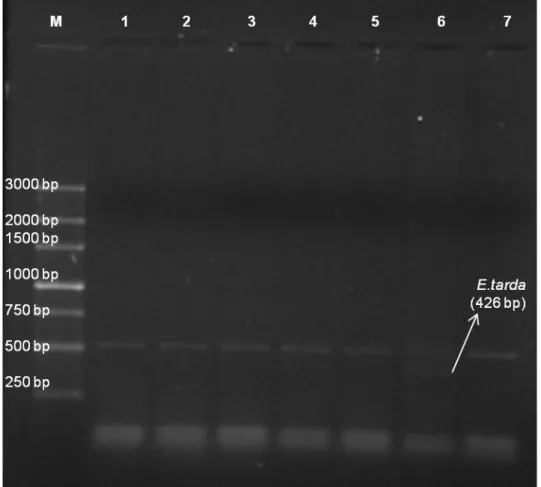
This detection method was also applied to liver and blood samples from experimentally infected fish. Negative controls produced no amplifications. All the samples and control reaction presented one band above 500 bp, believed to be a non-specific band resultant from the tissue interference with the multiplex reaction once it appears on the tissue control fish, previously analyzed and confirmed as a healthy fish (Figure 2.6).
Chapter II. Development of an m-PCR tool for detection of four bacterial pathogens on Aquaculture
44
Figure 2.6 – m-PCR applied to tissue and blood samples from A. regius. M – 250 bp ladder; Lane 1 – Tissue from control fish; Lane 2 – Tissue from an infected fish with E. tarda; Lane 3 – Tissue from an infected fish with V. alginolyticus; Lane 4 – Tissue from an infected fish with V. anguillarum; Lane 5 - Tissue from an infected fish with V. harveyi; Lane 6 - Blood sample from infected symptomatic fish infected with E. tarda; Lane 7 – Control of the reaction, without DNA.
4. Discussion and conclusion
Currently, aquaculture is one of the fastest growing food production systems in the world (Castro et al., 2014).
However, the life-history characteristics of the meagre (e.g., longevity, large size and age at maturity, large variability in annual recruitment, formation of spawning aggregations in coastal waters and estuaries) pose significant management and conservation problems for it making the meagre rank high among the world's most vulnerable species (Cheung et al., 2007; Prista, 2013). Despite this, the biology, ecology and fisheries of the meagre are poorly documented, particularly in European waters, and only recently has interest in aquaculture production, management of artisanal fisheries, and the conservation of data-poor fish resources resulted in some direct scientific research on this species (Quéméner, 2002; Prista, 2013).
Chapter II. Development of an m-PCR tool for detection of four bacterial pathogens on Aquaculture
45
The variability in fish supply and increase in consumer demand for fish products, alongside the fast growth rate and the good properties of the meat in meagre and putatively good biological properties for growth in captivity (Quéméner, 2002; Prista, 2013) led to a spark in aquaculture production in France during the late nineties (Quéméner, 2002; Monfort, 2010; Prista, 2013). According to Prista (2003), aquaculture production of meagre has expanded to seven other southern European countries, including Portugal, reaching 14 000 t/year and being worth nearly 48 million USD/year (FAO, 2012b; Prista, 2013).
Disease management and assessment of cultured aquatic animals is a major concern in commercial aquaculture (Shi et al., 2012). Molecular methods like PCR and m-PCR are always more reliable to overcome such problems, being the last one more efficient due to multiple detection of pathogens in an only reaction.
mPCR is generally thought to be less sensitive than single PCR because of competition for reaction reagents, especially if the assays differ in their amplification efficiencies or one or more of the target organisms is present in high numbers (Tapia-Cammas et al., 2011; Castro et al., 2014).
Products of various lengths present a challenge for developing optimal PCR conditions (primer annealing temperatures and similar MgCl2 concentrations) (Gonzalez et
al., 2004). In the present work, several reaction conditions were tested for the
development of this tool, including varying annealing temperatures (between 49ºC and 55°C) and the variation in the MgCl2 concentration (from 2 mM to 8 mM). Best results were obtained at 51°C with an MgCl2 concentration between 4 mM and 6 mM where it appears the bands of the four of the five pathogens tested (E. tarda, V. alginolyticus, V.
anguillarum and V. harveyi) (Figure 2.2).
One of the most critical steps in the study of bacterial fish diseases is the correct identification of the infectious agent (Avendaño-Herrera et al., 2004). The primers described here proved to be specific under the conditions assayed both in relation to 4 of the 5 target species (E. tarda, V. alginolyticus, V. anguillarum and V. harveyi), with only the specific target species showing amplification in the multiplex reaction (Figure 2.3). For this reason, sensitivity was tested only against these 4 target species and not with Phdp that showed a non-specific band above 1500 bp instead of a specific band of 267 bp.
The detection limit of this tool was determined using purified bacterial DNA and was obtained a result of 0.4 ng/µl for V. anguillarum; 0.5 ng/µl for E. tarda; 1.5 ng/µl for V.
harveyi and 5.6 ng/µl for V. alginolyticus (Figure 2.4). present in a mixture of these