POSTER
E WI M I D ' 0 9 – P O S T E R P R E S E N TAT I O N S
P O S T E R P R E S E N T A T I O N S
Churg-Strauss syndrome with a significant (P < 0.01). The frequency of CD4+CD25+ was highly in-creased in patients with late CSS. Although the ex-pression of CD25 and IL-5 level was comparable between groups.
Conclusions/Significance: These results indicate that IL-5 could be dominant in the CSS development and its future complication. These data suggest that a defective T-regulatory function may underlie the immune dysfunction in Churg-Strauss syndrome.
References
1. Gotlib J., Cross N.C., Gilliland D.G. Eosinophilic disor-ders: molecular pathogenesis, new classification, and modern therapy // Best. Pract. Res. Clin. Haematol. -2006. - Vol.19, N3. - P.535-569.
2. Guillevin L., Pagnoux C. Treatment of ANCAassociated vascularitides // Presse MANCAassociated. 2007. Vol.36, N5. -P.922-927
3. Lhote F. ChurgStrauss syndrome // Presse Med. -2007. - Vol.36, N5. - P.875-889
4. Osei-Kumah A, Smith R, Clifton VL.Maternal and cord plasma cytokine and chemokine profile in pregnan-cies complicated by asthma. Cytokine. 2008 Jul 16. 5. Pepper R.J., Fabre M.A., Pavesio C., Gaskin G., Jones
R.B., Jayne D., Pusey C.D., Salama A.D. Rituximab is effective in the treatment of refractory Churg-Strauss syndrome and is associated with diminished T-cell interleukin5 production // Rheumatology. 2008. -Vol.47, N7. - P.1104-1105.
6. Roufosse F.E., Goldman M., Cogan E. The demise of anti IL-5 for asthma, or not ( Review) // Orphanet J. Rare Dis. - 2007. - Vol.11(2). - 37p.
7. Simon D., Braathen LR., Simon H.U. Antiinterleukin5 therapy for eosinophilic diseases // Hautarzt. -2007. - Vol.58, N2. - P.124-127.
8. Tsurikisawa N, Saito H, Tsuburai T, Oshikata C, Ono E, Mitomi H, Akiyama K. Differences in regulatory T cells between Churg-Strauss syndrome and chronic eosinophilic pneumonia with asthma // J Allergy Clin Immunol. - 2008. - V.122(3). - P.610-616.
P2 – THEINVOLVEMENT OF THECOMPLEMENTSYSTEM IN THEDEVELOPMENT OFAUTOINFLAMMATORYDISEASES
Mkrtchyan G.M., Hovhannisyan L.P., Khazan N., Boyajyan A.S.
Institute of Molecular Biology of Armenian National Academy of Sciences, Yerevan, Armenia
P1 – COMPROMISED FUNCTION AND INCREASED FREQUENCY OFIL- 5 ANDT-REGULATORY CELLS IN PATIENT WITHCHURG-STRAUSS SYNDROME WITH DIFFERENT DEGREES OF EOSINOPHILIA
Lishchuk-Yakymovych K., Chopyak V.,
Potomkina H., Synenkyy O., Pukalyak R., Valchuk I.
Lviv National Medical University named after Danylo Ha-lytsky, Dept. of Clinical Immunology and Allergology, Lviv; Lviv Oblast Clinical Hospital, Rheumatology Dept., Lviv, Ukraine
Introduction: Churg-Strauss syndrome is a rare di-sorder characterized by hypereosinophilia and systemic vasculitis occurring in patients with asth-ma and allergic rhinitis Regulatory T cells (Tregs) are essential in the control of tolerance. Evidence impli-cates Tregs in human autoimmune conditions. Aim: Here we investigated their role in Churg-Strauss syndrome with different stage of eosinop-hilia and IL-5 level.
Patients and Methods: Under our supervision the-re wethe-re 30 patients (16 women and 14 men) in age from 19 to 78 years. In all patients preliminary was diagnosed systemic vasculitis and then was verified Churg-Strauss syndrome accordant to the criteria of American College of Rheumatology (1990). Patients were subdivided as having CSS with light eosinop-hila (absolute eosinophil count > or = 500 cells/mi-croL) (n = 12) or CSS with severe eosinophilia (abso-lute eosinophil count > or = 1500 cells/microL) (n = 18). Further subdivision was made between ear-ly CSS (5,0±2,3 years) (n = 14) and late CSS (15,0±3,5 years) (n = 16) based upon the duration of disease. 31 controls were studied for comparison.
CD3+ cells were isolated using FACS and subse-quently studied for the expression of CD4, CD8, CD25, CD71, CD HLA-DR using flow cytometry. IL-5 as selective cytokine for eosinophils production was performed using ELISA.
Results: Increased level of IL-5 with severe eosi-nophilia in patients with CSS was correlated with higher CD71 and CD HLA-DR surface expression with a significant (P < 0.05). And cell surface anti-gen CD25 was correlated with some increasing of IL-5 level with light eosinophilia in patients with
P3 – A WHOLEBLOODASSAY TOASSESS THEEXVIVO
RESPONSIVENESSOFBLOODPDC, BDCA1+ AND
BDCA3+ DENDRITICCELLSUBSETSTOTLRLIGANDS
Braudeau C., Josien R., Neel A., Rimbert M.
CHU Nantes, INSERM U643 and Laboratory of Immunology, Nantes, France
Blood dendritic cells (DC) encompass plas-macytoid DC (pDC) and several subsets of so-cal-led conventional DC (cDC): BDCA1+ DC (CD1c), BDCA3+ DC (CD141, thrombomodulin) and CD16+ DC which also express CD14. Here we set up an whole blood assay to assess DC responsiveness to various TLR ligands. Heparanized blood was incu-bated with various TLR ligands in the presence of brefeldin A and then stained with CD45, Lin cock-tail, HLA-DR, CD11c and CD123 mAb followed by intracellular cytokine staining (IL-12p40, TNF-α and IFNα). cDC were separated with BDCA1 and BDCA3 mAb whereas CD16+DC were excluded from this analysis. We demonstrate that, as expec-ted, cDC (CD11c+CD123-) responded to to TLR 1, 2, 3, 4, 5, 6 and 8 but not 7 and 9 and produce IL12-p40 and TNFα but not IFNα whereas pDC (CD11c -CD123+) respond only to TLR 7 and 9 by producing IFNα and TNF-α but not IL-12 although we could consistently detect 1-2% of IL-12p40 producing pDC upon TLR7/8 triggering. BDCA1+DC respon-ded to TLR2/6, 3, 4, 5, 7/8 and slightly to TLR1/2 and produced IL-12p40 and TNF-α. In contrast, BDCA3+DC responsiveness appeared restricted to TLR1/2 (IL-12p40 but not TNF-α) and 3 (IL-12p40 and TNF-α) with a limited responsiveness to TLR9 ligands (IL-12 but not TNF-α). Of note, a higher frequency of BDCA3+ DC produced IL-12p40 as compared to BDCA1+cells.
Therefore this short and rather simple assay ap-pears suitable to assess the responsiveness of blood DC subsets to TLR ligands in patients.
P4 – NEGATIVEREGULATIONOFHUMAN
OSTEOCLASTOGENESIS BYIL-1β ANDINHIBITION OF
OSTEOCLASTOGENESIS INRHEUMATOIDARTHRITIS
SYNOVIALMACROPHAGES
Ji J.D.,1Lee B.,2Kim T.,2Jun J.,2Yoo D.,2Woo J.,1 Choi S.J.,1Lee Y.H.,1Song G.G.,1Park-Min K.,3 Ivashkiv L.B.3
1. Division of Rheumatology, College of Medicine, Korea University, Seoul
2. The Hospital for Rheumatic Diseases, Hanyang University, Seoul, Korea
E WIMID'09 – P O S T E R P R E S E N TAT I O N S
Introduction: Familial Mediterranean fever (FMF) is the most prevalent member of autoinflammatory diseases worldwide, occurring mainly in Mediterra-nean populations. FMF is defined as illnesses cau-sed by primary dysfunction of the innate immune system. It is characterized by unexplained recurrent attacks of inflammation, which respond favorably to colchicine treatment. Although the possibility of multiple immunological mechanisms has been stu-died, however the actual molecular mechanism in-volved in the development of the inflammatory res-ponse occurring in FMF is unresolved.
Aim: The present study evaluates the role of com-plement system in the pathogenesis of FMF and the influence of colchicine therapy on these reac-tions.
Patients and Methods: As the indicators of the in-flammatory response, the hemolytic activities of al-ternative (AH50) and classical (CH50) pathways of the complement and the activities of the key com-plement components C3 were determined in the se-rum of 52 FMF patients with and without colchici-ne treatment. The control group consisted of 26 sex-and age-matched healthy volunteers without FMF positive family history. A hemolytic assay was based on the standard 50% complement hemolysis test. Results: In comparison to healthy subjects, signi-ficant increase of the CH50 and the C3 were detec-ted in the serum of colchicine-free patients. But in the serum of patients who were receiving regular colchicine treatment the differences of these para-meters were not significant. In the serum of colchi-cine-free FMF patients the differences of these pa-rameters were significantly higher, than in patients, who received regular colchicine treatment.
However, a decrease of the hemolytic activities of complement by alternative pathway was found in both cases.
Conclusion: Finally, we assessed the effect of col-chicine treatment on the decrease of the serum he-molytic activity of complement classical pathway. By contrast, we observed a significant difference between treated and untreated patients, sugges-ting that the therapeutic effect of colchicine oc-curs by inhibiting the activation of complement system by classical pathway. Altogether, our results suggest that regular colchicine treatment results in suppression of hyperactivation of the comple-ment system in patients with FMF and brings it to a normal level. In addition, these results raises the possibility that colchicine might be potential im-munomodulating drug.
3. Arthritis and Tissue Degeneration Program, Hospital for Special Surgery, New York, NY, USA
Introduction: IL-1β is a key mediator of bone re-sorption and cartilage destruction in rheumatoid arthritis. IL-1β indirectly stimulates osteoclasto-genesis via increased RANKL expression in the stromal/osteoblastic cells, and directly stimulates the resorbing activity of the osteoclasts formed. Recently, we found that TLR ligands inhibit the ear-ly steps of human osteoclast differentiation by ac-ting directly on osteoclast precursors. TLRs and IL-1R share a cytosolic domain termed Toll-IL-IL-1R (TIR) domain and common intracellular signaling molecules such as MyD88, IRAK, and TRAF6. Aims: In this study, we examined the direct effects of IL-1β on osteoclastogenesis in primary human peripheral blood (PB) monocytes and rheumatoid arthritis (RA) synovial macrophages.
Methods: In vitro osteoclastogenesis assays were performed using normal peripheral blood mo-nocytes and RA synovial fluid macrophages. Gene expressions were analyzed using real-time PCR. Results: IL-1β strongly inhibited human osteoclas-togenesis as assessed by generation of TRAP+ mul-tinucleated cells and induced a dramatic decrease in RANK, TREM2 and BLNK mRNA in human PB osteoclast precursors. These inhibitory effects on osteoclastogenesis and expression of osteoclast-related genes were reversed by IL-1 receptor anta-gonist (IL-1RA).
Primary transcript analysis showed that IL-1β inhibits RANK gene transcription. RANK expres-sion is dependent on M-CSF, and IL-1β inhibited M-CSF induced RANK expression through down-regulation of M-CSF receptor. Similar to normal PB osteoclast precursors, treatment with IL-1β strongly suppressed the osteoclastogenesis and the expressions of RANK, TREM2 and BLNK in RA synovial macrophages. Compared with normal PB osteoclast precursors, osteoclast differentiation from RA synovial macrophages was strongly inhi-bited.
Conclusion: These results show that IL-1β inhibits osteoclastogenesis by suppression of several osteoclast-related gene expressions in human osteoclast precursors. Inhibition of osteoclast-re-lated genes and M-CSF receptor expression by IL-1β likely serve as a homeostatic mechanism that suppresses excessive bone destruction in inflammatory diseases such as rheumatoid ar-thritis.
P5 – EFFECTS OFMORBIDOBESITY ANDTREATMENT
WITHGHRELIN ONT CELLFUNCTION
Van der Weerd K.,1,2Van Hagen P.M.,1,2Dik W.A.,2 Schrijver B.S.,2Kiewiet R.M.,1 Van Aken M.O.,3 Van der Lelij A.J.,1 Van Dongen J.M.,2Staal F.J.T.2
1. Dept. of Internal Medicine and
2. Dept. of Immunology, Erasmus MC, Rotterdam 3. Dept. of Internal Medicine, HagaZiekenhuis, Den Haag, The Netherlands
Close links exist between metabolism and immu-nity for example demonstrated by the state of chro-nic low-grade inflammation in obesity. Many hor-mones, cytokines, transcription factors and bioac-tive lipids can function in both metabolic and im-mune roles. Interestingly, ghrelin, an orexigenic hormone involved in energy balance, and long term regulation of food intake is known to be ex-pressed in the immune system and has immuno-regulatory effects. In mice it has been demonstra-ted that treatment with ghrelin results in increased thymocytes numbers and thymic output. However, until now no human data are available.
Here, we studied the effects of two different forms of ghrelin, unacylated ghrelin (UAG) and acylated ghrelin (AG) on the human immune sys-tem in individuals suffering from morbid obesity and controls, focusing on the T cell compartment. The T cell compartment was defined phenotypi-cally using flow cytometry and T cell receptor sion circle (TREC) analyses. TRECs are circular exci-sion products formed by deleted DNA during T cell receptor gene rearrangements. TRECs are known to have a high over-time stability, but they can not mul-tiply and consequently are diluted during T cell pro-liferation. Therefore TREC analysis can give infor-mation about thymic output and T cell proliferation. Although treatment with either one of the ghre-lin forms had no significant effects on thymic out-put or T cell proliferation, we did find a highly sig-nificant increase (p<0.001) in thymic output and T cell proliferation in individuals suffering from mor-bid obesity compared to lean controls.
These new data suggest that morbid obesity is associated with significant changes in the T cell compartment.
P6 – IDENTIFICATION OFSPECIFICPHENOTYPIC
MARKERS FORHUMANPOLARIZEDMACROPHAGES
Ambarus C.,1Hamann J.,2Krausz S.,1Tak P.P.,1 van Eijk M.3
1. Clinical Immunology and Rheumatology 2. Experimental Immunology
3. Medical Biochemistry, Academic Medical Center – University of Amsterdam, Amsterdam, The Netherlands
The concept of macrophage polarization describes the heterogeneity of activated macrophages un-der specific microenvironmental conditions. Ba-sed on their pro- or anti-inflammatory functions in mice, two major types of macrophages (M1 and M2) have been described. According to this para-digm, we aim to assess type 2 inflammation in spondyloarthritis (SpA) versus type 1 inflamma-tion in rheumatoid arthritis (RA). Here, we inves-tigated the expression of cell surface molecules on in vitro differentiated human macrophages in or-der to identify reliable markers for characterizati-on of polarized human macrophages in vivo.
Monocytes were isolated from peripheral blood of healthy donors and patients with RA and SpA and differentiated in vitro for 4 days in the presen-ce of IFN-γ, LPS, TNF-α, IL-4, or IL-10.
In a second set of experiments monocytes were maturated in the presence of GM-CSF or M-CSF and subsequently polarized with Th1 and Th2 cytokines, respectively. Expression of CD14, CD16, CD32, CD64, CD80, CD86, TLR2, TLR4, CD 163, CD200R and CD206 was analyzed by flow cyto-metry.
M1 macrophages are prototypically induced by IFN-γ (M1a) or by TNF-α or bacterial components like LPS (M1b). In our analysis, IFN-γ induced se-lective up-regulation of CD64 and CD80 while LPS strongly up-regulated CD14. M2 cells develop in the presence of IL-4 or IL-13 (M2a), immune com-plexes (M2b), or IL-10, TGF-β, or glucocorticoids (M2c). IL-4 strongly up-regulated CD200R expres-sion whereas IL-10 up-regulated the expresexpres-sion of CD163 and, to a lesser extent, CD16. In contrast to mouse macrophages, CD206 was not a specific marker for IL-4 polarized macrophages in humans. Additionally, CD86 was up-regulated by both IFN--γ and IL-4. TLR2 and TLR4 did not display a spe-cific expression profile. The expression of spespe-cific phenotypic markers by polarized cells was similar for monocytes in healthy controls, RA, and SpA. However, the phenotype defined here upon pola-rization of fresh peripheral blood monocytes only partially holds true when polarizing cells which were already matured with GM-CSF and M-CSF. Especially LPS becomes a potent inducer of both M1 and M2 markers in these conditions.
Prelimi-nary data on macrophages isolated from synovial fluid of patients with SpA indicate that different populations of macrophages with distinct phe-notypes are present during peripheral arthritis.
Upon polarization in vitro the phenotype of the-se cells becomes homogenous and the pattern of biomarker expression is similar to that of healthy blood monocyte-derived macrophages.
Our data indicate that CD64 and CD80 are spe-cific surface markers for human in vitro-polarized M1a macrophages and CD14 for the M1b phe-notype. CD200R specifically characterizes the M2a and CD163 and CD16 the M2c macrophages. The-se phenotypic markers allow us to classify polari-zed human macrophages in inflamed tissue and to study the function and signalling pathways of spe-cific macrophage subsets.
P7 – B CELLABLATIVETHERAPY INREFRACTORY
SIGHT-THREATHENINGSCLERITIS
van Bilsen K.,1Missotten T.,3Baarsma G.S.,3 Kuijpers R.W.,2van Laar J.A.M.,1van Hagen P.M.1,3
1. Internal Medicine
2. Ophthalmology at Erasmus Medical Center, Rotterdam
3. The Rotterdam Eye Hospital, Rotterdam, The Netherlands
Introduction: Scleritis is a rare chronic ocular vas-culitis of scleral vessels leading to a substantial amount of morbidity and even blindness. Oral ste-roids are widely used to treat scleritis and some patients will require intensive immunosuppressi-ve treatment to achieimmunosuppressi-ve long-term control of dise-ase. The use of those drugs is limited by adverse ef-fects such as life-threatening infections and malig-nancies. Moreover, a significant number of patients does not respond.
Aim: We describe two patients with refractory an-terior scleritis responding to B cell ablative therapy (rituximab).
Methods: A case series of two patients with therapy refractory anterior scleritis.
Results: Patient 1: A 36-year old female with relap-sing polychondritis developed scleritis of the right eye, with photophobia and deteriorating vision. During a period of 18 months she received several immunosuppressives including oral steroids, me-thotrexate, mycophenolate sodium (MPS), high dose solumedrol, infliximab, cyclophosphamide, and intravenous immunoglobulins (IVIG).
Al-though the other symptoms responded, the scle-ritis persisted.
This patient received two doses of 1000 mg ri-tuximab with a two-week interval whilst tapering MPS and IVIG. Two months hereafter the scleritis improved and pain resolved. This was accompani-ed by an significant raccompani-eduction of peripheral B lym-phocyte for at least 6 months.
Patient 2: A 39-year old man was diagnosed with bilateral idiopathic scleritis. Treatment in course of years included high dose steroids, diafenylsulfon, azathioprine, methotrexate, cyclophosphamide and adalimumab. The scleritis only briefly respon-ded and the patient became dependent on high doses of steroids, with unacceptable adverse ef-fects. Ten years after onset of disease, treatment with two doses of 1000 mg rituximab and subcu-taneous immunoglobulins was initiated. Within 3 months the inflammation resolved completely and steroids could be stopped. Peripheral B lympho-cytes were undetectable for at least 10 months and recovered after 17 months. To date (2 years after ri-tuximab therapy) no relapse occurred.
Conclusions: Clinical activity of scleritis in both patients with severe therapy refractory scleritis di-sappeared after B cell ablative therapy. Rituximab may be a new promising therapeutic tool in the treatment of refractory scleritis.
P8 – HISTONEDEACETYLASEINHIBITORSSUPPRESS
INFLAMMATORYCYTOKINEPRODUCTION BY
RHEUMATOIDARTHRITISFIBROBLAST-LIKE
SYNOVIOCYTESVIA INHIBITION OFNF-κB NUCLEAR ACCUMULATION
Grabiec A.M., Sanders M.E., Tak P.P., Reedquist K.A.
Div. of Clinical Immunology and Rheumatology, Academic Medical Center/University of Amsterdam, The Netherlands
Background and Objectives: The accumulation and persistence of activated fibroblast-like syno-viocytes (FLS) contributes significantly to the pa-thology of rheumatoid arthritis (RA).
Under inflammatory conditions, signaling path-ways responsible for cellular activation are tightly regulated by the reversible acetylation and dea-cetylation of histones, transcription factors and structural proteins. Histone deacetylase (HDAC) inhibitors (HDACi) have demonstrated potent the-rapeutic effects in animal models of chronic
in-flammatory disorders.
Aim: The purpose of this study was to examine the effects of HDACi on the activation status of RA FLS and to analyze the molecular mechanism of poten-tial anti-inflammatory effects of HDACi.
Methods: RA FLS were treated with IL-1β or TNFα in the absence or presence of the HDACi trichosta-tin A (TSA) and suberoyl bis-hydroxamic acid (SBHA). IL-6 and IL-8 production was measured by ELISA. The activity of NF-κB p65 and p50 subunits was measured in FLS nuclear extracts by an ELISA-based activity assay. Nuclear accumulation of p65 and p50 was analyzed by immunoblotting of FLS nuclear fractions. The phosphorylation status of IκBα, and MAP kinases p38 and ERK was assessed by immunoblotting.
Results: Both TSA and SBHA potently and dose-de-pendently blocked IL-1-induced IL-6 and IL-8 pro-duction by RA FLS. 250 nM TSA suppressed production of IL-6 and IL-8 by 60% and 75%, respectively (p < 0.001), while 50 µM SBHA reduced the secretion of IL-6 by 30% (p < 0.05) and IL-8 by 70% (p < 0.01). TSA treatment did not affect the phosphorylation status of IκBα following IL-1 stimulation, but induced a 60% reduction in activity of NF-κB subunits p65 (p < 0.05) and p50 (p < 0.01) in nuclear fractions of IL-1-stimula-ted FLS. The suppression of p65 and p50 activity in HDACi-treated FLS was associated with decre-ased p65 and p50 protein nuclear accumulation. At the same time TSA and SBHA failed to affect the phosphorylation status of p38 and ERK MAP ki-nases.
Conclusions: We demonstrate that inhibition of HDAC activity in RA FLS efficiently blocks produc-tion of inflammatory cytokines IL-6 and IL-8. The inhibition of cytokine production by HDACi is as-sociated with suppression of nuclear accumulati-on of p65 and p50 NF-κB subunits. Therapies tar-geting HDAC activity may be useful in suppressing inflammation in RA.
P9 – VAGUSNERVEACTIVITYPOTENTIATES
TGF-BETA SIGNALING ANDINDUCESTOLERANCE IN
PERITONEAL ANDINTESTINALMACROPHAGES
de Jonge W.1, Verseijden C.1, Boeckxstaens G.2
1. Dept. of Gastroenterology and Hepatology, Academic Medical Center, Amsterdam, The Netherlands
2. Gastroenterology, KU Leuven, Leuven, Belgium
(VNS) has been shown to ameliorate intestinal in-flammation via the peripheral release of acetylcho-line. Macrophages express nicotinic acetylcholine receptors (nAChR), and nAChR activation blunts inflammatory cytokine production. However, in-testinal macrophages are intrinsically tolerant and produce little cytokines, possibly due to high TGFβ1 levels in the intestinal mucosa. Therefore we investigated whether the anti-inflammatory ef-fect of VNS rests on altered macrophage TGFβ sig-naling and responses to luminal bacteria.
Methods: To assess the effect of VNS on bacterial uptake in vivo, mice were gavaged 2*10exp7 heat-killed FITC-labelled enterococcus feacum bacteria, after which the right cervical vagus nerve was elec-trically stimulated for 20min 1 and 5 V at 5Hz fre-quency.
Bacterial translocation and uptake in the intes-tinal mucosa was assessed by histological exami-nation 3 hrs thereafter. The effects of nAChR acti-vation on NF-kB actiacti-vation in a kB luciferase repor-ter system. Cytokine release, and smad 3, 4 and 7 transcripts were analyzed by ELISA and Light Cy-cler RT-PCR.
Results: In vivo, VNS led to increased mucosal up-take of orally administered commensal e.faecium
by phagocytes residing in the intestinal mucosa (0.2+/-0.1 vs 11.2+/-1.2 bacteria/villus in sham vs VNS resp.). Immune-histochemical confocal anal-ysis of the small intestinal mucosa indicated that bacteria were mainly taken up by CD11b+/CD11c-/F4-80+ intestinal macrophages. NAChR beta2/al-pha4 was expressed in isolated F4/80+ peritoneal as well as lamina propria macrophages. Activation of this nAChR by nicotine pretreatment (20 min, 1000nM) enhanced phagocytosis of e.faecium
(2.3-fold;p<0.05; compared to control) in lamina pro-pria macrophages. Nicotine reduced the activati-on of NF-kB and the transcriptiactivati-onal activatiactivati-on of p65 by LPS in a dose-responsive fashion down to 25% of vehicle.
In addition, nicotine (1000nM) reduced expres-sion of the inhibitory smad7 in activated macrop-hages down to 42% of control, while expression of Smad3 and 4 were not affected.
Conclusions: We conclude that vagus nerve acti-vation potentiates TGFβ signaling in intestinal and peritoneal macrophages, which may contribute to their tolerant phenotype as found in intestinal tis-sue. This mechanism can explain for the anti-flammatory effect of VNS in various models of in-testinal inflammation.
P10 – ANTIIL-17A THERAPYINHIBITS
TNF-MEDIATEDBONELOSS BYMODULATION OF
T CELLBALANCE
Polzer K.,1Koenders M.,2Baum W.,1Patakas A.,3 McInnes I.,4Joosten L.,2Schett G.,1
van den Berg W.,2 Zwerina J.1
1. Dept. of Internal Medicine 3 and Institute for Clinical Immunology, University of Erlangen-Nuremberg, Erlangen, Germany
2. Dept. of Rheumatology, Radboud University, Nijme-gen, The Netherlands
3. Centre for Biophotonics, Strathclyde Institute for Pharmacy and Biomedical Sciences, University of Strathclyde, UK
4. Centre for Rheumatic Diseases, University of Glasgow, Scotland, UK
Objective: Immune activation is the major driver of local and systemic bone loss. Pro-inflammatory cytokines, in particular TNF, link immune activa-tion with bone loss. Recently, T cells have been im-plicated as regulators of bone turnover. TH1 and TH2 associated lymphokines such as IL-12, IFNγ and IL-4 are strong inhibitors of osteoclastogene-sis, whereas IL-17A produced by TH17 cells incre-ases osteoclast differentiation. In this study, we blocked IL-17A in a murine TNF-mediated arthri-tis model.
Methods: Human TNF transgenic mice were trea-ted with an anti-IL-17A antibody for 4 weeks. Mice were clinically and histologically assessed for signs of inflammation, cartilage damage and bone da-mage. T-cell balance was evaluated with quantita-tive mRNA analysis of T-cell associated genes and measurement of serum cytokines. Moreover, we analysed the effects of IL-17A blockade in hTNFtg mice devoid of IL-1 signalling.
Results: Despite only minor effects on inflamma-tion IL-17A blockade effectively reduced local and systemic bone loss based on reduced osteoclast differentiation in vivo. These effects were due to a shift to bone- protective T-cell responses including TH2 differentiation, induced IL-4 and IL-12 expres-sion and increase in foxp3-expressing lympho-cytes. When blocking IL-17A in IL-1-/-TNFtg mice, arthritis was virtually abrogated and no osteoclasts and bone erosions formed. Moreover, no shift in T cell lineages was observed in IL-1-/-TNFtg mice tre-ated with 17A and no additional benefit of IL-17A blockade on bone mass was found.
Conclusion: We thus conclude that IL-17A regula-tes bone mass in conjunction with IL-1 through
suppression of bone regulatory pathways of T cell mediated adaptive immunity.
P11 – INVESTIGATION OF THEMIGRATORY AND
PATHOGENICPROPERTIES OFBYSTANDERT CELLSIN
VITRO ANDINVIVO
Bryant J., Ahern D.J., Brennan F.M.
Kennedy Institute of Rheumatology, Imperial College London, UK
Introduction: Bystander lymphocytes generated
in vitro by cytokine stimulation (IL-2, IL-6, TNFα)
were previously shown to induce TNFα producti-on from mproducti-onocytes in a cproducti-ontact-dependent man-ner. These cytokine activated T cells (Tck) resem-ble T cells isolated from rheumatoid arthritis syno-vial tissue in phenotype and by the signalling path-ways they activate in monocytes. The Tck are therefore used as a surrogate model for studying the RA T cell.
Aim: The aim of this project is to extend the phe-notypic data and effector function studies of the Tck and investigate whether this data reflects a functional migratory and pathogenic potential in vitro and in vivo.
Methods: Lymphocytes were obtained by elutria-tion and CD4 populaelutria-tion enriched using positive selection. HUVECs were isolated from umbilical cords of consented patients undergoing scheduled C sections. Cell phenotype was determined by flow cytometry. In vitro chemotaxis and
transendothe-lial migration assays determined the migratory po-tential of the Tck. Tck effector function was explo-red using co-culture with monocytes or endothe-lial cells followed by ELISA analysis.
Results: Phenotypic data showed an upregulation of CXCR3, CXCR4, CCR5 and CCR6 on the Tck com-pared to matched resting T cells. However the che-motaxis assays showed functional migration of Tck only to SDF-1, ligand of CXCR4. Transendothelial migration assays showed that Tck spontaneous and stimulated migration was significantly higher than matched resting or antiCD3/CD28 stimulated T cells. Use of blocking antibodies indicated a role for CD29, CD49d, CD11a, CD18 and CXCR4 in this process. Studies to further explore this migration, the effector function of the migrated cells and mi-croarray analysis of the HUVECs are ongoing. Conclusion: The inflammatory phenotype of the Tck results in higher transendothelial migration compared to resting or antigen activated T cells;
this role in vivo is currently being explored using
the RA/SCID mouse model.
References
1. Brennan FM, Smith NM, Owen S, Li C, Amjadi P, Gre-en P, Andersson A, Palfreeman AC, Hillyer P, Foey A, Beech JT, Feldmann M. Arthritis Res Ther. 2008;10(2):R36.
2. Brennan FM, Hayes AL, Ciesielski CJ, Green P, Foxwell BM, Feldmann M. Arthritis Rheum. 2002 Jan;46(1):31-41.
P12 – THEPEGCOMPONENT OFCERTOLIZUMAB
PEGOLINHIBITSDEGRANULATION BYSTIMULATED
MASTCELLS
Bracher M., Lamour S., Nesbitt A.
UCB, Inflammation Discovery, Slough, UK
Introduction: The administration of some conven-tional injectable TNF inhibitors is associated with severe injection-site pain (ISP). ISP may be linked to the inflammatory mediators released upon mast cell degranulation. In contrast to conventional TNF inhibitors, certolizumab pegol (CZP) lacks an Fc re-gion, and consists of a Fab' linked to a 40 kDa PEG moiety. A low incidence of ISP has been observed in patients receiving CZP in clinical trials in patients with rheumatoid arthritis and Crohn’s disease (CD). Aim: To determine if the PEG moiety of CZP inhi-bits non-immune-stimulated mast cell degranula-tion and may be responsible for the low incidence of ISP with CZP.
Methods: Mast cells were cultured in vitro from stem cells over an 8–12 week period using the me-thod of Saito et al (1). Mast cell degranulation, as measured by β hexosaminidase release, was sti-mulated by addition of compound 48/80. Titra-tions of CZP, PEG, and a mixture of PEG and naked Fab' at a PEG concentration of 45 mg/mL were in-cubated with mast cells and a fixed amount of compound 48/80 to determine the effect on mast cell degranulation. Mast cell viability was assessed using the Promega Cell titer 96 Aqueous One Solu-tion cell proliferaSolu-tion assay.
Results: Compound 48/80 stimulated mast cell de-granulation although the absolute level varied between cell preparations. PEG (45 mg/mL) inhi-bited mast cell degranulation stimulated by 20µM and 200µM compound 48/80 by 66% (n=3) and 57.5% (n=4) respectively (P< 0.001). CZP (100 mg/mL), PEG alone (45 mg/mL) and the mixture of PEG (19.8 mg/mL) and naked Fab' (23.9mg/mL) all
inhibited the majority of mast cell degranulation. None of the reagents affected overall cell viability. Conclusion: PEG inhibited compound 48/80–sti-mulated degranulation of mast cells. The concen-trations at which an effect is observed are what might be expected at the injection site but not systemically. This beneficial effect of PEG on mast cells may explain the low level of ISP observed with CZP in clinical trials in RA and CD. However, the ex-act mechanism behind this ex-activity is unclear and warrants further investigation.
Reference
1. Saito H, et al. Nature Protocols 2006;1:2178–2183.
P13 – PLATELET-DERIVEDGROWTHFACTOR-BB:A
STIMULUS FORCYTOKINEPRODUCTION BYORBITAL
FIBROBLASTS INGRAVES' OPHTHALMOPATHY
van Steensel L.,1Paridaens D.,2Dingjan G.M.,1 van Hagen P.M.,1Hooijkaas H.,1Dik W.A.1
1. Dept. of Immunology, Erasmus MC, Rotterdam 2. Rotterdam Eye Hospital, Rotterdam, The Netherlands
Introduction: Graves’ ophthalmopathy (GO) is characterized by infiltration of immune cells into the orbit, a process in which cytokines play a cen-tral role. Orbital fibroblasts are believed to play an important role in GO and are potent producers of cytokines upon different stimuli.
Recently, we showed increased expression of the PDGF-B chain in GO orbital tissue and a stimula-tory effect of the dimeric PDGF-BB molecule on fi-brosis in GO. PDGF-BB has also been described to activate the NF-kB pathway, which is well recogni-zed for its role in regulating cytokine production. Aim: The current study was conducted to determi-ne whether PDGF-BB induced orbital fibroblasts to produce pro-inflammatory cytokines associated with GO and whether the NF-kB signaling is invol-ved herein. In addition we tested whether orbital fibroblasts were unique in their reaction upon PDGF-BB stimulation compared to fibroblasts from other anatomical locations.
Methods: Orbital, lung and skin fibroblasts were stimulated with PDGF-BB and 1β, 6, 8, IL-16, CCL2, CCL5, CCL7 and TNF-α production was determined by ELISA.
Involvement of NF-κB activation through PDGF-signaling was investigated by Electropho-retic Mobility Shift Assay (EMSA), specific NF-κB inhibitors and the PDGF-receptor kinase inhibitor
imatinib mesylate.
Results: IL-6, IL-8, CCL2, CCL5 and CCL7 produc-tion by orbital fibroblasts was enhanced by PDGF-BB stimulation while IL-16, IL-1ß and TNF-α pro-duction was not affected. PDGF-BB induced NF-κB activity in orbital fibroblasts and both NF-κB inhibitors and imatinib mesylate reduced PDGF-BB induced cytokine production. Further-more, similar, but less vigorous effects of PDGF-BB on cytokine production were observed in lung and skin fibroblasts.
Conclusion: PDGF-BB induces production of pro-inflammatory cytokines via the NF-kB pathway and orbital fibroblasts are special in that they ex-cel in this function over fibroblasts from other ana-tomical locations. These data imply a role for PDGF-BB in regulating orbital inflammation in GO and identifies the PDGF-signaling cascade as a the-rapeutic target in GO.
P14 – DECREASEDGLUCOCORTICOIDSENSITIVITY IN
EARLYARTHRITIS ANDESTABLISHEDRHEUMATOID
ARTHRITIS
Quax R.A.M.,1Koper J.W.,1Weel A.E.,2 Huisman M.A.,3Luime J.J.,4van Zeben D.,3 de Jong P.H.P.,4de Jong F.H.,1Lamberts S.W.J.,1 Hazes J.M.W.,4Feelders R.A.1
1. Dept. of Internal Medicine, Erasmus MC, University Medical Center, Rotterdam
2. Dept. of Rheumatology, Maasstad Hospital, Rotterdam 3. Dept. of Rheumatology, Sint Franciscus Gasthuis, Rotterdam
4. Rheumatology, Erasmus MC, University Medical Center, Rotterdam, The Netherlands
Introduction: Apart from a blunted hypothalamic-pituitary-adrenal axis activity, alterations in gluco-corticoid (GC) sensitivity might play a role in the susceptibility to develop rheumatoid arthritis (RA). GCs exert their effects by transrepression or tran-sactivation of target genes.
We have assessed GC sensitivity of peripheral blood mononuclear cells (PBMC) using a bioassay in patients with early arthritis and established RA. Patients: A multicenter stratified randomized sin-gle-blind controlled trial is currently being perfor-med in patients 18 years or older with recent-on-set arthritis (tREACH-study)[3].
Patients with a high probability (>70%) of per-sistent arthritis were selected according to a pre-diction model (N=41). In an independent cohort of
patients with established RA and active disease (FLARE-study), additional patients were recruited (N=16). Patients in the tREACH were treatment-naïve. Patients in the FLARE-study were not trea-ted with GC for at least 3 months.
Methods: PBMC were obtained from all patients and healthy controls. Cellular GC-sensitivity was determined in vitro using a bioassay measuring
GC-specific transactivation of the GC-induced leu-cine zipper (GILZ) and transrepression of the in-terleukin-2 (IL-2) gene using qRT-PCR. Half maxi-mal effective concentration (EC50) was used as a read-out for in vitro GC-sensitivity. Differences
between the different groups were tested by ana-lysis of variance (ANOVA).
Results: IL2-EC50 values (mean ± SD) were higher in patients with RA (8.43 nM ± 0.39, p<0.001) and patients with recent-onset arthritis and high chan-ce of developing persistent arthritis (8.82 nM ± 1.72, p<0.001) when compared to healthy indivi-duals (3.48 nM ± 0.71).
Patients with low (DAS44 <2.4), moderate (DAS44≥2.4 and <3.7) and high disease activity (DAS44≥3.7) had similar IL2-EC50 values. GILZ-EC50 values did not differ between patients and healthy controls.
Conclusions: Patients with established RA had a decreased in vitro cellular GC-sensitivity at the
le-vel of GC-regulated gene transcription as measu-red by the IL-2 bioassay.
Interestingly, also in patients with recent-onset arthritis (<1 yr) a diminished GC-sensitivity was observed. This might imply that an altered GC-sen-sitivity plays a role in the pathophysiology of RA. Although (severe) inflammation has been reported to influence GC-sensitivity we did not find diffe-rences in GC-sensitivity when patients were stra-tified according to their disease activity.
P15 – THECARTILAGE-SPECIFICMOLECULE,
MELANOMAINHIBITORYACTIVITYPROTEIN,
CONTRIBUTES TOCOLLAGEN-INDUCEDARTHRITIS
Baeten D.L.P.,1Bosserhoff A.,2Cantaert T.,1Tak P.P.,1 Yeremenko N.1
1. Division of Clinical Immunology and Rheumatology, Academic Medical Centre/University of Amsterdam, Amsterdam, The Netherlands
2. Institute of Pathology, University of Regensburg, Regensburg, Germany
Introduction: Melanoma inhibitory activity (MIA)
is a small chondrocyte-specific protein with un-known function, which is regulated similarly as collagen type II [1]. MIA-deficient mice (MIA-/-) have a normal phenotype and only minor micro-architectural alterations of the cartilage [2]. We re-cently demonstrated that the immunodominant epitopes of MIA are actively presented in an HLA-DR4-restricted manner in the inflamed rheuma-toid arthritis (RA) joint [3].
Aim: To assess the potential role of MIA as autoan-tigen, we investigated here collagen-induced arthri-tis (CIA) in wild type C57BL/6 (WT) and MIA-/-mice. Materials and Methods: 12 weeks old wild type C57BL/6 (N=33) and MIA-/-(N=24) female mice were immunized intradermally at the base of the tail with chicken type II collagen emulsified in an equal volume of complete Freund’s adjuvant; this procedure was repeated as a boost 21 days later. The severity of the arthritis was assessed using a semi-quantitative scoring system (0 to 4). For flow cytometric analysis splenocytes were collected at day 10 after primary immunization and restimu-lated in vitro with anti-CD3/CD28, collagen type II or MIA.
Results: MIA-/-mice developed markedly reduced
incidence of arthritis compared to WT mice (50% versus 87.9%), which resulted in a significantly
lo-wer clinical arthritis score.
Phenotyping of splenocytes showed higher per-centages of B cells (p<0.001) and CD4+ T cells (p<0.0001) in MIA-/-mice. Furthermore MIA-defi-cient mice had fewer CD8+ T cells (p<0.0001) in their spleen than WT animals. Ex vivo restimulati-on of isolated after primary immunizatirestimulati-on lym-phocytes with MIA protein showed a significantly increased proliferation in WT versus MIA-/-mice. Accordingly, there was a significant increase of FoxP3-expressing CD25+CD4+regulatory T cells (p<0.05) upon restimulation with MIA protein in MIA-/-mice.
Conclusion: MIA contributes to collagen-induced arthritis. MIA-deficient mice are partially protec-ted against CIA. The inhibition of lymphocyte pro-liferation and increase of regulatory T cells upon restimulation with MIAin vitro support a role for
MIA as autoantigen. Ongoing analyses are investi-gating how this translates into bone and cartilage damage in these mice.
References
1. Vandooren et al. Ann Rheum Dis 2008 2. Moser et al. Mol Cell Biol 2002
P16 – HEMORHEOLOGICALCHANGESASSOCIATED WITHSYSTEMICLUPUSERYTHEMATOSUS AND
RHEUMATOIDARTHRITIS
Santos M.J.,1,2Vinagre F.,2Capela S.,3Canhão H.,1,3 Canas da Silva J.,2Saldanha C.,4Fonseca J.E.1,3
1. Rheumatology Research Unit, Instituto Medicina Molecular da Faculdade de Medicina de Lisboa 2. Rheumatology Dept., Hospital Garcia de Orta, Almada 3. Rheumatology and Bone Metabolic Diseases, Hospital Santa Maria, Lisboa
4. Biochemistry, Faculdade de Medicina de Lisboa, Portugal
Introduction: The increased cardiovascular (CV) risk associated with inflammatory diseases such as Systemic Lupus Erythematosus (SLE) and Rheu-matoid Arthritis (RA) still remains to be fully explai-ned. Hemorheological changes have been associa-ted with myocardial infarction and stroke in the ge-neral population. Although hemorheological para-meters are altered in inflammatory states and may add to CV risk, data concerning SLE and RA pa-tients is scarce.
Aim: To evaluate hemorheological parameters in SLE and RA and to determine the effect of disease activity in these parameters.
Patients and Methods: Whole blood viscosity, ery-throcyte deformability at low and high shear rates, plasma viscosity, erythrocyte aggregation, plasma fibrinogen and hematocrit were determined in wo-men with SLE and RA, without previous CV events and compared with healthy controls. Disease ac-tivity, as well as the presence of comorbidities and medication was assessed.
Results: One hundred and sixty six women, aged 48.7 years (SD -13.6) were evaluated (64 with SLE, 57 with RA and 45 controls). Hypertension was more common among SLE and RA patients, but otherwise the groups were well balanced for the presence of obesity, diabetes, dislipidemia, and smoking. A significant decrease in erythrocyte de-formability in the shear stress range 0.6-6 Pa was found in SLE and RA patients compared to healthy controls.
Conversely, erythrocyte aggregation measured at 5 and 10 seconds was significantly higher in SLE and RA. The difference remained significant after adjusting for hematocrit, age, corticosteroid use and comorbidities. No significant differences in other rheological parameters were observed. Al-though we could not depict a significant relation between hemorheological changes and disease ac-tivity, a trend toward lower red cell deformability
in active SLE (39.2% in patients with SLEDAI>4 vs 41.2% in those with SLEDAI ≤ 4) was detected. Conclusion: SLE and RA patients present some al-terations in hemorheological parameters, which are more pronounced in lupus patients. Taking into consideration that changes in erythrocyte defor-mability and aggregation compromise the ability of red blood cells to pass through capillaries and impair tissue oxygen supply, the abnormalities found in SLE and RA may be clinically relevant and may contribute to circulatory disorders in these patients. SLE RA Control n=64 n=57 n=45 Deformability % 0.6 Pa 4.73 (2.1)* 4.38 (1.96)# 5.61 (1.92) Deformability % 6 Pa 40.8 (4.6)* 41.4 (4.2)* 42.8 (3.7) Aggregation 5’ 11.9 (2.4)§ 11.9 (2.3)* 10.8 (1.9) Aggregation 10’ 18.6 (3.9)§ 18.5 (3.9)§ 16.8 (2.9) Aggreg. adjusted 19.7 (4.2)§ 19.4 (4.1)* 17.8 (3.4) for Hct 45%
Differences statistically significant versus control group *p=0.02; #p=0.002; §p=0.01
P17 – ANTIDKK-1 TREATMENTPROTECTS
TNF-TRANSGENICMICEFROMINFLAMMATORY
OSTEOPOROSIS
Ruiz G.,1Polzer K.,1Baum W.,1Axmann R.,1 Krönke G.,1Schett G.,1Zwerina J.1
1. Dept. of Internal Medicine 3, University of Erlangen-Nuernberg, Germany
Introduction: Chronic inflammation is a major risk factor for systemic bone loss leading to osteo-porotic fracture and substantial risk of morbidity and mortality. Inflammatory cytokines are thought to play a key role in the pathogenesis of inflamma-tion and the Wnt signalling pathway was shown to regulate joint destruction in animal models of arthritis.
Study Aim: To analyze whether stimulation of the Wnt pathway with an anti-DKK1 antibody alters in-flammatory osteopenia in an animal model of arthritis.
Methods: Six weeks old human TNF transgenic (hTNFtg) mice were treated with the following agent during four weeks: Vehicle (PBS), an anti-TNF antibody (10 mg/kg three times a week), a rat antibody to mouse DKK-1 (10 or 30 mg/kg three ti-mes a week) or a combination of anti-TNF and anti-DKK-1. Systemic bone mineral density was analyzed by micro computed tomography and
bone histomorphometry. Calcein labelling was used for dynamic histomorphometry. In vitro, iso-lated osteoblasts were stimuiso-lated with TNF and analyzed functionally.
Results: Treatment with anti-TNF partially reversed systemic bone loss. Despite leaving synovial inflam-mation untouched, anti-DKK1 treatment dose de-pendently inhibited TNF-induced bone loss. The combination of anti-TNF and anti-DKK1 was the most efficient treatment strategy. In contrast to anti-TNF, anti-DKK1 treatment leads not only to a decrease in osteoclast numbers but also an incre-ase in bone formation, having a protective effect on bone morphology. In the present study, we could demonstrate the importance of DKK-1 as regulator of bone metabolism in TNF induced osteopenia. Conclusions: These data indicate that modulation of the Wnt pathway essentially influences TNF-me-diated bone loss. Despite TNF-meTNF-me-diated inflam-matory arthritis, systemic bone is protected by treatment with an anti-DKK1 antibody.
References
1. MacDonald B, Joiner D, Oyserman S et al. Bone Mass is Inversely Proportional to DKK-1 Levels in Mice. Bone 2007; 41:331-39.
2. Diarra D, Stolina M, Polzer K, Zwerina J et al. Dick-kopf -1 is a Master Regulator of Joint Remodeling. Nature Medicine 2007; 13: 156-63.
P18 – TLR2 PROMOTESTH2/TH17 RESPONSESVIA
TLR4 ANDTLR8 BYHUMANDENDRITICCELLS BY
ABROGATINGTHETYPE1 INTERFERON
AMPLIFICATIONLOOP
Broen J.,1Lubberts E.,2Radstake T.,1
Santegoets K.,1van Bon L.,1van den Berg W.,1 van Riel P.,1Wenink M.1
1. Dept. of Rheumatic Diseases, Radboud University Nijmegen Medical Center, Nijmegen;
2. Dept. of Rheumatology, Erasmus Medical Center, Rotterdam, The Netherlands
Background: While the function of most Toll-like Receptors (TLRs) seems clearly aimed at removing invading bacteria and viruses the precise function of TLR2 is less clear.
Aim: We aimed to delineate the role for TLR2 in the modification of the phenotype and functionality of human Dendritic Cells (DCs) and its interaction with other TLRs.
Patients and Methods: Monocyte-derived DCs were cultured from 15 healthy controls and in
vi-tro experiments were performed to investigate the effect of TLR2 activation on other TLRs.
Results: Both P3C (TLR2/1) and P2C (TLR2/6) dose-dependently inhibited LPS (TLR4) and R848 (TLR7/8) induced cytokine production, particu-larly IL-12p70, but not Flagellin (TLR5) and even enhanced Poly(I:C) (TLR3) mediated cytokine pro-duction. This was not due to hindrance of the bin-ding of the TLR ligands as was shown by using sta-bly transfected HEK-TLR4 cells. TLR2 inhibits the type 1 interferon (IFN) amplification loop of TLR4 and 7/8, which is crucial for the induction of IL-12p70, by abrogating the production of type 1 IFNs, inhibiting the phosphorylation of STAT1 and strongly reducing the transcription of IRF1 and IRF8. In line with this, an increased expression of SOCS1 was observed. Furthermore, the inhibitory effect of TLR2 on the release of TNFα but not of IL-12p70 was mediated by PI3K.
Erk or IL-10 did not play a role in the suppressi-on by TLR2. In MLR experiments TLR2 co-activati-on of the DCs in additico-activati-on to stimulatico-activati-on with LPS and R848 led to a significant shift from Th1 to Th2 and Th17 prone responses compared to the stimu-lation with LPS and R848 alone. This was shown to be due to the inability of TLR2 co-stimulated DCs to produce IL-12p70. We found that IL-6 and IL-1 were essential in DC facilitated Th17 differentiation. Conclusions: Here, we report that TLR2 is able to dampen TLR4 and 7/8 induced cytokine produc-tion by DCs which led to a shift from Th1 to Th2 and Th17 cell differentiation. This puts TLR2 in the middle of the immune network deciding whether the effector response against microorganisms or in autoimmunity is mainly Th1 or Th17 mediated.
P19 – THEINHIBITORYFCIIB RECEPTORDAMPENS
TOLL-LIKERECEPTOR4 MEDIATEDIMMUNE
RESPONSES AND ISSELECTIVELYUPREGULATED ON
DENDRITICCELLS FROMRHEUMATOIDARTHRITIS
PATIENTS WITHQUIESCENTDISEASE
Bonvini E.,1Koenig S.,1Radstake T.,2Santegoets K.,2 van den Berg W.,2van Riel P.,2Wenink M.2
1. Macrogenics Inc, Rockville, USA
2. Dept. of Rheumatic Diseases Radboud University Nijmegen Medical Center Nijmegen, The Netherlands
Introduction: Despite extensive research little is known about the exact pathogenesis of Rheumatoid Arthritis (RA), let alone the mechanisms at work aimed at dampening the chronic inflammatory response.
Aim: In the present study we aimed to study whe-ther RA patients who are able to successfully dis-continue anti-rheumatic treatment have phe-notypically and functionally different dendritic cells (DCs) compared to healthy controls and RA patients in need of anti-rheumatics.
Patients and Methods: All patients in our well do-cumented cohort who were not on DMARD therapy for more than 2 years were selected (DMARD(-) RA, N=16) and matched with RA patients in need of DMARD therapy (DMARD(+) RA, N=16). Additio-nally ten healthy controls were included. Monocytes were isolated and cultured into monocyte-derived DCs and in vitro experiments were performed. Results: DCs from RA patients able to successfully halt DMARD therapy expressed very high levels of the inhibitory FcγRIIb compared to RA patients on DMARDs or healthy controls. All other markers were equally expressed on DCs from DMARD(-) RA and DMARD(+) RA patients and healthy con-trols. The expression of FcγRIIb on DCs was negati-vely correlated with disease activity in DMARD(-) RA patients. Only DCs from DMARD(-) RA patients were able to inhibit pro-inflammatory TLR4 res-ponses when co-stimulated with ICs, lowering the release of TNFα and IL-12p70 and dampening the ability to induce T cell proliferation. In addition the production of Th2 cytokines by T cells as well as the presence of regulatory T cells was markedly enhanced. By the use of blocking antibodies we demonstrate that FcγRIIb is crucial for the inhibi-tory effect of ICs on TLR4 responses. The inhibiinhibi-tory effect of FcγRIIb is mediated via the PI3K/Akt path-way and is characterized by the increased phosphorylation of SHIP and Akt and the decrea-sed degradation of IkBα.
Conclusion: Our data collectively indicate that FcγRIIb is decisive in the modulation of the TLR4 mediated immune response, which might be cru-cial in generating sustained low RA disease activity.
P20 – GENERATION OFANTI-NAG-2 MONOCLONAL
ANTIBODY FROMPATIENTS' MEMORYB CELLS:
IMPLICATIONS FOR ANOVELTHERAPEUTICSTRATEGY INSYSTEMICSCLEROSIS
Traggiai E.,1*Lunardi C.,2*Bason C.,2Dolcino M.,1 Corrocher R.,2Puccetti A.1,3
1. Dept. of Clinical and Experimental Immunology, Institute G. Gaslini, Genova
2. Dept. of Clinical and Experimental Medicine, University of Verona,
Policlinico G.B. Rossi, Verona
3. Dept. of Experimental Medicine, University of Genova, Genova, Italy
*These authors contributed equally to this study
Aim: We have previously reported that antibodies directed against the Cytomegalovirus-derived pro-tein UL94 cross-react with the cell surface tetraspa-nin NAG-2 molecule inducing apoptosis of endo-thelial cells and activation of fibroblasts in patients with systemic sclerosis (SSc). We aimed at genera-ting a non functional monoclonal antibody direc-ted against NAG-2 from patients’ memory B cells. Methods: Direct and competitive ELISA methods have been used to evaluate the binding of antibo-dies from scleroderma patients’ and controls’ sera to the NAG-2 peptide.
Immunoglobulin G memory B cells were sor-ted, EBV transformed and cloned to obtain NAG-2 specific monoclonal antibodies. Endothelial cells and fibroblasts were cultured under standard con-ditions and used for functional assays.
Results: Anti-NAG-2 purified antibodies obtained from patients’ Immunoglobulins induce endothe-lial cells apoptosis and fibroblast proliferation. Pa-tients’ Immunoglobulins depleted of the anti-NAG-2 fraction do not exert such functional activity. The-refore the NAG-2 molecule represents a potential novel candidate for therapeutic intervention in SSc. Here we describe the generation of a human noclonal antibody directed against the NAG-2 mo-lecule. Such monoclonal antibody does not retain any functional property and is able to block the ef-fect of serum pathogenetic anti-NAG-2 antibodies. Conclusions: The majority of SSc patients present antibodies directed against tetraspanin NAG-2 and mediate both endothelial cells apoptosis and fi-broblast proliferation, features of the disease. The anti-NAG-2 human monoclonal antibody we have obtained blocks signal transduction and therefore may be a potential candidate for a new treatment in SSc, a disease where the current biological the-rapies have little or no efficacy.
References
1. Lunardi C, Bason C, Navone R, Millo E, Damonte G, Corrocher R et al. Systemic sclerosis IgG autoantibo-dies bind the human cytomegalovirus late protein UL94 and induce apoptosis in human endothelial cells. Nat Med 2000; 6: 1183-1186.
2. Tachibana I, Bodorova J, Berditchevski F, Zutter MM, Hemler ME. NAG-2, a novel transmembrane-4 su-perfamily (TM4SF) protein that complexes with
inte-grins and other TM4SF proteins. J Biol Chem 1997; 272: 29181-29189.
3. Lunardi C, Dolcino M, Peterlana D, Bason C, Navone R, Tamassia N et al. Antibodies against human cyto-megalovirus in the pathogenesis of systemic sclero-sis: a gene array approach. PLoS Medicine 2006; 3 (1):e2.
4. Traggiai E, Becker S, Subbarao K, Kolesnikova L, Ue-matsu Y, Gismondo MR et al. An efficient method to make human monoclonal antibodies from memory B cells: potent neutralization of SARS coronavirus. Nat Med 2004; 10: 871-5.
5. Lanzavecchia A, Bernasconi N, Traggiai E, Ruprecht CR, Corti D, Sallusto F. Understanding and making use of human memory B cells. Immunol Rev 2006; 211: 303-9.
P21 – CYTOKINENETWORK IN THEFIRST6 WEEKS OFRHEUMATOIDARTHRITISONSET
Cascão R.,1*Moura R.A.,1*Canhão H.,1,2Sousa E.,1,2 Mourão A.F.,1,3Rodrigues A.M.,1,2Polido-Pereira J.,1,2 Viana Queiroz M.,1,2Rosário H.S.,4Souto-Carneiro M.M.,5Graça L.,6Fonseca J.E.1,2
1. Rheumatology Research Unit, Instituto de Medicina Molecular, Faculdade de Medicina da Universidade de Lisboa, Lisboa
2. Rheumatology Dept., Hospital de Santa Maria, Lisboa 3. Rheumatology Dept., Hospital Egas Moniz, Lisboa 4. Microvascular Biology and Inflammation Unit, Instituto de Medicina Molecular, Faculdade de Medicina da Universidade de Lisboa, Lisboa 5. Center for Neurosciences and Cell Biology, Autoimmunity Group, Coimbra
6. Cellular Immunology Unit Instituto de Medicina Molecular, Faculdade de Medicina da Universidade de Lisboa, Lisboa, Portugal
*These authors contributed equally to this work
Introduction: Rheumatoid arthritis (RA) is a chro-nic inflammatory disease mainly characterized by synovial hyperplasia and joint destruction. Al-though the etiopathology of this autoimmune di-sease is not completely understood, it is known that it is associated with a misregulation of both the cellular immune system and the cytokine network. Nevertheless, little is known about the blood cytokine milieu in the first few weeks of RA. Aim: The objective of this study was to determine cytokines’ concentration in blood (serum) sam-ples from patients with undiagnosed polyarthritis with less than 6 weeks of duration that later on evolved into RA (very early RA, VERA) or into other conditions (non-RA, VEA).
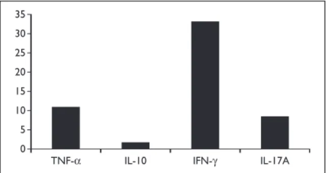
Patients and Methods: Levels of a panel of cytoki-nes and chemokicytoki-nes (1, 2, 4, 6, 8, IL-10, IL-12 (p70), IL-17A, IL-18, IL-22, IL-23, IFN-γ, Leptin, MCP-1, MIP-1α, OPG, TGF-β and TNF) were measured in serum samples of patients and controls by FlowCytomix assay kit. IL-21, APRIL and BAFF levels were determined by ELISA. Results: VERA patients have increased levels of IL-1β, IL-6, IL-8, IL-17A, IL-22, APRIL, MCP-1 and Leptin at baseline in comparison with healthy con-trols. Interestingly, IL-1β, IL-8, Leptin and BAFF were higher in VERA as compared to VEA patients. Both VERA and VEA patients have reduced levels of TGF-β when compared to controls. No statistically significant differences could be observed for IL-4, IL-12, IL-18, MIP-1α or OPG. Regarding IL-2, IL-10, IL-23, IFN-γ and TNF the level of production was un-detectable or very low in all early arthritis patients. Conclusion: In very early arthritis the increased production of serum IL-1β, IL-8, IL-17A and MCP-1 could support neutrophil’s and macrophage’s ac-tivation and migration towards the synovium. The elevated IL-6 and APRIL levels generated during the inflammatory process could stimulate and ac-tivate B cells towards the synovial membrane. Fur-thermore, the increase of IL-1β and IL-6 might be related to the early driving of Th17 differentiation observed in these patients. These observations might be of interest for targeting selection of very early arthritis patients.
P22 – CLINICAL ANDDIAGNOSTICVALUE OF
RIBOSOMALP AUTOANTIBODIES INSYSTEMICLUPUS
ERYTHEMATOSUS
Bahloul Z.,1Fourati H.,2Hachicha J.,3Haddouk S.,2 Marzouk S.,1Masmoudi H.,2Samoud S.4
1. Dept. of Medicine, Hedi Chaker University Hospital, Sfax
2. Dept. of Immunology, Habib Bourguiba University Hospital, Sfax
3. Dept. of Nephrology, Hedi Chaker University Hospital, Sfax
4. Dept. of Immunology, Farhat Hache University Hospital, Sousse, Tunisia
Introduction: Although antibodies to ribosomal P antibodies have only been recognized since 1985, many recent research developments have served o highlight the special significance of these auto-antibodies.
sen-sitivity and specificity, as well as the clinical rele-vance of ribosomal P autoantibodies (anti-P) in a large cohort of systemic lupus erythematosus (SLE) patients.
Patients and Methods: Anti-P were evaluated in the serum of 200 Tunisian SLE patients at disease onset and 130 various control subjects by a sensi-tive immunodot assay. A complete laboratory eva-luation and clinical examination were performed in each SLE patient. During the follow-up, the pa-tients were regularly monitored for clinical para-meters. Global SLE activity was measured by the European Consensus Lupus Activity Measure (ECLAM).
Results: The sensitivity and specificity of anti-P testing for SLE were 23.5 % and 98.4%, respectively. 14/47 (29.8 %), 27/47 (57.4 %), and 5/47 (10.6 %) anti-P positive samples were negative for anti-ds-DNA, anti-Sm, or both antibodies, respectively.
The anti-P positive patients showed more acti-ve disease activity and a much higher prevalence of arthritis. An association between IgG anticardi-olipin antibodies and anti-P was also found. Howe-ver, anti-P were not associated with neuropsychia-tric manifestations or lupus nephritis.
Conclusion: This study does not seem to confirm the described association of anti-P with neu-ropsychiatric manifestations of SLE. However, it supports the anti-P association with arthritis and disease activity as well as the presence of anticar-diolipin antibodies.
Based on our study and other related studies, we propose that, akin to Sm and antidsDNA, anti-P antibodies detected by one agreed method may be considered for inclusion as a criterion for the classification of SLE.
References
1. Elkon KB, Parnassa AP, Foster CL. Lupus autoantibo-dies target ribosomal P proteins. J Exp Med 1985; 162: 459-71.
2. Sato T, Uchiumi T, Ozawa T, et al. Autoantibodies against ribosomal proteins found with high fequency in patients with systemic lupus erythematosus with active disease. J Rheumatol 1991; 18: 1681-1684. 3. Bonfa E, Golombek SJ, Kaufman LD et al. Association
between lupus psychosis and anti-ribosomal P pro-tein antibodies. N Engl J Med 1987; 317: 265-271. 4. Mahler M, Kessenbrock K, Szmyrka M et al.
Interna-tional multicenter evaluation of autoantibodies to ri-bosomal P proteins. Clin Vaccine Immunol 2006; 13: 77-83.
5. Ghirardello AL, Doria A, Zampieri S, Gerli R, Rapizzi E, Gambari PF. Anti-ribosomal P protein antibodies
detected by immunoblotting in patients with con-nective tissue diseases: their specificity for SLE and association with IgG anticardiolipin antibodies. Ann Rheum Dis 2000; 59: 975-981.
6. Yoshio T, Hirata D, Onda K, Nara H, Minota S. Antiri-bosomal P protein antibodies in cerebrospinal fluid associated with neuropsychiatric systemic lupus erythematosus. J Rheumatol 2005; 32: 34-39. 7. Chandran V, Upadhyaya SD, Haroon N, Aggarwal A,
Misra R. Lack of clinical association with antibodies to ribosomal P proteins in Indian patients with syste-mic lupus erythematosus. J Rheumatol 2006; 33: 1987-1989.
8. Caponi L, Chimenti D, Pratesi F, Migliorini P. Anti-ri-bosomal antibodies from lupus patients bind DNA. Clin Exp Immunol 2002; 130: 541-547.
P23 – DENDRITICCELLCHANGES INRA SYNOVIAL
TISSUE AFTERINFLIXIMABTREATMENT
Lebre M.C., Oliveira A.S.F., Ramos M.I., Wijbrandts C.A., Aarrass S., Tak P.P.
Div. Clinical Immunology/Rheumatology, AMC/ /University of Amsterdam, Amsterdam, The Netherlands
Purpose: Tumor necrosis factor-alpha (TNF-α) blockade using infliximab, a chimeric anti-TNF-α antibody, is an effective treatment for psoriatic arthritis (PsA) and rheumatoid arthritis (RA). It has previously been shown that infliximab reduces the synovial infiltrate very early after initiation of tre-atment. Dendritic cell (DC) subsets are critically in-volved in RA pathogenesis. It is at present unknown whether anti-TNF therapy exerts its effects in part via an effect on synovial DC subsets, taking into ac-count that myeloid DC (mDC) might have a more inflammatory role in RA compared to plas-macytoid DC (pDC). Therefore, we investigated whether infliximab treatment changes the num-bers of synovial DC subsets (mDC and pDC). Methods: Thirteen patients with active RA were ran-domized to received either a single infusion of infli-ximab (3 mg/kg) (n=7) or placebo (n=6) intrave-nously. All patients were subjected to an arthrosco-pic synovial biopsy immediately before initiation, and 48 hour as well as 28 days after initiation of tre-atment. Immunohistochemical analysis was per-formed to analyze the inflammatory infiltrate (CD3 and CD68) and CD1c+(mDC and CD304+pDC.
Stained tissue sections were quantified by digi-tal image analysis.
Results: There was a highly significant reduction of synovial CD1c+mDC 28 days after initiation of in-fliximab treatment (from [mean±SEM] 6782±3304
cells/mm2to 602±141 cells/mm2, P=0.0047). We found already a clear trend 48h after infusion (1469±426 cells/mm2, P=0.1375). In contrast, the-re wethe-re no statistically significant changes in the numbers of CD304+synovial pDC after infliximab treatment (at baseline: 4609±1413 cells/mm2, at 48h: 3687±426 cells/mm2, at 28d: 1787±494 cells/mm2; P=0.5338 and P=0.1014, respectively). Conclusions: These findings suggest that inflixi-mab treatment exerts a prominent effect on the findings suggest more inflammatory mDC subset compared to pDC, resulting in immunomodula-tion and resoluimmunomodula-tion of inflammaimmunomodula-tion.
P24 – SPLENICDC SUBSETSDURING
COLLAGEN-INDUCEDARTHRITIS INMICE:AROLE FOR
INFLAMMATORYCONVENTIONALDC?
Lebre M.C.,1Bevaart L.,2Ramos M.I.,1Aarrass S.,1 Koepke J.,2Tak P.P.1
1. Div. of Clinical Immunology/Rheumatology, AMC/University of Amsterdam, Amsterdam 2. Arthrogen BV, Amsterdam, The Netherlands
Purpose: Dendritic cells (DC) play a pivotal role in the orchestration of T cell immunity and toleran-ce due to their ability to stimulate naive T toleran-cells and direct effector cell function.
Immunomodulated and tolerogenic DC could be used to ameliorate arthritis. Therefore, in order to gain insight into the characteristics of DC sub-sets in murine collagen-induced arthritis (CIA) we analyzed the frequencies and phenotype of con-ventional (c)DC and plasmacytoid (p)DC in a mu-rine C57Bl/6 CIA model during different stages of the arthritis process.
Methods: Arthritis was induced in female C57Bl/6 mice on day 0 (i.d. injection of a chicken colla-gen/CFA emulsion at the base of the tail) and on day 21 i.d. injection with this emulsion was repe-ated. Mice were inspected daily from day 20 on for signs of arthritis by two independent observers. Clinical scores were assigned using an established method. At different time points (at days 20, 30, 41 and 63 after CIA induction), mice were sacrificed and spleens were collected. The frequency and phenotype of cDC and pDC was assessed by FACS using specific antibodies: CD11c (total cDC), CD8α (to distinguish 2 cDC populations: CD8α+and CD8α-) and PDCA-1 together with B220 and Ly-6C (pDC). In addition, isolated cDC and pDC were sti-mulated for 48h with LPS or CpG, respectively, and
cell-free supernatants were analyzed for the con-tents of inflammatory cytokines using a cytokine-bead assay (CBA).
Results: As expected clinical signs of arthritis star-ted at day 22 (mean±SEM, 0.125±0.125). On days 30, 41 and 63 the mean clinical scores were 1.000±0.555, 2.563±0.922 and 6.500±0.945 respec-tively. At all the time points studied, the frequen-cies of splenic cDC (total CD11c+, CD8α+or CD8α) exceeded significantly those of pDC (PDCA-1+B220+Ly-6C+) except CD8α-at days 20 and 30. Wi-thin CIA mice the frequencies of CD8α-increased significantly (compared to day 20) starting from day 30 while the frequencies of CD8α+and pDC were significantly increased on day 63 only. When all the DC subsets from CIA mice were compared to those present in mice without CIA the frequen-cies of total CD11c, CD8α+, CD8α-and pDC were significantly increased on day 63. Interestingly, wi-thin CIA mice activated cDC produced significantly higher levels of IL-6 on days 41 and 63 while TNF-α was increased only on day 63 (compared to day 20). In contrast, IL-6 and TNF-α-derived from pDC decreased on day 63.
Conclusions: The observation that cDC subsets and their inflammatory cytokines are significantly increased during the development of CIA and that pDC-derived inflammatory cytokines are decrea-sed suggests an inflammatory role for cDC and an regulatory role for pDC in the arthritic process. Thus, cDC rather than pDC may represent an im-portant therapeutic target in arthritis.
P25 – MODULATINGTLR RESPONSES INSYSTEMIC
SCLEROSISVIAHEMEOXYGENASE-1
van Bon L.,1Scharstuhl A.,2Pennings B.C.W.,2 Hu-ijbens R.J.F.,1Wenink M.H.,1Santegoets K.C.M.,1 Vonk M.,1van den Berg W.,1Wagener F.A.D.T.G.,2 Radstake T.R.D.J.1
1. Dept. of Rheumatology
2. Dept. of Pharmacology and Toxicology, Radboud University Nijmegen Medical Centre, Nijmegen, The Netherlands
Background: Systemic sclerosis (SSc) is an autoim-mune disease characterized by fibrosis of the skin and the internal organs. The etiology is still un-known but prominent features are vascular injury and chronic inflammation resulting in fibrosis. Ac-cumulating evidence suggests a role for Toll-like re-ceptor (TLR) mediated activation of dendritic cells